Optic Nerve
Introduction
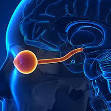
The optic nerve is the second cranial nerve (CN II) that transmits visual information. The optic nerve has only afferent (sensory) fibres and, like all cranial nerves, is paired. During embryogenesis, the optic nerve develops in the retina, exits the orbit via the optic canal, and travels throughout the central nervous system.
The optic nerve’s synaptic targets include the suprachiasmatic nucleus (SCN), the lateral geniculate nucleus (LGN), the pretectal nucleus, the superior colliculus, and the primary visual cortex. The stimulation of these various structures produces different functions. Additionally, the optic nerve acts as an afferent limb for the pupillary light and accommodation reflexes.
The optic nerve originates in the back of the eye. This particular pair of cranial nerves is the second in a series. The optic nerve uses electrical impulses to transfer visual information to the retina to the brain’s vision centres.
The optic nerve is composed of ganglionic or nerve cells. It contains over a million nerve fibres. Our blind spot is caused by the lack of specialised photosensitive (light-sensitive) cells, or photoreceptors, in the part of the retina where the optic nerve exits the eye.
Anatomy
The retinal ganglion cells’ axons (nerve fibres) make up the majority of the optic nerve. The optic disc, also known as the nerve head, is where the axons of the retinal ganglion cells leave the eye.
The nerve head is visible as a white circular structure in the back of the eye. There are no photoreceptors in this structure. As a result, humans possess a natural blind spot.
The lamina cribrosa, a structure that allows nerve fibres to pass through multiple holes and enter the extraocular (outside of the eyeball) space, is the mechanism by which nerve cells exit the nerve head. As the fibres pass through, they are covered in a type of insulation known as myelin. Oligodendrocytes, a type of glial cell, insulate nerve fibres.
Embryology
The optic stalk appears during the fourth week of gestation and eventually develops into the optic nerve. The lumen of the optic stalk connects the diencephalon of the forebrain proximally and the optic vesicle distally. The lateral walls of the optic vesicle invaginate, forming the optic cup that eventually develops into the retina. Invagination also occurs on the inferior surface of the optic stalk and vesicle, resulting in the choroidal fissure. The hyaloid artery and vein are located within this fissure and will eventually fuse to form the central retinal artery and vein.
The optic nerve is a CNS structure that develops from the diencephalon. The optic nerve is a CNS structure that is myelinated by oligodendrocytes and encased in three layers of meningeal tissue. As previously discussed, myelination begins centrally and ends at the lamina cribrosa.
Almost all optic nerve fibres will be myelinated during or soon after birth. The three meningeal layers surrounding the optic nerve are connected to the three meningeal layers of the brain. As a result, increased intracranial pressure can spread from the intracranial subarachnoid space to the perineural subarachnoid space, resulting in papilledema visible on fundoscopy.
Anatomical Course of the optic nerve
The anatomical course of the optic nerve describes how special sensory information is transmitted from the retina of the eye to the brain’s primary visual cortex. It is divided into intracranial and extracranial components, which are located outside the cranial cavity.
Extracranial
The optic nerve develops while the axons of retinal ganglion cells converge. These cells receive impulses from the eye’s photoreceptors (rods and cones).
Following its formation, the nerve exits the bony orbit via the optic canal, which runs through the sphenoid bone. It enters the cranial cavity and runs along the middle cranial fossa (near the pituitary gland).
Intracranial (the Visual Pathway)
The optic chiasm exists whenever the optical nerves of each of the eyes meet in the middle cranial fossa. At the chiasm, fibres from the nasal (medial) half of each retina cross over to the contralateral optic tract, whereas fibres from the temporal (lateral) halves remain ipsilateral:
- The left optic tract contains fibres from both the left temporal (lateral) and right nasal (medial) retinas.
- The right optic tract contains fibres from both the right temporal and left nasal retinas.
Before arriving at the lateral geniculate nucleus (LGN), a system of relays in the thalamus where fibres synapse, each optic tract is connected to its corresponding cerebral hemisphere.
Axons from the LGN then carry visual information along a pathway known as optic radiation. The route itself is separated into:
- Fibres coming from superior retinal quadrants—which correspond to the inferior visual field quadrants—are transported by upper optic radiation. It travels from the parietal lobe to the visual cortex.
- Fibres enter the inferior retinal quadrants (corresponding to the superior field of vision regions) when optic radiation levels are lower. It travels through the temporal lobe, via Meyers’ loop, to the visual cortex.
When in the visual cortex, the brain processes sensory information and reacts appropriately.
Blood supply
The ophthalmic artery (OA) is the primary artery that supplies the optic nerve and is the first branch of the internal carotid artery (ICA). The OA emerges from the ICA just distal to the cavernous sinus and flows through the optic canal. Within the optic canal, the OA runs inferior and lateral to the optic nerve. The OA has numerous branches, but the two primary ones are the central retinal artery (CRA) and the posterior ciliary arteries (PCAs).
The CRA is the first branch of the OA and runs within the optic nerve’s dura mater. The CRA then penetrates the optic nerve approximately 12 mm posterior to the globe and travels anteriorly to perfuse the inner layers of the retina. The OA results in several PCAs. The PCAs travel anteriorly and penetrate the sclera, perfusing the optic nerve and posterior uveal tract.
Function
The typical function of the optic nerve is to deliver signals from the eye to the brain, acting as a messenger that helps us understand what we see.
When light enters the eye, the cornea attempts to focus it directly on the retina to produce the clearest image. The retina detects light and uses it to generate impulses or currents. The optic nerve receives the current and transports it to the brain, where it is registered as an image.
While the optic nerve appears to have a simple function, it is an essential component of our ability to make sense of our surroundings. It is so important that scientists are working to develop a machine that can perform the same function as the optic nerve to restore vision to people who have had their optic nerves damaged.
The optic nerve also controls the light reflex and the accommodation reflex.
Pupillary Light Reflex.
The optic nerve is the afferent limb of the pupillary light reflex. When light enters the eye, photoreceptors become activated and transmit the signal to the retinal ganglion cells. The nasal retinal fibres transmit the stimulus to the contralateral pretectal nucleus, while the temporal retinal fibres transmit it to the ipsilateral pretectal nucleus.
The nerve fibres in the two pretectal nuclei turn bilaterally on neurons in the Edinger-Westphal nucleus, triggering the reflex’s efferent limb. The branches of these preganglionic parasympathetic neurons innervate the ciliary ganglion by travelling along the periphery of the oculomotor nerve (CN III).
The short ciliary nerves are composed of the ciliary ganglion’s postganglionic parasympathetic axons. The short ciliary nerves stimulate the sphincter pupillae to constrict, resulting in miosis. Because both the pretectal nuclei and the Edinger-Westphal nuclei are activated bilaterally, stimulating one eye with light causes pupillary constriction in the ipsilateral eye (direct response) and in the contralateral eye (consensual response).
Accommodation Reflex
The accommodation reflex is triggered by shifting focus to a nearby object. The optic nerve serves as the afferent limb of this reflex. Unlike the pupillary light reflex, an afferent stimulus must be relayed through the visual pathway to the primary visual cortex and visual association areas. The neurons situated in the visual association regions stimulate the Edinger-Westphal nucleus and the oculomotor nucleus in the midbrain, activating the reflex’s efferent limb.
Similar to the pupillary light reflex, pupillary constriction is caused by the activation of the previously described two-neuron system. In addition to stimulating the sphincter pupillae, the short ciliary nerves innervate the ciliary muscle.
The ciliary muscle contracts, causing the zonular fibres attached to the lens to relax. This relaxation causes the axial thickness to increase, resulting in higher refractive power. Finally, the somatic fibres of the oculomotor nerves stimulate the medial rectus muscle.
Contraction of the medial rectus muscles causes the eyes to adduct, which coincides with convergence. Thus, the accommodation reflex consists of miosis, an increase in lens refractive power, and convergence.
Causes
Such conditions may affect the optic nerve and impair vision.
- Glaucoma: Fluid buildup in the front of your eye causes pressure on the optic nerve. The pressure harms your optic nerve. Glaucoma is the leading cause of blindness among adults over the age of sixty.
- Anterior ischemic optic neuropathy is caused by a loss of blood flow to the optic nerve. The condition results in sudden vision loss.
- Congenital abnormalities: Some babies are born with differences in their optic nerve(s), which can cause poor vision.
- Optic atrophy occurs when the optic nerve does not receive enough blood. Trauma, strokes, hydrocephalus, infections, and brain tumours can all cause optic atrophy. Certain cases are inherited.
- Optic nerve coloboma is an inherited condition in which one or both optic nerves do not develop normally.
- Optic nerve drusen: This condition develops when protein and calcium deposits (drusen) accumulate on the optic nerve.
- Gliomas are tumours (growths) of the optic nerve. They are usually benign (not cancerous). These tumours frequently affect people with an inherited condition known as neurofibromatosis type 1 (NF1).
- Optic nerve meningiomas: Although rare and benign, these slow-growing tumours can cause severe vision loss.
- Optic neuritis occurs when infections or autoimmune diseases such as multiple sclerosis irritate or inflame the optic nerve.
- Papilledema occurs when the optic nerve swells due to pressure around the brain caused by a traumatic brain injury, brain tumours, meningitis, or another condition.
- NMO, also known as Devic’s disease, is a condition in which the immune system mistakenly attacks the optic nerves and spinal cord.
Symptoms
Optic nerve problems cause a variety of symptoms depending on the underlying condition. The symptoms could be temporary or permanent. You might experience:
- Colour blindness.
- Eye pain.
- Headaches.
- Nausea and vomiting.
- Night blindness.
- Partial or total vision loss.
- Peripheral (side) vision loss.
- Tinnitus, or ringing in the ears.
Assessment
The optic nerve is not assessed separately from other cranial nerves involved in vision (such as CN III, IV, and VI). Still, particular tests for the optic nerve consist of visual acuity, colour perception, visual fields, pupillary light reflexes, accommodation, and funduscopic examination.
Treatment
The treatment of optic nerve, chiasma, and optic radiation damage is determined by the cause. However, treatments for optic nerve damage may not restore lost vision. In most cases, precautions are taken to prevent further damage and worsening of symptoms. For example:
Glaucoma is caused by increased pressure inside the eye, so medications for glaucoma are designed to reduce the pressure to the point where the disease process stops. Although glaucoma can be treated with surgery, lasers, and oral medications, the majority of cases are treated with topical medication in the form of eye drops.
Oral and intravenous steroids are used to treat diseases like optic neuritis, which causes inflammation. Additionally, if the cause of the optic neuritis is identified, the underlying condition will be treated.
Diseases of the optic chiasm are frequently treated with neurosurgery and medication or hormones. Depending on the severity of optic chiasm disease, such as a pituitary adenoma, simple observation may be sufficient.
Vascular accidents, or strokes, are more difficult to treat unless the condition is detected quickly. Occasionally, blood thinners are prescribed. Surgery may be required if the disease is caused by aneurysms.
Surgical requirements
Fenestration of the optic nerve sheath is a widely accepted treatment for benign intracranial hypertension. Optic nerve sheath meningioma and optic nerve glioma may require surgical intervention in some cases.
Protection
To care about your vision, take the following steps:
- Get regular eye examinations.
- Maintain a healthy weight by exercising and eating a nutritious diet.
- Diabetes and high blood pressure are two conditions that can impair vision and nerve function.
- Seek help for quitting smoking. Smoking heightens the risk of optic nerve damage and other vision issues.
- Wear sunglasses and eye protection (goggles or safety glasses) when participating in sports or activities that may harm your eyes.
Summary
The optic nerve is the second cranial nerve (CN II) that transmits visual information. It originates in the back of the eye and uses electrical impulses to transfer visual information to the retina and the brain’s vision centres. The optic nerve is composed of ganglionic or nerve cells and contains over a million nerve fibers. The optic disc, or nerve head, is where the axons of retinal ganglion cells leave the eye. The optic nerve is myelinated by oligodendrocytes and encased in three layers of meningeal tissue.
The optic nerve’s anatomical course describes how special sensory information is transmitted from the retina to the brain’s primary visual cortex. Blood supply to the optic nerve is provided by the ophthalmic artery (OA), which is the first branch of the internal carotid artery (ICA).
The optic nerve is responsible for delivering signals from the eye to the brain, helping us understand our surroundings. It controls the pupillary light reflex and the accommodation reflex, which are triggered by shifting focus to a nearby object.
Various conditions can affect the optic nerve, impairing vision, such as glaucoma, anterior ischemic optic neuropathy, congenital abnormalities, optic atrophy, optic nerve coloboma, optic nerve drusen, gliomas, optic nerve meningiomas, optic neuritis, papilledema, and NMO. Symptoms can include colour blindness, eye pain, headaches, nausea, vomiting, night blindness, partial or total vision loss, peripheral vision loss, and tinnitus.
Treatment for optic nerve damage is determined by the cause, but precautions are taken to prevent further damage and worsening of symptoms. For example, glaucoma can be treated with surgery, lasers, and oral medications, while optic neuritis can be treated with neurosurgery and medication.
Protection for vision includes regular eye examinations, maintaining a healthy weight, quitting smoking, and wearing sunglasses and eye protection.
FAQs
What nerve has been damaged in the optic?
Optic neuritis develops when swelling (inflammation) damages the optic nerve, a bundle of nerve fibres that transmits visual information from the eye to the brain. Optic neuritis can cause pain with eye movement and temporary vision loss in one eye.
Which is the most common optic nerve disorder?
Glaucoma is the most common optic nerve disorder, affecting over 3 million people each year in the United States.
How many optic nerves exist in the eye?
The junction of the two optic nerves is called the optic chiasm. The optic nerve from each eye splits at this point, and half of the nerve fibres from each side cross over to the other.
What is the location of the optic nerve?
The axon to the retinal ganglion cells converges to create the optic nerve. These cells receive impulses from the eye’s photoreceptors (rods and cones). Following its formation, the nerve exits the bony orbit via the optic canal, which runs through the sphenoid bone.
Which nerve connects the eye and brain?
The optic nerve is a bundle of over 1 million nerve fibres that transmit visual signals. You have one that connects the back of each eye (the retina) to your brain. Loss of vision may occur from damage to the optic nerve.
What is the main function of the optic nerve?
Visual images are created by relaying messages from your eyes to your brain via the optic nerves. They play a critical role in your vision. Each optic nerve consists of millions of nerve fibres.
References:
- Smith, A. M., & Czyz, C. N. (2022, November 7). Neuroanatomy, Cranial Nerve 2 (Optic). StatPearls – NCBI Bookshelf. https://www.ncbi.nlm.nih.gov/books/NBK507907/#:~:text=The%20optic%20nerve%20is%20the,all%20cranial%20nerves%20is%20paired.
- Professional, C. C. M. (n.d.). Optic Nerve. Cleveland Clinic. https://my.clevelandclinic.org/health/body/22261-optic-nerve
- Optic nerve. (2018, January 21). Healthline. https://www.healthline.com/human-body-maps/optic-nerve#1
- Optic Nerve. (n.d.). Physiopedia. https://www.physio-pedia.com/Optic_Nerve
- The Optic Nerve – Visual Pathway – Chiasm – Tract – TeachMeAnatomy. (2022, December 16). TeachMeAnatomy. https://teachmeanatomy.info/head/cranial-nerves/optic-cnii/
- Bedinghaus, T. (2023, May 28). The Anatomy of the Optic Nerve. Verywell Health. https://www.verywellhealth.com/optic-nerve-anatomy-4686150
- Sprabary, A. (2021, February 16). Optic nerve: Anatomy, function and conditions. All About Vision. https://www.allaboutvision.com/eye-care/eye-anatomy/optic-nerve/
2 Comments