Deep Brain Stimulation: 5 Key Benefits Explained
Deep brain stimulation (DBS) involves putting electrodes into particular areas of the brain. Electrodes provide electrical impulses that modify brain activity in order to treat medical conditions. Electrical impulses can also affect cells and chemicals in the brain, causing medical issues.
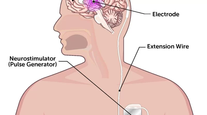
A pacemaker-like device controls the degree of electrical activity during deep brain stimulation. The device is put beneath the skin in the upper chest region. This gadget is attached to the electrodes in the brain via an under-the-skin connection.
Deep brain stimulation is widely used to treat a variety of disorders, including:
- Parkinson’s disease.
- Essential tremor.
- Dystonia is a group of conditions that cause muscle contraction, including Meige syndrome.
- Epilepsy.
- Tourette syndrome involves repeated actions and sounds.
- Obsessive–compulsive disorder.
Deep brain stimulation is also being investigated as a potential therapy for:
- Long-term pain and discomfort
- Cluster headaches.
- Dementia.
- Depression.
- Addiction.
- Obesity.
What is deep brain stimulation?
- Deep brain stimulation involves placing electrodes in a specific area of the brain to activate the symptoms being treated.
- The electrodes are placed on the left and right sides of the brain through tiny holes in the skull.
- The electrodes are connected by lengthy cables that run beneath the skin and down the neck to a battery-powered stimulator beneath the chest.
- When activated, the stimulator sends electrical pulses to inhibit abnormal nerve impulses that cause tremors, stiffness, and other symptoms.
The components of the DBS system—
An electrode, also known as a lead, is a short, insulated wire that is introduced into a specific location of the brain by a tiny incision in the skull.
The extension cable is also insulated and extends beneath the skin on the head, neck, and shoulders, connecting the electrode to the internal pulse generator (IPG).
The third component of the system, the IPG, is usually implanted beneath the skin in the upper chest.
Anatomy
The DBS system consists of electrodes placed near specific deep-brain areas, which are then connected to a pacemaker-like device (pulse generator) inserted into the chest wall via a subcutaneous wire. A computer then sends stimulation settings to the pulse generator, which changes the amplitudes, frequencies, and pulse width appropriately. DBS typically targets the subthalamic nucleus, globus pallidus interna, and ventral intermediate nucleus of the thalamus.
The precise mechanism of deep brain stimulation’s therapeutic effects is unknown, but some studies suggest that high-frequency stimulation (HFS) in the subthalamic nucleus reduces the firing rate of STN neurons, thereby suppressing symptoms in neurologic conditions such as Parkinson’s. However, several DBS imaging and physiologic investigations suggest that the final consequence of deep brain stimulation is an increase in the firing rate of the targeted neurons.
The figure on the right is a reconstruction of bihemispheric DBS electrodes surgically implanted into the subthalamic nucleus (orange), the most frequent target structure for Parkinson’s disease therapy. Other subcortical structures include the red nucleus (green), substantia nigra (yellow), internal and external pallidum, and striatum (red).
Uses of the DBS system:
Deep brain stimulation is commonly used to treat a variety of disorders, including:
- Parkinson’s disease.
- Essential tremor.
- Dystonia
- Epilepsy
- Obsessive–compulsive disorder
Deep brain stimulation is also under investigation as an essential therapy for:
- Tourette Syndrome
- Huntington’s Disease and Chorea
- Chronic pain.
- Cluster headache.
Symptoms-
Each illness kind has a unique set of symptoms that patients may experience. Frequent ones are:
Dystonia
- Involuntary muscle contractions occur during certain tasks. For example, writing.
- Muscle contractions worsen with tension, weariness, or worry.
Epilepsy
- Temporary disorientation.
- A gazing spell.
- Loss of consciousness.
- Uncontrollable jerking motions in the arms and legs
- Emotional reactions such as dread and worry.
- Tremors occur during ordinary tasks like writing or drinking.
Obsessive-Compulsive Disorder-
- Fear of germs and pollution.
- Aggressive feelings against others or oneself
- Having certain objects in perfect or symmetrical order.
- Intensive cleaning or handwashing
- Compulsive counting.
- Checking items frequently (for example, the oven is turned off, the door is locked, etc.).
Parkinson’s disease causes
- Tremors.
- Bradykinesia is the slowing down of movement.
- Stiffness
- Abnormal walking.
Testing & Diagnosis-
- A multidisciplinary team of executives, which includes a neurologist, neurosurgeon, neuropsychologist, and psychiatrist, may care for patients.
- Patients with Parkinson’s disease or tremors may be examined for motor symptoms both on and off medication to establish the severity of their illness.
- Electroencephalography, a more advanced sort of testing, can be used to detect epilepsy.
- A few patients may be subjected to neuropsychological testing during their examination.
- Patients with obsessive-compulsive disorder must take the Yale-Brown Obsessive Compulsive Scale (YBOC) test.
Before surgery, patients go through the following:
Blood and urine testing.
This aids in detecting poisons and abnormalities.
MRI and CT scans.
Imaging may enable clinicians to target the appropriate brain location for symptom alleviation.
Medical clearance.
Criteria for DBS System-
- Symptoms significantly reduce quality of life.
- Despite obtaining the appropriate drug dose, symptoms are not under control.
- Side effects from present drugs cannot be tolerated.
Surgery
How to use the DBS system-
The DBS system is composed of four parts:
- One or more insulated cables known as leads, or electrodes, are placed in the brain.
- Anchors to secure the leads to the cranium. The neurostimulator delivers an electric current.
- The stimulator is similar to a heart pacemaker.
- It is most commonly found beneath the skin below the collarbone; it may also be found elsewhere in the body.
- In certain cases, another thin, insulated wire called an extension is used to link the lead to the neurostimulator.
Stage 1
- A small amount of hair on the head is likely shaved.
- The head is secured in a specific frame with microscopic screws to keep it motionless during the surgery.
- Where the screws come into contact with the scalp, numbing medication is used.
- Sometimes the treatment is performed in the MRI machine, with a frame on top of the head rather than around it.
- The surgeon injects numbing medication into the scalp at the region where the skin will be opened, then drills a small aperture in the skull and inserts the lead into a specific area of the brain.
- If both sides of the brain are being treated, the surgeon creates an aperture on either side of the skull and inserts two leads.
- Electrical impulses may need to be delivered via the lead to guarantee that it is connected to the area of the brain producing the symptoms.
- People may ask questions, read cards, and describe visuals.
- The individual may also be requested to move their legs or arms.
- These are to ensure that the electrodes are in the appropriate places and that the anticipated impact is achieved.
Stage 2
Stage 2 involves general anesthesia, which allows for pain-free sleep. The time of this round of surgery depends on where the stimulator will be inserted in the brain.
- The surgeon creates a tiny hole (incision) just below the collarbone and inserts the neurostimulator.
- (Sometimes it is located beneath the skin in the lower chest or abdominal area).
- The extension wire is inserted beneath the skin of the head, neck, and shoulder and attached to the neurostimulator.
- The incision is closed.
- The device and cables are located beneath the skin and may seem like a little bump.
- Once connected, electric pulses go from the neurostimulator to the extension wire, the lead, and finally into the brain.
- These tiny pulses interfere with and disrupt the electrical impulses that generate symptoms in some disorders.
Advantages of surgery-
- Depending on the symptoms, it can be conducted on either one or both sides of the brain.
- The effects are reversible and can be adjusted to each patient’s specific situation.
- Stimulation settings can be adjusted to reduce potential negative effects and increase efficacy over time.
- The gadget may provide continuous symptom management 24 hours a day.
- Patients who have had DBS may be able to engage in other therapies, such as stem cell or gene therapy, when they become available.
Risk factors of surgery
Deep brain stimulation involves making small holes in the skull to insert electrodes into brain tissue, as well as performing surgery to implant the device that contains the batteries beneath the skin in the chest.
Complications of surgery-
- Misplacement of Leads
- Bleeding in the brain.
- Stroke Infection
- Breathing issues
- Nausea
- Heart issues.
- Seizure
Side effects after surgery-
- Possible adverse effects following surgery
- Seizure Infection
- Headache
- Confusion
- Difficulty focusing
- Stroke
- Hardware problems, such as a corroded lead wire.
- Nonpermanent pain and edema at the insertion site.
- A few weeks following the operation, the device will be turned on, and the process of determining the ideal settings for the patient will begin.
- Some settings can create negative effects, although these often improve with additional modifications to patients’ devices.
- Because there have been uncommon reports that DBS treatment impairs the movements required for swimming, the Food and Drug Administration recommends visiting a doctor and following water safety precautions before swimming.
FAQs
How effective is deep brain stimulation?
Patient satisfaction remained high (92.5% were happy with DBS, 95% would suggest treatment, and 75% considered it offered symptom relief).
How does deep brain stimulation feel like?
During typical usage, most people experience little to no feeling. For those who do, it is described as a little tingling feeling down an arm or leg, or a faint feeling in the face that goes away. This is more likely in people who use DBS for critical tremors, as the device may be switched off before bedtime.
What are the disadvantages of DBS?
Concentrating is difficult. Stroke. Hardware issues, such as a damaged lead wire. Pain and edema at the insertion site are only temporary.
References:
- https://www.mayoclinic.org/tests-procedures/deep-brain-stimulation/about/pac-20384562
- https://www.physio-pedia.com/Deep_Brain_Stimulation