Pseudarthrosis (Nonunion Fracture)
Introduction
Pseudarthrosis, also known as a nonunion fracture, is a condition in which a broken bone fails to heal properly, resulting in persistent movement at the fracture site that resembles a false joint.
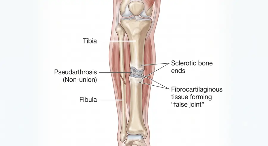
Pseudoarthrosis, sometimes referred to as a “fake joint,” occurs when a bone fracture fails to unite and instead produces fibrous or fibrocartilaginous tissue between the bone ends, giving the appearance and restricted movement of a joint.
It is regarded as a nonunion and a late consequence of the fracture healing process. Inadequate stabilisation, poor blood flow, infection, or other systemic and local causes might disrupt the normal bone repair process, leading to the disease.
At the affected spot, pseudoarthrosis may result in discomfort, abnormal movement, and functional impairment. Long bones like the clavicle and tibia are where it is most frequently seen. While management typically necessitates surgical intervention such as bone grafting and internal fixation to restore stability and encourage appropriate bone healing, diagnosis is mostly based on clinical examination and radiographic findings.
Pathophysiology:
- Bone healing often takes place through callus development and remodelling following a fracture.
- This process ends before full union in pseudoarthrosis.
- Instead of bone bridging at the fracture site, cartilage or fibrous tissue is present.
- A little “joint-like” hollow may form between the rounded, sclerotic (hardened) ends of the bone.
Etiology
Numerous causes that impede the normal healing process can result in pseudoarthrosis. Among them are:
- Repeated micro-movements or insufficient stabilisation
- The fracture site’s poor vascularity
- Osteomyelitis infection
- Loss of bone or severe comminution
- Inappropriate surgical methods
- Systemic diseases like starvation, smoking, and diabetes
- Extended usage of corticosteroids or NSAIDs
Causes
A delayed or unsuccessful healing process following a bone fracture is the most frequent cause of bone fracture nonunion.
Surgical failure: Following a spinal fusion procedure meant to fuse vertebrae, it may also happen.
Congenital conditions: Congenital pseudarthrosis of the tibia, which is linked to neurofibromatosis type 1, is one example of a rare congenital ailment.
Contributing variables: Smoking, diabetes, advanced age, obesity, and certain medications are additional risk factors.
Symptoms
- Ongoing discomfort at the fracture site.
- swelling.
- A feeling of instability or fake movement.
- Decreased movement and clicking noises.
Clinical Features:
- Chronic pain at the fracture site
- Unusual mobility (movement at the fracture site)
- Deformity or swelling
- Failure to heal after several months
- potential limb instability or shortening
Types:
- Hypertrophic pseudoarthrosis.
- Hypertrophic pseudoarthrosis.
- The bone ends are well vascularized and show excessive callus formation.
- Indicates good healing potential if stabilised properly.
- Cause: usually instability at the fracture site.
Atrophic pseudoarthrosis:
- Limited callus development and poor vascularity.
- Shows a low capacity for biological healing.
- Cause: infection or inadequate blood flow
Common Sites:
- Tibia (most common)
- Clavicle
- Humerus
- Radius/ulna
- Spinal vertebrae (in spinal fusion surgeries)
Risk Factors:
- Inadequate immobilization
- Infection at the fracture location
- Poor blood supply
- Bone loss or a big fracture gap
- Malnutrition or smoking
- Systemic illnesses, such as diabetes
- Use of specific drugs (such as steroids and NSAIDs)
Symptoms:
- Chronic pain at the fracture site
- Abnormal movement (mobility where bone should be solid)
- Tenderness or swelling
- X-ray healing that is either missing or delayed
Complications:
- Chronic pain
- Deformity
- Limb shortening
- Functional disability
Diagnosis:
- X-ray: shows nonunion with rounded bone ends, gap, and possible “pseudo-joint” appearance
- CT scan: gives a detailed view of bone healing and the gap
- MRI: evaluates vascularity and soft tissue around the site
Treatment:
Depends on type and cause:
Conservative (rare):
Only in hypertrophic cases with minor mobility:
- Immobilisation
- Bone stimulator
- Ultrasound therapy.
Surgical:
- Debridement of fibrous tissue
- Grafting bones (autograft or allograft)
- Stability is ensured by internal fastening (plates, rods, nails).
- BMPs, or bone morphogenetic proteins, aid in healing
- Revision fusion surgery in instances involving the spine
Non-surgical:
Bone stimulators: These devices are used for at least 20 to 60 minutes per day to encourage healing using pulsed electromagnetic or ultrasonic waves. Taking care of the root causes, Healing can be improved by controlling diseases like diabetes or anaemia.
Prognosis:
- Excellent when properly managed surgically.
- Chronic discomfort, deformity, or loss of function may result from untreated conditions.
Prevention:
- Appropriate immobilisation and fracture fixation
- Early infection detection
- Maintaining a healthy blood supply and eating well
- During the healing process, abstain from smoking and excessive movement.
FAQs
What is meant by pseudoarthrosis?
When a fracture or surgical fusion fails to heal properly, an aberrant “false joint” made of fibrous tissue rather than solid bone results, a disease known as pseudoarthrosis. This “false joint” is frequently identified by imaging tests like X-rays or CT scans that reveal a lack of bone fusion. It can cause discomfort, swelling, and instability.
How to Avoid Pseudarthrosis
Quit Smoking: Osteoblast function can be enhanced by quitting smoking.
Weight Loss: Achieving optimal bone fusion can also be facilitated by working towards a healthy weight.
What distinguishes pseudoarthrosis from nonunion?
The final stage of nonunion is pseudarthrosis, which is characterised by the formation of a fake joint that is filled with fluid and lined with pseudosynovial cells. If the bone ends are not joined within two years, a pseudarthrosis usually develops.
What side effects may pseudoarthrosis cause?
Instability, chronic discomfort, rod fracture, and restricted mobility can result from pseudoarthrosis. Surgeons can use a variety of techniques to treat pseudoarthrosis and rod breakage. Using multiple-rod structures during spinal fusion procedures is one method.
What side effects may pseudoarthrosis cause?
Instability, chronic discomfort, rod fracture, and restricted mobility can result from pseudoarthrosis. Surgeons can use a variety of techniques to treat pseudoarthrosis and rod breakage. Using multiple-rod structures during spinal fusion procedures is one method.
What is the therapy success rate for pseudoarthrosis?
The type of non-union (infected versus uninfected, atrophic versus hypertrophic) has a significant impact on how pseudarthrosis is managed. The success rate for surgical treatment of pseudarthrosis ranges from 75 to 100%.
What is a pseudoarthrosis bone scan?
A helpful method for diagnosing pseudarthrosis is SPECT-CT. Intense uptake may occur at several levels, such as around screws or cages and in the vertebral endplates. As in our situation, the lack of uptake surrounding a broken screw shouldn’t be regarded as a false negative.
References
- Hellicar, L. (2023, March 29). Pseudarthrosis: Symptoms and treatment. https://www.medicalnewstoday.com/articles/pseudoarthrosis.
- Kotsifaki A, Kalouda G, Maroulaki S, Foukas A, Armakolas A. The Genetic and Biological Basis of Pseudoarthrosis in Fractures: Current Understanding and Future Directions. Diseases. 2025 Mar 3;13(3):75. doi: 10.3390/diseases13030075. PMID: 40136615; PMCID: PMC11941250