Knee Joint Mobilization Technique
Introduction
Knee joint mobilizations, sometimes referred to as nonthrust manipulation, are manual physical therapy procedures. Depending on the patient’s presentation, an oscillatory manual force may be delivered to the patellofemoral, tibiofemoral, or proximal tibiofibular joints during mobilization in several hand positions or grips.
The application of the four types of mobilization grades (Maitland’s Mobilizations) is determined by the degree of movement and resistance, which vary depending on the treatment’s goal.
- Grade I: At the start of the joint’s range of motion, mobilizations in this grade have a small amplitude and sluggish oscillations. Light pressure is used during these mobilizations.
- Grade II: Throughout the joint’s whole range of motion, mobilizations in this grade have a significant amplitude and sluggish oscillations. Movements before attaining joint resistance are referred to as grades I and II. Pain modulation is the aim of these two grades.
- Grade III: This grade is characterized by sluggish oscillations and large-amplitude mobilizations that are concentrated in the middle to end of the range of motion.
- Grade IV: At the conclusion of the range of motion, mobilizations have a tiny amplitude and slow oscillations.
Indications
People with a range of motion affected by a condition may benefit from the knee mobilizations technique. They can be employed in other situations where knee joint therapy is required, such as knee stiffness, pain, and hypo mobility, even though they are frequently used in post-operative hypo mobility and knee osteoarthritis (OA).
Knee mobilizations have been utilized in many trials to treat knee disease. The two in OA with the longest follow-up, up to a year, are:
- Knee OA: Deyle et al. (2000): In 83 patients with knee OA, manual therapy and exercise were compared to a placebo ultrasonography. Patients were monitored for a year after receiving treatment twice a week for four weeks. At four weeks, the manual treatment and exercise group showed a statistically and clinically significant improvement over the placebo ultrasonography group, and these gains persisted at one year. The proximal tibiofibular joint, patellofemoral joint, tibiofemoral joint, and surrounding soft tissue were all mobilized as part of the authors’ impairment-based method. As needed, mobilizations were also administered to the ankle, hip, and lumbar spine.
- Knee OA: Deyle et al. (2005): 134 participants with knee OA were compared to a home exercise program and manual treatment. Pain and function improved with both therapies, but at the one-year mark, the manual therapy and exercise group were more satisfied with the overall results of their treatment and were less likely to be taking medication for their OA. They employed the same methods as in the initial experiment.
A recent systematic review by Weleslassie et al. examined the efficacy of knee mobilizations in knee OA and comprised 15 RCTs with 704 participants. In most studies, there was a significant improvement in both pain reduction and functional improvement compared to the control groups.
Additionally, knee mobilizations for Patellofemoral Pain Syndrome (PFPS) were used in several trials.
Patellar mobilizations and patellar tapping significantly improved the functional outcome of the knee and significantly reduced discomfort in 50 PFPS patients, according to one randomized trial.
- According to the study, treating PFPS with a 12-session regimen that includes both patellar mobilization and tapping in addition to traditional therapy is successful in reducing pain and enhancing knee function.
- Joint mobilizations have been linked to a reduction in aberrant pain mechanisms, and when paired with their biomechanical effects, they may be used as an additional intervention in the treatment of PFPS, according to the findings of another study.
Contraindications
Joint mobilization should not be used in the following situations:
- Joint replacement arthroplasties
- Any fracture next to the joint that hasn’t fully healed
- Acute inflammatory (septic arthritis)
- Bone disease (Osteomyelitis)
- Bacterial infection
- Malignancy and neoplastic disease
- Physiologically unstable joint
Tibiofemoral Articulation
The tibial plateau is concave, whereas the femoral condyles are convex.
- Resting position: 25 degrees of knee flexion when at rest
- The treatment: It is large; the tibial plateaus
- Stabilization: There is stability in the femur bone.
Tibiofemoral joint distraction
- Indications: Pain management, general patient mobility, and initial treatment.
- Patient posture: A stabilizing belt is used to secure the patient’s thigh to the table while they are in a prone position.
- Hand placement and mobilizing force: To administer a distraction force (along the tibia’s long axis), the therapist leans back along the tibia’s line while holding the afflicted leg just proximal to the malleoli.
An alternate position involves the patient either supine or sitting with one leg dangling off a table.
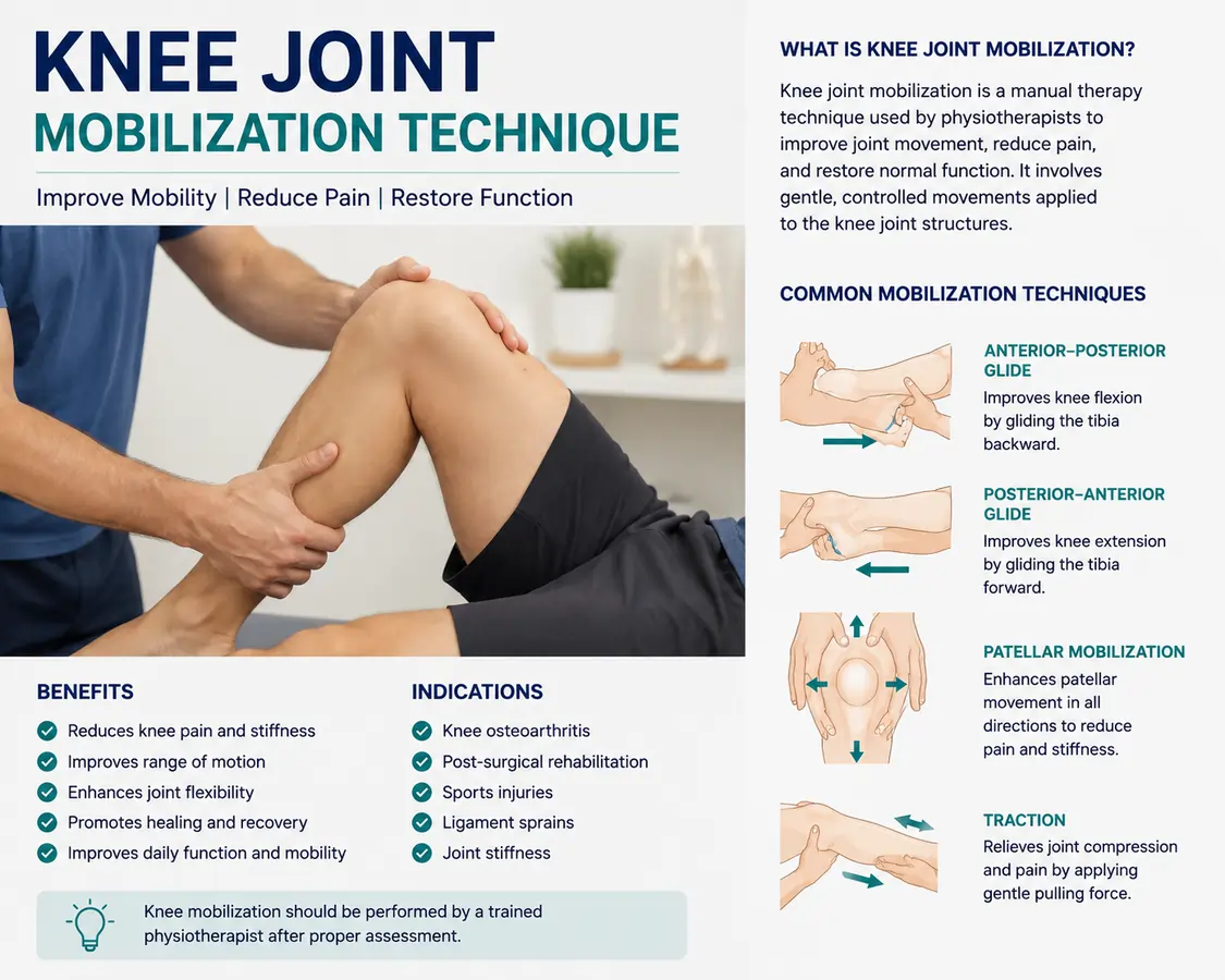
Anterior Glide
Increased knee extension is the indication.
- Patient Position: The drawer test position is crook-lying.
- Hand placement and mobilizing force: As the therapist leans back, the fingers on the posterior tibia provide the mobilizing force.
- Alternatively, place the patient in a prone position. The knee joint is first maintained in a resting posture before being advanced to the full range of motion. To prevent the patella from being compressed, a little pad is applied to it. One hand is used to grasp the distal tibia, and the proximal hand’s palm is positioned over the proximal tibia on one side. Over the tibial plateau, the force is applied medially and laterally.
Posterior Glide
Increasing knee flexion is the indication.
- Position of the Patient: The patient lies supine with a prop positioned beneath the distal femur and the knee slightly flexed.
- Hand positioning and mobilizing force: The actual mobilization is carried out by a force that is perpendicular to the tibia’s line.
Although this technique is commonly used in the closed-packed position, it may be performed with the knee flexed near to the level of limitation, which is similar to the posterior drawer test for the PCL.
Patellofemoral Joint
For normal knee flexion, the patella can glide distally on the femur, and for normal knee extension, it can glide proximally.
Rotational Glides
- Indication: Knee flexion is increased internally. Knee extension is increased by external rotation glides.
- Patient posture: With the patient in a supine posture, these glides can be executed at different positions in the knee’s typical range of motion.
- Hand placement and mobilizing force: The patient’s heel is grasped by the mobilizing hand, while the distal femur is grasped by the stabilizing hand. To apply rotational motion to the rotating tibia rather than to other joints farther away, the ankle is maximally dorsiflexed. Depending on the intended mobilization (internal or external rotation) and at the range where constraint may be visible, the foot is rotated either medially or laterally.
Patellofemoral Glides
Increased patellar mobility is indicated.
- Position of the patient: The patient is lying supine with their knee slightly bent.
- Placement of hands and force mobilization:
- Medial Glide: The therapist glides the patella medially by applying light pressure from the lateral (outside) patellar border.
- Lateral Glide: The therapist glides the patella laterally by applying light pressure from the medial (inside) patellar border.
- Superior Glide: To enhance knee extension, the therapist gently presses forward from the inferior (lower) patellar border.
- Inferior Glide: To increase knee flexion, the therapist gently presses down from the superior (upper) patellar border
FAQs:
What are joint mobilization techniques?
Joint mobilization is a manual therapy technique in which a physical therapist directs your joint’s motion by applying pressure with their hands. They accomplish this by using one hand to stabilize the joint and the other to press, twist, or tug it in a way that prevents it from moving correctly on its own, either softly or more violently.
What is a grade 4 joint mobilization?
A small-amplitude passive movement at the start of the range with no tissue resistance was considered grade I mobilization. A small-amplitude passive movement against tissue resistance at the end of range was known as grade IV mobilization.
Who performs joint mobilization?
Osteopathic doctors, chiropractors, and licensed physical therapists are the main practitioners of joint mobilization, a manual therapy approach. These skilled practitioners expand joint capsules, ease discomfort, and restore range of motion using deliberate, hands-on passive movements.
Is joint mobilization suitable for everyone?
Although mobilization can be used to treat a wide range of injuries and ailments, it is not advised for all diseases. Severe infection or inflammation are two joint diseases that actually make it impossible to perform mobilization treatments.
References
- (2026). Knee Mobilizations. Physiopedia. https://www.physio-pedia.com/index.php?title=Knee_Mobilisations&lang=en
- Maitland G., Hengeveld E., Banks K. (eds.). Maitland’s peripheral manipulation. 4th ed. Oxford: Butterworth-Heinemann, 2005.
- Deyle G., Henderson N., Matekel R., Ryder M., Garber M., Allison S. Effectiveness of manual physical therapy and exercise in osteoarthritis of the knee. A randomized, controlled trial. Ann Intern Med. 2000 Feb 1;132(3):173-181.