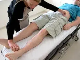
Hip Examination
Introduction
Hip Examination is a systematic clinical assessment used to evaluate the structure, function, and movement of the hip joint.
It aids in determining whether hip pain, stiffness, weakness, or mobility issues are caused by musculoskeletal disorders, arthritis, trauma, or imbalances in the muscles. In order to support a precise diagnosis and direct the right course of therapy, a thorough hip examination usually consists of the patient’s history, observation, palpation, range of motion testing, muscle strength assessment, gait analysis, and special orthopedic tests.
Anatomy
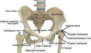
The largest bone in the human skeleton that bears weight is the hip joint. It is encircled by tendons, ligaments, and muscles and is also known as a joint with a ball and socket. The hip joint is made up of the pelvis and the thigh bone, sometimes known as the femur.
The range of movement and weight-bearing capacity of the hip joint will be negatively impacted by any injury or illness.
The following components make up the hip joint:
- Joints and bones
- Joint capsule and ligaments
- Tendons and muscles
- Blood vessels and nerves supplying the hip’s bones and muscles
Hip bones:
Also referred to as the innominate bones, pelvic bone, or coxal bone. A hip bone ( coxae) constitutes a bilateral, irregularly shaped component of the skeletal pelvis. The ischium, pubis, and ilium are the three minor bones that make up this composite structure. The pubis or pubic bone makes up the front section of the hip bone, the ischium is situated posteroinferiorly, and the ilium is the biggest and most superior region of the bone.
Ligaments:
The hip joint’s ligaments work to make it more stable. They fall into two categories: extracapsular and intracapsular.
- Inside the capsule:
The head of the femur ligament - Outside the capsule:
The iliofemoral ligament
The pubofemoral ligament
Ischiofemoral ligament
Arteries and Nerves:
To facilitate hip mobility, the brain sends impulses to the muscles through the hip nerves. They also return sensory information to the brain, including temperature, touch, and pain.
The sciatic nerve in the rear of the femur or the femoral nerve at the front are the two primary nerves of the hip area. The obturator nerve, a lesser nerve, also supplies the hip.
Blood vessels supply the lower limbs in addition to these nerves. One of the biggest arteries in the body is the femoral artery, which emerges deep within the pelvis and is palpable in front of the upper thigh.
Muscles and Motions:
The following is a list of the hip joint motions that can be performed, along with the primary muscles involved in each action:
- Flexion: rectus femoris, sartorius, pectineus, and iliopsoas
- Extension: biceps femoris (the hamstrings); semimembranosus; semitendinosus; gluteus maximus
- Abduction: Gluteus medius, gluteus minimus, piriformis, and tensor fascia lata
- Adduction: pectineus, gracilis, brevis, and adductor magnus
- Lateral rotation: Biceps femoris, gluteus maximus, and piriformis, with help from the obturators, gemilli, and quadratus femoris.
The tensor fascia lata and the anterior fibers of the gluteus medius or gluteus minimus are involved in medial rotation.
If the knee is flexed, which reduces the hamstring muscles and expands the range of flexion, it determines how much flexion at the hip is possible.
The iliofemoral ligament and the joint capsule restrict hip joint extension. During extension, these structures tighten to prevent additional movement.
Typical conditions affecting the hip joint:
- Hip strains: Damage or rupture to the muscles.
- Bursitis: Inflammation of the fluid-filled sac in the joint.
- A hip dislocation occurs when the bone in your thigh pops out of its socket.
A broken femur occurs when one of the hip joint’s bones is broken or fractured.
Your hip joint may become weak due to certain medical issues. You may be more vulnerable to harm or side effects like stiffness or discomfort. The following are typical conditions that impact your hip joint:
- Osteoarthritis: Joint inflammation that results in pain and swelling.
- Osteoporosis: Weak bones that are weak and prone to breaking are known as osteoporosis.
- Hip dysplasia: Occurs when the bones in your thighs do not fit together properly in your pelvis.
The following are typical signs of hip joint disorders:
- Swelling and pain.
- Restricted range of motion.
Clinical Significance:
Congenital Hip Dislocation
Hip developmental dysplasia (DDH) is the cause of congenital hip dislocation. It happens if the region of the acetabulum becomes shallow due to improper fetal development.
Typical clinical characteristics consist of:
- Restricted hip joint abduction
- Disparity in limb length: the affected limb is shorter
- Unbalanced skin folds on the thighs or glutes.
- The most common treatment for DDH is a Pavlik harness. This supports the hip joint’s normal development and keeps the femoral head in the acetabular fossa. When harness treatment is ineffective, surgery is recommended.
Acquired Hip Dislocation
Because of the hip joint’s strength and stability, acquired dislocations are comparatively rare. Usually brought on by trauma, they can also develop as a side effect after hemiarthroplasty or total hip replacement.
Acquired hip dislocations come in two primary varieties: anterior and posterior.
In a posterior dislocation (90%), the femoral head is forced posteriorly, tearing the inferior and posterior parts of the joint capsule where it is weakest.
The affected limb medially rotates and gets shorter.
The probability of injury to the sciatic nerve, which passes posteriorly to the hip joint, is 10–20%. This is frequently linked to fractures of the posterior wall and anterior femoral head.
Rarely, traumatic extension, abduction, and lateral rotation can result in anterior dislocation. Concerning the acetabulum, the head of the femur is shifted anteriorly and (typically) inferiorly.
Subjective Assessment of Patient Admission:
The first step in an examination is a patient consultation. Where a doctor asks the patient to describe their presenting symptoms. According to research, a patient’s medical history is crucial in making an accurate diagnosis of hip pain and, in certain situations, may be more reliable than objective tests and measurements.
As it helps a clinician form hypotheses regarding the process of injury, the type of harmed structures, and the degree of injury or even damage, taking a patient’s history is a crucial part of a subjective assessment. Information is obtained from the patient during the interview, including the location, type, and 24-hour frequency of the discomfort, activities that cause the pain, things that aggravate and relieve the pain, etc.
A recent systematic review that has appeared in the Archives for Physical Medicine and Rehabilitation states that hip crepitus is an excellent sign of intra-articular hip pathology, and that thigh/groin pain and persistent back/buttock discomfort are more accurate signs of hip OA compared to stand-alone tests.
To help a doctor rule out inherited illnesses, a patient’s past medical history as well as their social and family background are crucial. Any hip-specific surgical histories are also crucial, such as the patient who underwent hip joint replacement treatment and is currently experiencing hip joint pain.
Documentation may include a patient’s past drug use, current drugs, and any allergies they may have. It is possible to thoroughly evaluate and document the situation’s effects on the patient’s functional state and Activities of daily living (ADLs).
It’s important to think about whether a fall in an aged person was caused by a simple trip or by something else, including a seizure, a cardiovascular trigger, a stroke, or unconsciousness. Additional tests, such as an ECG and a brain CT scan, may be necessary to check the neurological or cardiovascular systems.
Particular Points to Remember:
Warning Signs:
- The pain started suddenly.
- Any trauma
- Any edema or abnormality
- A failure to weight-bear
- Any visible pulsations in the groin, lumps or small bumps there, and night pain
- Vomiting or even constipation
- Haematuria
- A fever
- Neurological symptoms of the lower limbs, such as tingling, numbness, or weakness
- History of steroid use
- Enlargement of the testicles
- Unintentional weight loss, appetite loss, and night sweats
- History of Malignancy
Observation of the Objective Examination:
A doctor does an objective examination after a patient interview is over. An examiner examines and records the following after administering the Pain VAS (visual analog scale) to ascertain the patient’s baseline degree of pain:
The alignment or malformation of the bones
- Walking style: A clinician needs to be on the lookout for a variety of abnormal gait patterns linked to hip diseases, including Lurching, Trendelenburg, Arthrogenic, and Antalgic gait.
- Weight-bearing.
- Additionally, posture and balance.
- Standing: the lumbar region, iliac crest level (to rule out SIJ/pelvic dysfunction)
- Supine lying: Leg length (both perceived and actual), and the leg’s external rotation.
- For example, color changes, rash, inflammation, infection, and bruises.
- Muscle bulk, muscle spasms, and muscle loss.
- Skin alterations, scarring, and swelling (wounds).
Palpation:
During palpation, the examiner makes sure to:
Vascular System: Capillary refill and distal pulses.
Sensation (Neurological Examination): Skin sensation and power of peripheral nerves.
The temperature, inflammation, and Infection
Swelling: Extracapsular Tenderness, Synovial Thickening, and Effusion
The lumbar spine
The ASIS, pubic rami, symphysis pubis, PSIS, SIJ, ischial tuberosity, sacrum, coccyx, and iliac crest make up the pelvis.
The greater trochanter
The inguinal ligament
Knee and femoral triangle (hip joint).
Manual for Neurologic Assessment Muscle Testing:
It is important to grade and record the strength of the muscles that surround the hip joint, including the internal and external rotators, abductors, adductors, flexors, and hip extensors.
- Straight Raise Leg Test
- Testing for Dermatome
- Skin sensation test
Movement examination:
A goniometer is used to measure and record the active and passive ranges of movement at the hip joint. These measurements act as reference points and aid in assessing the effectiveness of the intervention.
Special Test:
Sign of Trendelenburg:
Goal
Finding the hip abductors’ weakness is the aim of this test.
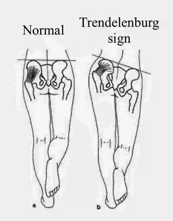
This Trendelenburg sign can be used to evaluate various mechanical, neurological in nature, or even spinal abnormalities, such as a congenital hip dislocation or hip subluxation, in addition to detecting the weakness of the abductors of the hip of a standing leg.
Method
- For 30 seconds, the patient is asked to get up on one leg without slanting to one side; if balance is an issue, the patient may grasp onto something. When a patient is in a single-leg stance, a physiotherapist watches to check if the pelvis remains level.
- If the pelvis lowers towards an unsupported side while unilaterally bearing weight, the Trendelenburg test is affirmative.
Faber Test:
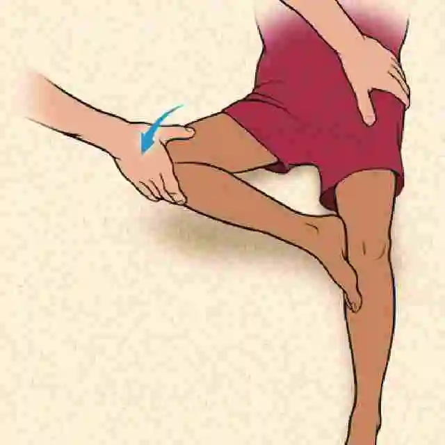
A Faber (Patrick’s) test is an acronym for external rotation, flexion, and abduction. Together, these three movements provide the clinical discomfort provocation test, which aids in determining the presence of disorders affecting the lumbar, hip, and sacroiliac regions.
Method
- A supine position is adopted for the patient. A lateral ankle rests over an ipsilateral thigh proximal to the knee when a leg is in the figure-4 posture, which includes both hip flexion and abduction. The external rotation, abduction, and posterior force are then gently applied to a contralateral knee when an end range of motion is reached while supporting the opposite side of the pelvis at the anterior superior iliac spine. To check for discomfort aggravation towards the end of the range of motion, a few more oscillations with a smaller amplitude may be used. A test that replicates a patient’s pain or even restricts range of motion is considered positive.
Interpretation
The clinical diagnosis can be guided by the following results from the positive Faber test:
Hip Impingement of the (femoral acetabular impingement), Hip Labral Tear, Hip Loose Bodies, Hip Osteoarthritis, Posterior Hip discomfort on External Hip Rotation Action, Iliopsoas Strain or Bursitis, Intraarticular Hip Disorder, and Sacroiliac Pain in the Joint on External Hip Rotation Motion are all examples of these conditions.
Ober’s Test:
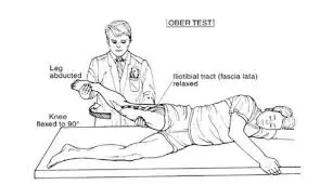
This Ober’s test primarily evaluates the iliotibial band (ITB) and the tight, constricted, or even inflammatory Tensor Fasciae Lata (TFL). The Renne test and Noble’s test, two other tests frequently used to identify iliotibial band syndrome, should not be confused with Ober’s test.
Position for testing:
How to carry out this test:
- The patient should first lie sideways with the side that is affected facing up.
- To eliminate a lumbar curve, the hip and bottom knee may then be flexed.
- After that, stand behind the patient and securely support the greater trochanter and pelvis to stop them from moving in any direction.
- Using the other hand, grasp the distal end of the patient’s affected leg and flex it at the knee to the correct angle.
Test:
- Abduct and extend the hip joint.
- Adduct your hip and slowly lower your leg towards the table until your range of motion is limited.
- Make sure the hip doesn’t flex or internally twist during the exam so that the pelvis can be stable. For a thigh to decrease in both internal rotation and flexion would “give in” to a tight TFL and result in an inaccurate length measurement.
Findings:
A test is considered negative if a leg can adduct with a thigh lowering slightly to a horizontal and a patient does not feel any discomfort when the iliotibial band is normal.
The test is considered positive when the iliotibial tract is tight, a leg stays in an abducted position, and the patient has lateral knee pain.
Log Roll Test:
A log roll test can be used to isolate a patient’s pathology to the hip rather than outside of it. It evaluates for pathology within the hip joint.
Put one hand over the mid-thigh and rotate the hip passively both internally and externally toward the ends of the joint’s range of motion while the patient is in the supine posture. Examine the range of motion on either side.
Intra-articular pathology is abnormal if pain occurs.
Clicking: Labral tear of the acetabular region.
Ligamentous laxity: greater range of motion.
The most accurate test for hip discomfort is a log roll. With little strain on the nearby muscle or nerve, this shifts the surface of the joint of the femoral head concerning an acetabulum.
Thomas Tests:
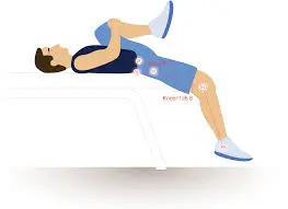
The primary use for the Thomas Test is to assess hip flexor flexibility.
Psoas syndrome, patellofemoral pain syndrome, lower back pain, osteoarthritis, and rheumatoid arthritis are among the disorders that can have impaired hip range of motion as an underlying cause.
Frequently linked to runners, dancers, and gymnasts who experience “stiffness” in their hip joints and a “snapping” sensation when flexing their waists.
Method
The patient is supine, with their hips and knees flexed, and the lower gluteal folds at the base of a table. The patient can use their hands to hold their legs in a flexion position.
A physical therapist ensures that a patient has a normal pelvic tilt in their lower back.
The patient flexes an unaffected leg while lowering the injured limb gradually and allowing it to extend as much as possible.
Interpretation
The Thomas test: When the test is positive, the structures that follow can be taken into consideration.
Sign Affected Structures
Knee extension: rectus femoris and quadriceps
Hip flexion muscles
Hip abduction: iliotibial band and tensor fascia lata
Lateral rotation of the tibiofemoral biceps
Ely’s Test:
Goal
Rectus femoris tightness or spasticity can be evaluated with the Duncan-Ely or Ely’s test.
Method
- The patient is relaxed and lying prone. Beside the patient, at the side of the leg that will be examined and checked, is a physiotherapist. A leg at the heel can be held with one hand while the other rests on the lower back. Rapidly flex your knee passively. The heel may then come into contact with the buttocks. To compare, test both sides. A test is therefore considered successful if a patient experiences discomfort or even numbness in their legs or back, a hip on the tested side lifts from a table, and a heel can’t reach their buttocks.
FADDIR Test:
One of the patients is resting supine.
- An examiner passively bends the knees and hips while holding the affected leg at the knee and close to the heel using one hand.
The hip is then adducted, internally rotated, and hyper-flexed. Kendall test: - A modified version of the Kendall Test, the Thomas Test is a popular orthopedic test used to measure the rectus femoris muscle length of the quadriceps.
- Place the patient in an upright position using both of their legs off a table to conduct the test. The patient should next be instructed to bring and hold one knee to the chest.
Modified Ober Test:
For flattening the lower back, the patient is first placed in a side-lying position with their underleg bent at the hip and knee.
The examiner then maintains a lateral trunk in proximity to a table and stabilizes the pelvis.
Note: A hip abduction is the same as a downward lateral tilt, which would “give in” to the tight TFL.
Test: An examiner puts out their leg. The limb must not be internally rotated on any side.
Findings: A thigh falls roughly 10 degrees beyond a horizontal when the knee is straight, and the pelvis is in its typical posture. This suggests that the length is typical.
Tensiofascial late and a constricted iliotibial tract are suggested by a leg that does not drop.
FAQs
What is the hip’s anatomy?
The point where the hip connects a leg to the body’s trunk is called the hip joint. The thigh bone, also known as the femur, and the pelvis, which is composed of the ischium, pubis, and ilium, make up this structure. The acetabulum forms the hip joint’s socket, whereas the femoral head forms the hip joint’s ball.
The hip joint has how many ligaments?
The iliofemoral, pubofemoral, and ischiofemoral ligaments are the three main ligaments that make up the hip joint capsule.
Which physical component is referred to as the hip?
The region on either side of the pelvis is called the hip. There are three parts to the pelvic bone: the ilium. The pelvis’s wide, flared section.
What is the hip exam Thomas test?
The person being tested lies on their side or back during the test. To try to smooth out the lumbar spine bulge, the patient is instructed to draw the non-testing leg towards the chest. When the patient feels the stretch in their groin and the stretched leg can be lifted off the treatment table, the test is considered successful.
What is a hip physical examination?
Monitoring of walking, location, and inspection in the lying down, lateral, and frequently prone positions are necessary for an appropriate hip assessment. Gait and station, probing of often bothersome areas, strength, and range-of-motion tests, and fundamental impingement testing are typically included in the basic examination.
The FABER test: what is it?
By transmitting forces through the joint, the test evaluates the hip. The femoral-acetabular joint is stressed when the positions of flexion are coupled with excessive pressure; if irritation or disease is present, this can cause pain.
When does a positive Thomas test result occur?
When the contralateral leg exhibits noticeable hip flexion, as shown by a space between it and the tabletop, the TT is considered positive, indicating hip flexion contracture.
What is Faber’s full form?
Although it has been used to evaluate a combined hip range of motion (ROM), the flexion Abduction External Rotation (FABER) test is usually employed as a provocation special test.
References:
- Hip Surgeon Frisco, TX | Hip Surgery Frisco, TX | Hip Pain Treatment Frisco, TX. (n.d.). https://www.kennethestreramd.com/hip-anatomy-orthopaedic-surgeon-texas.html
- Hip bone. (2023, October 30). Kenhub.
- Battista, C., MD. (n.d.-b). Hip physical exam – adult – recon – orthobullets.
- Hip Region Exam, Approach to. (n.d.). Stanford Medicine 25. https://stanfordmedicine25.stanford.edu/the25/HipRegionExam.html