Metacarpal Bones
Introduction
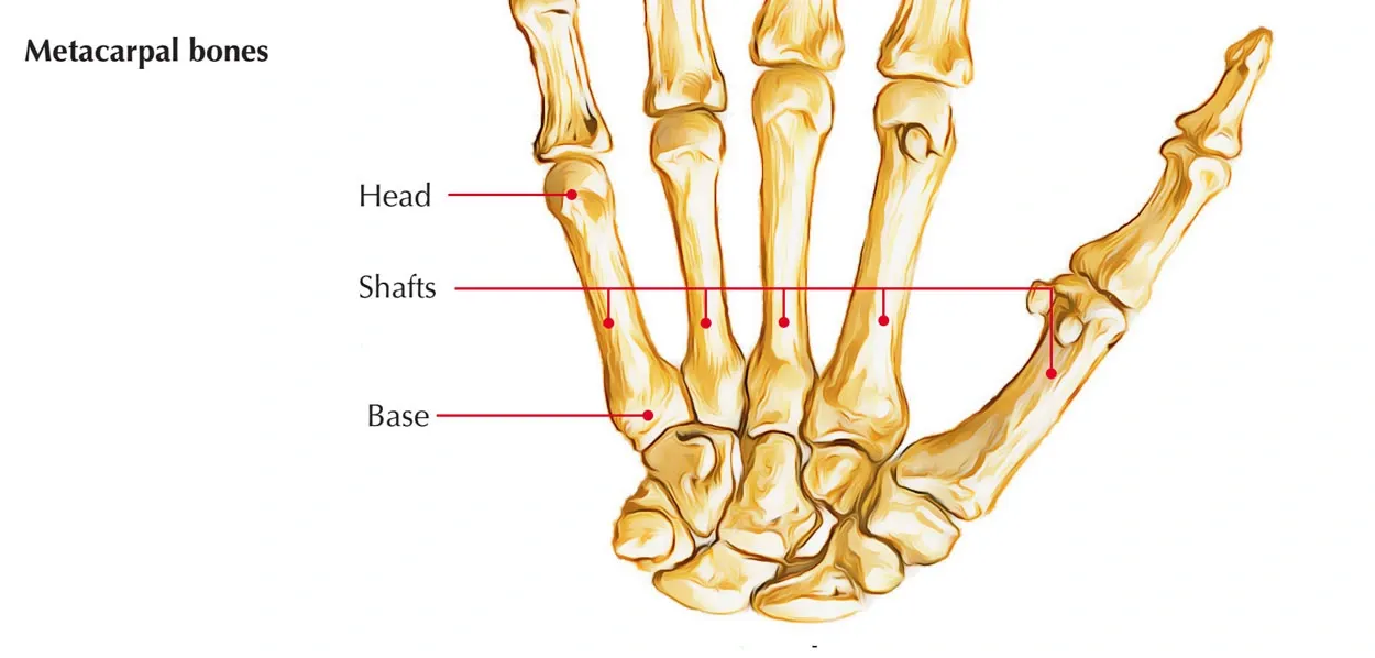
The five hand bones that lie between the phalanges and the carpus are called the metacarpus. The metacarpal bones are short, but because they share structural traits with long bones, they are categorized as long bones. Each metacarpal bone has a large proximal base, a distal head, and a shaft.
The proximal ends (bases) of the metacarpal bones articulate with the carpal bones, whereas the distal ends (heads) articulate with the proximal phalanges. Numbers 1 through 5 are used to identify them as they move in the radio-ulnar direction.
Five long bones in the hand, known as metacarpal bones, are situated between the phalanges (fingers) and the wrist (carpal bones). The palm is made up of several bones, which also help the hand move and have overall structure. From the thumb (first metacarpal) to the little finger (fifth metacarpal), each metacarpal has a name and number. They are made up of a head, shaft, and base. The head forms joints with the proximal phalanges, while the base articulates with the carpal bones.
Structure and Anatomy
The palm’s framework is made up of five long bones called metacarpal bones. From the thumb (first metacarpal) to the little finger (fifth metacarpal), they are numbered. The base, shaft, and head are the three primary components of each metacarpal.
General Features
Base
Shape: Each metacarpal has a large, flat proximal end.
Articulations: At the carpometacarpal joints, the base articulates with the carpal bones.
Intermetacarpal joints allow adjacent metacarpals to articulate with one another.
Shaft
Shape: The long, somewhat curved shaft is convex on the dorsal surface and concave on the palmar surface.
Features: gives ligaments and muscles places to attach.
The shaft has a nutrient foramen that permits blood vessels to enter.
Head
- Shape: The knuckle is formed by the rounded distal end of each metacarpal.
- Articulations: At the metacarpophalangeal joints, it articulates with the proximal phalanges.
- Features: For joint mobility, the smooth head is covered in articular cartilage.
Individual Metacarpals
First Metacarpal (Thumb)
Size and Shape: The thickest and shortest metacarpal.
Base: allows for considerable motion at the first carpometacarpal joint by articulating with the trapezium (saddle joint).
Head: forms the joint with the thumb’s proximal phalanx.
Second Metacarpal
Size and Shape: The longest metacarpal.
Base: mostly articulates with the trapezoid, but also with the third metacarpal, trapezium, and capitate.
Head: attaches to the index finger’s proximal phalanx.
Third Metacarpal
Size and Shape: Not quite as long as the second metacarpal.
Base: articulates with the second and fourth metacarpals as well as the capitate.
Notable Feature: features a noticeable styloid structure for ligament attachment on its dorsal surface.
Head: joins the middle finger’s proximal phalanx to form a joint.
Fourth Metacarpal
Size and Shape: narrower than the third metacarpal and shorter.
Base: articulates with the third and fifth metacarpals as well as the hamate.
Head: attaches to the ring finger’s proximal phalanx.
Fifth Metacarpal (Little Finger)
Size and Shape: The thinnest metacarpal in size.
Base: articulates with the fourth metacarpal and the hamate.
Head: forms the joint with the little finger’s proximal phalanx.
Articulations
Carpometacarpal Joints
Proximal articulation between the distal carpal bones and the bases of the metacarpals.
With a broad range of motion, the first carpometacarpal joint (thumb) is a saddle joint.
Intermetacarpal Joints
Interosseous ligaments maintain the articulation of neighboring metacarpal bases.
Metacarpophalangeal Joints
Distal articulation between the proximal phalanges and the metacarpal heads. Flexion, extension, abduction, and adduction are all made possible by these condyloid joints.
Vascular Supply
- Arteries: supplied by ulnar and radial artery branches.
- Nutrient Foramina: situated on the shafts, which permit blood vessels to enter and supply the bone with nutrients.
Muscular Attachments
Extrinsic Muscles:
The bases and shafts are where tendon attachments of muscles such as the flexor digitorum profundus and superficialis and the extensor digitorum are found.
Intrinsic Muscles:
The sidewalls of the shafts are where the interossei muscles are attached.
Function
Support and Stability -The hand’s structural framework is provided by the metacarpal bones, which form the palm and join the fingers and wrist. During rest and motion, they preserve the stability and form of the hand.
Facilitation of Movement– Through their articulations with the phalanges at the metacarpophalangeal joints, the metacarpals allow the thumb and fingers to move. The thumb’s opposable movements, which are necessary for gripping and precise activities, are made possible by the first metacarpal.
Force Transmission: When gripping, pulling, or lifting, the metacarpals transfer pressures from the fingers to the wrist.
Muscle Attachment
The intrinsic and extrinsic hand muscles link to the metacarpals, enabling coordinated hand movements and fine motor skills.
Formation of Arches
The metacarpals support the hand’s longitudinal and transverse arches, which improve the hand’s capacity to grasp items and withstand mechanical pressures
Physiology
The metacarpal bones support the structures of the hand in a number of important ways. They provide the palm its rigid structure and constitute its solitary hard tissue.
The development of the metacarpophalangeal joints with the phalanges and the carpometacarpal joints with the carpals is another crucial role of the metacarpal bones. The hand may flex, extend, abduct, and adduct at the wrist thanks to carpometacarpal joints. An especially special saddle joint that gives the thumb and first metacarpal additional movement is the first carpometacarpal joint. To provide the fingers with a complete range of motion, the phalanges at each metacarpophalangeal joint can flex, extend, abduct, and adduct.
The metacarpals are where a number of significant muscles attach. The metacarpals are the origin of the dorsal and palmar interossei muscles, which move the fingers. The wrist can be extended by inserting the extensor carpi radialis longus, extensor carpi radialis brevis, and extensor carpi ulnaris. To oppose the thumb, the opponens pollicis muscle inserts on the first metacarpal, and to oppose the little finger, the opponens digiti minimi muscle inserts on the fifth metacarpal. Lastly, the thumb is adducted by the adductor pollicis longus inserting on the second and third metacarpals.
Clinical importance
These bones, particularly the first and fifth metacarpals, are prone to fracture, particularly in athletes.
A) Bennett’s Fracture, the initial metacarpal fracture
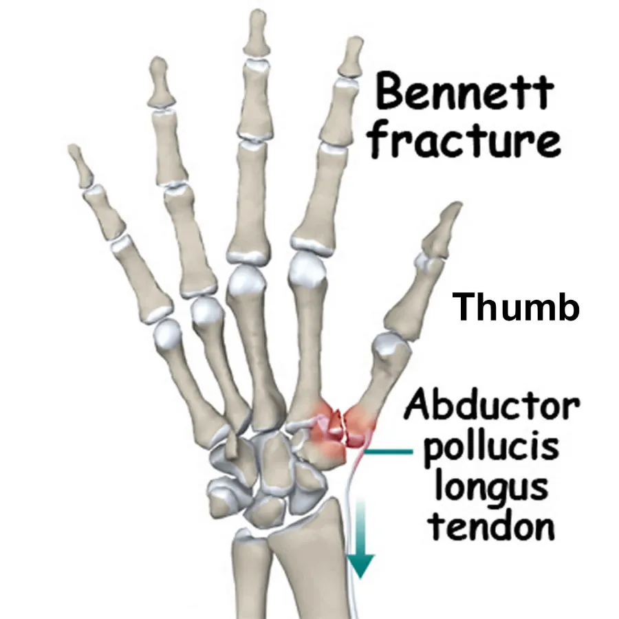
The first metacarpal base fracture that causes the thumb to split from the medial side of the hand is referred to as this condition.
Among the potential reasons for the fracture are: 1. The force imparted to the thumb during flexion is the primary cause of the fracture.
- The adjacent ligament injuries and the trapezium fracture are the other causes.
The symptoms - Tenderness and pain in the base of the thumb.
- Thumb movement is limited.
- A diminished capacity for pinch grasp.
Diagnosis
- To diagnose the fracture type and site, radiographs of the hand and thumb are taken, particularly during specific movements.
- CT scans are also used in the diagnosis to display the specifics of the fracture and the extent of the injury.
The therapy - A splint that is appropriate for the fracture condition is part of the treatment.
It is feasible to fix the bone by using wire and screw fixation procedures.
- Physical therapy under a specialist’s supervision and, for a specific amount of time, splint treatment or surgery are also part of the treatment.
B) The fifth metacarpal fracture (Boxer’s fracture)
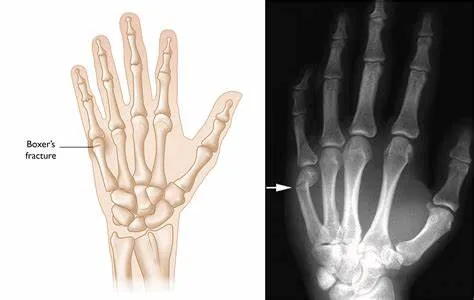
The fifth metacarpal neck fracture is the condition in question.
- Males are more likely than females to have it.
- Individuals aged 10 to 29.
- Boxers.
The potential reasons behind the fracture - The direct impact on a clenched grasp that targets the metacarpals is the primary cause of this fracture.
- Motorcycle accidents are a rare cause of fractures to this bone.
The symptoms - Dorsal hand pain, precisely where the fifth metacarpal is located.
- Distortion and swelling at the trauma site.
The diagnosis - A physical examination is part of the diagnosis to assess the extent of structural damage.
- Radiographs and CT scans are also included to determine the severity of the fracture and to confirm the condition of other bones and structures.
The therapy
A splint that is appropriate for the fracture condition and takes into account the extent of damage to nearby structures and how to treat them is part of the treatment.
FAQs
What is a metacarpal bone?
In land vertebrates, the term “metacarpal” refers to any of a number of tubular bones that connect the wrist (carpal) bones to each forelimb digit, which corresponds to the metatarsal bones of the foot. Many mammals’ metacarpals, which were once five in number, have changed and decreased significantly over time.
Can a broken metacarpal heal without surgery?
Non-operative treatment is also an option for patients whose fractures can be realigned by a process known as “closed reduction,” which involves manipulation rather than surgery. Strength and range of motion in the fingers can be enhanced with hand treatment.
What are the 3 parts of the metacarpals?
A metacarpal bone is composed of three parts: the head, shaft, and base.
The proximal end of the metacarpal bone serves as the base. Small articular facets on the base attach to nearby metacarpal bones.
Shaft: The metacarpal bone’s curving portion.
Head: The metacarpal bone’s distal end. The smaller, convex head articulates sequentially with the subsequent bone.
What is the main function of metacarpals?
The metacarpals, which act as a framework to connect the carpus to the phalanges, are essential to the hand’s form and functionality. Together, the metacarpals, the longest bones in the hand, form a concave arch in the palm. The head, neck, shaft, and base comprise the bony anatomy.
What is the other name for metacarpal?
The metacarpus, sometimes referred to as the “palm bones” in human anatomy, are appendicular bones that make up the middle portion of the hand between the carpal bones (wrist bones) and the phalanges (fingers), which articulate with the forearm.
References
- Metacarpal bones. (2023, November 3). Kenhub. https://www.kenhub.com/en/library/anatomy/the-metacarpal-bones
- Kranthi, & Kranthi. (2025b, January 28). Metacarpal bones – Anatomy, Diagram, Function, Location. Anatomy.co.uk | Learn Human Anatomy From Experts. https://anatomy.co.uk/metacarpal-
bones/ - The Metacarpals: Anatomy and 3D illustrations. (n.d.). Innerbody. https://www.innerbody.com/image_skelfov/skel22_new.html
- Metacarpal bones. (n.d.). https://www.mickeymed.com/article/metacarpalbones