Thoracic Spine Examination
What is a Thoracic Spine Examination?
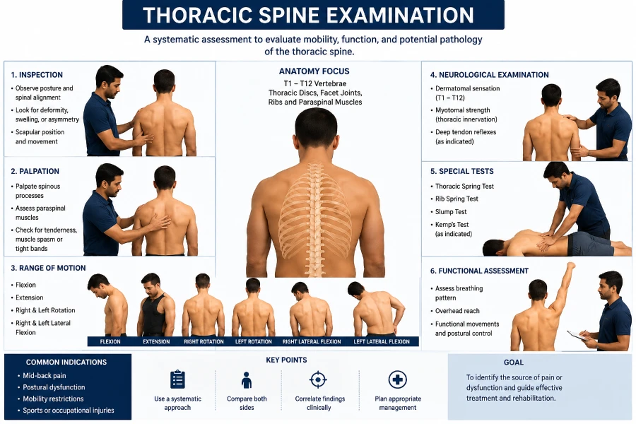
The thoracic spine examination assesses mobility, stability, and potential dysfunctions in the mid-back region. It includes inspection, palpation, range of motion testing, and special tests to evaluate posture, joint restrictions, and muscular imbalances.
This assessment helps identify conditions such as kyphosis, stiffness, or referred pain affecting overall spinal function.
Anatomy:
Your thoracic spine is the central portion of your spine. It stretches from the base of your neck to the base of your ribs. There are twelve vertebrae in it. Because of its excellent stability and rigidity, your thoracic spine is the least likely part of your spine to sustain damage.
The 33 separate, interconnecting bones that comprise your spinal column are called vertebrae. These bones allow you to twist and turn while protecting your spinal cord. Your vertebrae are supported and flexible by the disks that lie between the vertebral bones.
Additionally, the muscles, nerves, tendons, and ligaments surrounding the thoracic spine help in flexibility and mobility. It communicates with and receives information from the brain, which manages every bodily function.
The thoracic vertebrae are the source of nerves:
The T1 and T2 nerves supply your arms, hands, and upper chest. The T1 nerve is part of the brachial plexus, a network of nerves in your shoulders that relays movement and sensory data from your spinal cord to your hands and arms.
Your chest wall is where the T3 through T5 nerves enter. Together, these nerves control your breathing muscles, lungs, diaphragm, and rib cage.
The muscles in your back and abdomen are affected by the T6 through T12 nerves. These nerves help with coughing, posture, and balance when combined with particular muscles.
The organs affected by the thoracic spine are:
Your brain sends messages to your major organs through the branching nerves of the thoracic spine from the spinal cord, including your:
- Lungs
- Heart
- Liver
- Small intestine
- Your rib cage and thoracic spine work together to form a protective barrier for your heart and lungs.
What is the composition of the thoracic spine?
Vertebrae: Your spinal canal is made up of 12 of the 33 stacked vertebrae (small bones) that make up your thoracic spine. The spinal canal is a tunnel that encloses and protects the spinal cord and nerves from damage. The movement of these vertebrae allows for a wide range of mobility.
Facet joints: These spinal joints include cartilage, a slippery connective tissue that allows vertebrae to glide against one another. Facet joints provide you with stability and flexibility, but they also let you twist and turn.
The flat, round cushions that act as shock absorbers for your spine are called intervertebral disks. Each disk has a flexible outer ring encircling a soft, gel-like center.
Nerves and spinal cord: The spinal cord is a column of nerves that reside in the spinal canal. The vertebral column extends from your cranium to your lower back. 31 pairs of nerves emerge from neural foramina, or vertebral holes, along your spine. Twelve pairs of nerves branch off from your thoracic spine. These nerves allow communication between your brain and muscles.
Soft tissues: Your vertebrae are joined by ligaments to maintain the position of your spine. Your back is supported and moved by your muscles. Tendons facilitate movement by joining muscles with bones.
Disorders and Conditions:
- Strain or muscular pain
- Ligament sprains
- Trauma
- Overuse injuries
The following are more typical reasons for thoracic spine pain that directly affect your spinal column:
- The middle and lower back are the most common locations for spinal tumors, though they can form anywhere along the spine. Usually, metastatic cancer is the cause of them. The most prevalent sign of both benign (noncancerous) and malignant (cancerous) spinal tumors is back pain. A spinal tumor typically causes severe, throbbing pain that lasts through the night and occasionally interferes with sleep.
- Spinal fracture: Though less frequent than cervical and lumbar fractures, thoracic spinal (vertebral) fractures are more likely to occur in individuals with osteoporosis because their bones are weaker. A sudden twisting motion or sneeze might cause thoracic spinal fractures in people with osteoporosis.
The following are common disorders that affect the thoracic spine:
- Kyphosis
- Scoliosis in children and adolescents
- Thoracic radiculopathy
- (Osteophyte ) bone spurs
- Disk herniation
- Myelopathy
- Osteomyelitis
- Spinal cord injury
- Spinal stenosis
Indications of nerve injury to the thoracic spine:
- Loss or decreased sensation in your arms or legs.
- Difficulty in breathing.
- Loss of sensation in the rectal or genital area.
- Loss of bladder or intestinal control.
- Constipation.
Examination
History:
Symptoms of subjective assessment: distribution of Areas may follow the path of a rib or extend horizontally over the chest.
Once all other relevant areas had been cleared, tags were identified on the upper limbs, lower cervical spine (lower limbs), scapula, thoracic spine, sternum, and higher cervical spine.
VAS Type for Quality Intensity: maintaining a pain journal may be helpful for patients who experience chronic pain. Depth: unusual feeling, paresthesia, feeling numb, regular or sporadic. Could you check to see whether the severity of the symptoms varies if they are consistent? Persistent, continuous pain could be a sign of cancer. The disease is considered non-severe if the patient can maintain a position that reproduces the symptoms.
The ability to irritate.
The condition is considered severe if the patient cannot hold their position, and excessive pressure should not be used. All motions can be examined if the disease is considered non-irritable and the symptoms go away right away when caused.
The patient is agitated if symptoms go away in a few minutes, and just a few actions should be tried to prevent worsening of symptoms for a full day: Nighttime, morning symptoms (for example, stiffness and pain in the morning for a few minutes may indicate spondylosis, while stiffness and pain for a few hours may indicate inflammatory processes such as A.S.
Are pillows an obstacle to sleep? How soft or firm is the mattress?
Becoming better, staying the same, or getting worse.
Observation:
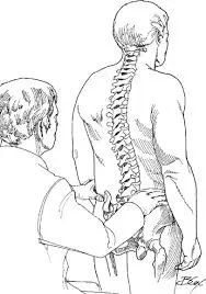
This should be objectively examined from the front, back, and sides when standing or sitting.
When examining the patient’s thoracic spine, the examiners search for abnormalities, including sagittal and frontal thoracic spine curvatures.
According to a study, there is a modest to large overall impression of inter-rater reliability for kyphosis postural observation and classification as excessive, normal, or decline.
Symmetry, Cervical/Thoracic/Lumbar curvatures, spine deviations, shoulder levels, shoulder girdle, GH joint position, pelvic level, limb position, muscle tone, and bulk (compare sides, hand dominance), soft tissues (skin color and quality, swelling, scars), gait (Trendelenburg gait, short leg gait), and attitudes and feelings are some of the things that need to be considered.
Palpation:
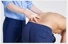
Palpation may reveal localized pain in some places or muscle spasms. The pain may be reproduced and made worse by pressure directly lateral to the spinous processes and manipulation or percussion over them. Reproducible pain felt when the spinous process is palpated could be a sign of a vertebral issue, such as a recent fracture. Muscle spasms may be indicated by pain felt in the paraspinal area, particularly if there are stiff back muscles.
Movement patterns:
Movement Patterns: To look for signs of change, the examiner might ask the patient to do the following movements: When the patient performs any activity that either worsens or alleviates symptoms, such as looking up at the sky, rising from a seated position, or raising one or both arms over the head, watch for any side-to-side deviations, compensations, or inconsistencies.
After a trial intervention, any of these movement patterns can be used as a symbol or similar sign for reevaluation.
Muscle Testing:
Similar variations were observed in the thoracic spine’s pain provocation, ranging from little agreement to significant agreement.
With a moderate level of agreement for intra-rater reliability and no agreement for inter-rater reliability, the rib cage has strict compliance. The greater the agreement, the better the intra-rater reliability and the better the inter-rater reliability.
Muscle strength:
Test of Latissimus Dorsi Muscle Length: Pectoralis Major, inter-rater reliability ranging from modest to substantial. Inter-rater reliability for Pectoralis Minor is moderate to high. Inter-rater dependability ranges from modest to substantial.
Strength of the Middle Trapezius Muscle: Lower Trapezius, Inter-rater reliability is nonexistent. Serratus Anterior: Inter-rater reliability is nonexistent.
Rhomboids: moderate reliability between tests and moderate to high dependability between raters.
Neurological Assessment of Thoracic Examination:
When you move your neck, do you feel any pain in your legs or lower back?
If so, perform a comprehensive neurological examination. Have you ever had intermittent problems with balance or coordination in your lower extremities, along with bilateral upper-extremity symptoms?
Reflexes of the upper motor neurons (Hoffman, Babinski, Clonus). Sensory Testing: If so, perform a comprehensive neurological assessment. T1 Nerve Root Level: first dorsal interossei dermatome (medial forearm). Tests of Neural Tissue Provocation in the Lower Thorax. Neurologic screening of the lower thorax in the upper quarter
Special Tests:
Passive Neck Flexion Test:
Use the Passive Neck Flexibility Test to find out how neural strain affects the patient’s symptoms.
Patient’s position: supine.
How to perform the test: To perform correctly, the patient actively bends in the upper cervical region while the examiner passively flexes the lower cervical spine. The test is successful if the thoracic spine reproduces pain or other neurological symptoms. Stretching is a common sensation.
The diagnosis’s accuracy is unknown.
Test Relevance: Nerves may cling to certain structures as they travel through the body’s numerous tissues. This test uses cervical flexion to induce strain to the spinal cord in an attempt to replicate these symptoms.
Straight Leg Raise Test:
Method
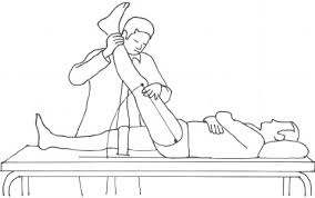
Passively, a typical straight leg raise is performed. The unaffected leg is examined first, followed by each leg in turn. While standing on the side being examined, the clinician places their distal hand on the patient’s heel and their proximal hand on the patient’s distal thigh (anterior) to maintain knee extension while the patient is in a supine position.
While keeping the patient’s knee fully extended, the physician raises the patient’s leg by the back of the ankle. Until the patient feels the same tension in the back or posterior thigh, the physician keeps slowly raising the patient’s leg by flexing the hip.
The examiner stops hip flexion when pain occurs and records the range of motion and pain location. One such outcome measure is the difference in range of motion between the affected and unaffected legs. It should be pointed out, nevertheless, that the inter-rater reliability for this is low.
Interpretation and Reasoning
A true positive SLR test is supposed to contain:
- Leg soreness that radiates below the knee.
- Hip flexion of 30 degrees, 60 degrees, or even 70 degrees from the horizontal plane causes pain. Neurological pain in the lower back and leg between 30 and 70 degrees of hip flexion is a sign of lumbar disc herniation at the L4-S1 nerve roots. Pain that occurs at more than 70 degrees of hip flexion may be caused by tightness in the hamstrings, gluteus maximus, or hip capsule, or by a problem with the hip or sacroiliac joints.
- If back pain is the primary complaint, the pressure on the spinal cord’s anterior theca is probably due to a disc herniation or a more central pathology. In a way, those who have plate prolapses have more targeted, smaller prolapses.
If the pain is often in the leg, it is more likely to be lateral, a condition that puts pressure on neurological tissue or even tissues.
It is more likely to cause pain in both areas if there is disc herniation or another problem that puts pressure between the extremes.
The SLR test may also show signs of nerve root irritation, such as facet joint cysts or hypertrophy. Intraspinal cancers and inflammatory radiculopathy can also result in a positive SLR test.
Prone Knee Bending Test:
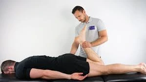
The purpose of the Prone Knee Bend Test is to find out whether the patient’s symptoms are due to neurological stress.
Patient’s Position: Prone.
How to perform the test: The examiner passively flexes the patient’s knee until it reaches the end range for 45 seconds. Rotating the hip is not recommended. A tight or strained quadriceps muscle or neural stress in the femoral nerve can both contribute to anterior thigh pain. Lumbar radiculopathy of the L2-L3 nerve roots may cause pain in one side of the lumbar region, buttocks, or posterior thighs.
The accuracy of the diagnosis is unknown.
Test Relevance:
Since the femoral nerve runs along the anterior portion of the lower extremity, knee flexion puts strain on it and its rootlets. The lower extremities may experience pain or other neurological symptoms if the femoral nerve becomes trapped in the tissues it passes through.
By putting strain on the whole nerve and its rootlets, the slump test can identify radicular pain or pain caused by spinal structural irritation. Although the L2-L3 nerve roots may be irritated by the innervation of the femoral nerve, the test should not be used alone for diagnosis.
Treat the underlying source of the patient’s deficiencies as you would normally. You should be able to use the location of the pain and symptoms to determine the pathology and determine the best course of treatment for the patient.
Halstead Test:
The Halstead Test is used to look for signs of developing thoracic outlet syndrome.
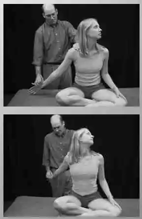
Technique:
The patient is standing or sitting. The radial pulse on the side being examined is continuously felt by the therapist. The therapist provides a downward distraction to the arm by externally twisting the upper extremity while still detecting the radial pulse, abducting the arm to 45 degrees, and extending the shoulder to 45 degrees. During the assessment, the patient is instructed to turn her head fully away from the side and stretch her cervical spine.
Roos Stress Test:

One diagnostic method for identifying TOS is the Roos Stress Test. Another name for it is the “elevated arm stress test,” or “EAST.”
Technique:
Starting Posture: The patient is placed in the frontal plane of the chest with both arms in the 90° abduction-external rotation position.
For three minutes, the patient is instructed to slowly open and close their hands.
The only symptoms of TOS that could appear are mild soreness and fatigue in the forearm muscles:
Neck and shoulder ache that progressively becomes worse and travels down the arm. The fingers and forearm become paralyzed when arteries are compressed. Reactive hyperemia happens when the limb is dropped under venous compression, and pallor happens when the arm is raised: Normal symptoms include cyanosis and edema, difficulty finishing the test, and the patient placing their arms on the floor in what appears to be agony.
Costoclavicular, military brace, or Eden’s test:
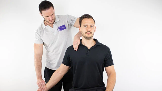
Finding thoracic outlet syndrome is the aim of the costoclavicular, military brace, or Eden’s test.
Technique :
The patient is upright. The examiner instructs the patient to elevate their chest in an exaggerated “at attention” position while pulling their shoulders back and down after feeling their radial pulse. When the pulse is missing or weakens, a positive test indicates that costoclavicular syndrome may be the cause. Additionally, it is advantageous if the client’s neurologic problems in the upper extremities worsen on that side. This test works well for patients who report symptoms while wearing a heavy coat or backpack.
Kehr’s Sign:
Put the patient in the supine position known as the Trendelenburg position, which involves elevating the lower extremities. Gently palpating the upper left quadrant of the abdomen will cause pain in the left shoulder.
Cervicothoracic tests: to determine the patient’s symptoms of neck pain.
Adson’s test:
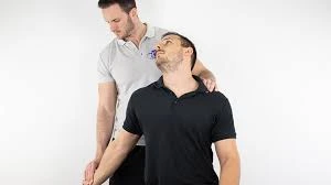
The Technique’s Beginning Point
- It is possible to have the patient sit or stand with their elbow fully extended.
- Whether the patient is standing or sitting, the arm is extended and abducted 30 degrees at the shoulder.
- The examiner holds the patient’s wrist and feels the radial pulse.
- The patient is then told to extend their neck, hold it, and turn their head toward the shoulder that is affected.
- The quality of the radial pulse is compared to the quality of the arm that is at the patient’s side during treatment.
- As part of a modified test, some doctors suggest that patients turn their heads away from the side that is being assessed.
Test Findings:
The test is considered successful if the radial pulse drastically diminishes or vanishes. To find out the patient’s normal pulse, the radial pulse on the other arm needs to be examined.
It is important to compare the positive side with the non-symptomatic side.
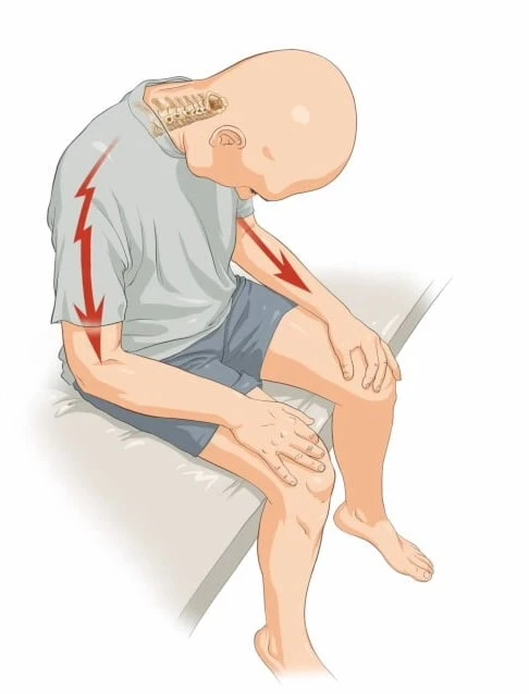
Slump Test:
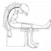
Technique:
- Many sources give different descriptions of the slump test. The reproduction of pain when the dura is stressed during testing.
- A patient may be sitting with their hands behind their back to create a neutral spine before the test starts. Slumping the patient forward at the lumbar and thoracic spines is the first step. Ask the patient to stretch their neck by placing their chin on their chest and then extending one knee as far as they can if they are not in any pain.
- Ask the patient to extend their neck into its natural position if extending their knee hurts. The test is deemed successful if the patient continues to experience pain when extending their knee.
- Request that the patient aggressively dorsiflex their ankle if extending their knee does not hurt. Encourage the patient to dorsiflex their knee while mildly flexing it if they are in pain. The test is considered successful if the pain returns.
- Repeat the test on the other side.
FAQs
Which 12 thoracic regions are they?
It is your spine’s longest segment. The twelve vertebrae that make up your thoracic spine are designated T1 through T12. The 33 separate, interconnecting bones that comprise your spinal column are called vertebrae. You can twist and turn due to these bones, which also assist in protecting your spinal cord from harm.
What signs indicate issues with the thoracic spine?
Tingling, weakness, or numbness. Although stenosis in the thoracic spine may compress the spinal cord and result in more severe pain radiating down the legs, compression of a nerve in the thoracic spine might generate pain or tingling that travels across the back into the chest.
What is the level of thoracic 7?
In the middle of the thoracic spinal column, the T7 vertebra is situated superior to the T8 and inferior to the T6. The centrum, sometimes called the vertebral body, is a broad, heart-shaped cylinder of bone that makes up the anterior part of the T7 vertebra.
What is the thoracic spine’s typical range?
The thoracic spine’s sagittal plane alignment is typically 35%, with a normal range of 20° to 50°.
Which thoracic spinal nerve is number twelve?
The thoracic segments of the spinal cord give rise to 12 pairs of spinal nerves. The intrinsic back muscles of the thoracolumbar area and the skin of the chest are primarily supplied by these nerves. Some of the skin covering the axilla and abdomen is also supplied by the thoracic nerves.
How is the thoracic spine examined?
Central and peripheral prone Thoracic and cervical spine. Palpate the upper and mid-thoracic region’s ribs 1–7. Motion of the ribs 1–7 from posterior to anterior accessory. Examine the cervical and thoracic spine’s medial grooves while seated to feel for variations in tissue texture. Check for variations in tissue texture on either side of the spinous.
What is a spine nerve test?
Nerve conduction tests are related to spinal diagnosis. Electromyography (EMG) is typically performed in conjunction with a nerve conduction investigation. Certain nerves are stimulated in the nerve conduction examination, and their capacity to transmit an impulse to the muscle is noted. The study can indicate the location of a neural route obstruction.
SLR is positive when?
When the straight leg is at an angle of between 30 and 70 degrees, the test is positive, and a herniated disk may be the origin of the sciatic pain, or more specifically, pain radiating down the leg (radiculopathy). If the test is negative, the source of the back pain is probably different.
What is the purpose of the slump test?
The slump test is a neural tension test used to help diagnose lumbar spine-based radicular pain or radiculopathy. Irritation of the nerve roots results in radiating pain, which usually manifests as shooting, stabbing, or acute pain along the nerve’s path.
References:
- Professional, C. C. M. (2024c, December 19). Thoracic spine. Cleveland Clinic. https://my.clevelandclinic.org/health/body/22460-thoracic-spine
- TeachMeAnatomy. (2024a, July 16). The thoracic spine – features – joints – ligaments – TeachMeAnatomy. https://teachmeanatomy.info/thorax/bones/thoracic-spine/
- Thoracic vertebrae. (2023, October 30). Kenhub. https://www.kenhub.com/en/library/anatomy/thoracic-vertebrae
- Moore, D. W., MD. (n.d.-b). Thoracic spine anatomy – spine – orthobullets. https://www.orthobullets.com/spine/2070/thoracic-spine-anatomy
- Yezak, M., DC. (n.d.). Thoracic spine anatomy and upper back pain. Spine-health. https://www.spine-health.com/conditions/spine-anatomy/thoracic-spine-anatomy-and-upper-back-pain
- Examination of the spine. (n.d.-b). Versus Arthritis. https://versusarthritis.org/about-arthritis/healthcare-professionals/training-and-education-resources/clinical-assessment-of-patients-with-musculoskeletal-conditions/the-musculoskeletal-examination-rems/examination-of-the-spine/
- Elsevier, Ltd. (2013). Clinical examination of the thoracic spine. In Clinical examination of the thoracic spine.