Clavicle Bone
Introduction
The long, thin bone that joins the shoulder girdle to the axial skeleton is called the clavicle, or collarbone. It supports the shoulder and makes arm motions easier with its S-shaped function as a structure. The medial and lateral ends of the shaft that make up the clavicle form significant articulations. As a location for muscle and ligament attachments, it enhances the upper limb’s stability and range of motion.
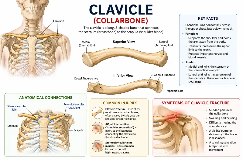
Given that it resembles an historic key in shape, the word “clavicle” is derived from the Latin word “clavicula,” which means “little key.” Additionally, when the arms are separated from the body, the bone spins like a key along its axis. Touching the area beneath your neck will allow you to feel its existence. Situated in the upper thorax, the clavicle runs horizontally between the scapula and the sternum. It joins the acromion of the scapula laterally at the acromioclavicular joint and articulates with the sternum’s manubrium medially at the sternoclavicular joint. Because it is subcutaneous and situated above the first rib, its length can be easily detected.
Anatomy of the Clavicle’s Components and Bony Features
The sternal and acromial ends are its two ends because it is a long bone. The shaft is the area that lies between the two ends. The long, thin, S-shaped clavicle, often known as the collarbone, is an essential component of the shoulder girdle. Because of its structure, it may support and move the upper limb while acting as a strut between the sternum and scapula.
- Medial or Sternal End
The sternal end, also known as the medial end, is the portion of the clavicle that faces the sternum. It has a rounded end that articulates with the sternum to produce the sternoclavicular joint. It is convex and curved.
i) The Sternal Facet
A triangular feature known as the sternal facet is located on the far side of the sternal end. At the sternoclavicular (SC) joint, it joins the clavicle to the sternum’s manubrium. It has an anterior base and a posterior tip. The inferior side is where the articular surface extends and articulates with the first rib’s costal cartilage.
- Lateral Acromial End
The acromial end or lateral end of the clavicle is the broad, flat area that faces the scapula. It is the clavicle’s thinnest and broadest section. The acromial facet, which articulates with the scapula, is present in this area. The anterior and posterior boundaries make up the lateral end. While the rear border is convex backward, the anterior border is concave forward.
i) The Acromial Aspect
The acromial facet is a tiny, round, flattened facet located at the very end of the acromial end. This facilitates the acromioclavicular (AC) joint’s formation by allowing the clavicle to articulate with the scapula’s acromion.
- The shaft
As previously stated, the shaft is the middle part of the clavicle, which is the area between the sternal and acromial ends. This area is where a number of muscles begin or attach.
Depending on its position and extent, the shaft can be separated into two areas. The largest part, or around two-thirds of the bone, is covered by the area on the side of the sternal end. The medial two-thirds is the name given to this area. Conversely, the remaining one-third of the bone is covered by the portion of the shaft that is located closer to the acromial end. The lateral third is the name given to this area.
These areas are home to a number of significant bone landmarks.
Costal Tuberosity (medial two-thirds)
The costal tuberosity is a roughened oval elevation on the inferior side of the sternal end. It serves as the costoclavicular ligament’s attachment point and is more than 2 cm long. The clavicle and the first rib’s costal cartilage are joined by this ligament.
Third lateral
(i) Conoid tubercle
The conoid tubercle is a rough, bumpy growth on the inferior surface of the bone. The conoid ligament, which is a component of the coracoclavicular ligament, attaches to it. The clavicle is connected to the scapula’s coracoid process by the coracoclavicular ligament. The conoid tubercle’s prominence is a helpful landmark for locating the clavicle’s inferior surface.
ii) Line of Trapezoids
It is a ridge or elevation that extends obliquely from the lateral end of the clavicle to the conoid tubercle. It offers a site of attachment for the trapezoid ligament, which is a component of the previously discussed coracoclavicular ligament.
Sulcus or subclavian groove
It is a horizontal indentation that extends from the costal tuberosity to the conoid tubercle along the shaft’s inferior surface. It serves as the subclavius muscle’s attachment point.
Articulations
a. Sternoclavicular Joint
A synovial saddle joint that connects the sternum’s manubrium to the clavicle’s sternal end.
Permits a variety of motions, such as rotation, depression, and elevation.
b. Acromioclavicular Joint
A planar synovial junction that connects the scapula’s acromion to the clavicle’s acromial end.
Transfers stresses from the upper limb to the axial skeleton and supports shoulder movements.
Blood Supply
The nutritional arteries might enter through the nutrient foramina, which are found on the shaft’s inferior surface.
The Arterial Supply
The clavicle receives blood from branches of the thoracoacromial and suprascapular arteries.
Ossification
Primary Ossification: Starting in the fifth or sixth week of fetal development, the clavicle becomes the first bone in the body to ossify.
The process of secondary ossification is finished by the age of 25 and takes place at the sternal end.
Muscular Attachments
The muscles that are connected to the clavicle are six in number. Two of these muscles are connected to the acromial end or lateral third of the clavicle, while the other four are connected to the sternal end or medial two-thirds.
The sternocleidomastoid muscle attaches to the sternal end’s superior surface.
The pectoralis major muscle attaches itself to the sternal end’s anterior surface.
The subclavius muscle is located at the inferior surface of the shaft’s subclavian groove. The muscle then spreads out to the medial and lateral sides.
The sternohyoid muscle is attached to the clavicle’s sternal end.
The trapezius muscle attaches to the acromial end of the bone along its rear surface.
The deltoid muscle is still connected to the acromial end of the bone along its anterior surface.
Function
The shoulder girdle’s support
By serving as a strut between the sternum and scapula, the clavicle keeps the shoulder away from the thorax and permits unrestricted upper limb movement.
Transmission of Force
It assists in distributing weights during lifting, pushing, and pulling actions by transferring mechanical forces from the upper limb to the axial skeleton.
Safeguarding Essential Structures
Underlying neurovascular structures such as the brachial plexus, subclavian artery, and vein are shielded from external harm by the clavicle.
Arm Movement Facilitation
The clavicle increases the shoulder’s range of motion by articulating with the scapula at the acromioclavicular joint, allowing for motions like raising and arm rotation.
Site of Muscle and Ligament Attachment
In addition to ligaments that maintain the acromioclavicular and sternoclavicular joints, the clavicle acts as an anchor point for muscles such as the pectoralis major, deltoid, trapezius, and sternocleidomastoid.
Nerve Supply
The subclavian, lateral pectoral, and supraclavicular nerves innervate the clavicle and clavicular joints.
The subclavius nerve (C5-C6) and the medial supraclavicular nerve (C3-C4) both directly innervate the sternoclavicular joint.
Ligaments
The coracoclavicular ligament and the acromioclavicular capsule and ligaments stabilize the acromioclavicular joint (AC).
The acromioclavicular ligament attaches to the lateral portion of the clavicle after emerging from the anteromedial border of the acromion. It is separated into anterior, inferior, and superior sections. It gives the AC joint horizontal stability.
The AC joint is vertically stabilized by the coracoclavicular ligament, which is made up of the conoid and trapezoid ligaments. The conoid ligament, which is conical in shape and almost vertical, is situated posteromedial to the trapezoid ligament and restricts superior migration of the clavicle, whereas the trapezoid ligament, which is larger and roughly quadrilateral in shape, inserts onto the distal clavicle along the trapezoidal line/ridge and limits posterior clavicular displacement and AC compression.
Fracture of the Clavicle
Forces from the upper limb are transferred to the axial skeleton via the clavicle. It is therefore more prone to breaking due to its relative size. A fall onto an outstretched hand or shoulder is the most frequent mechanism of injury.
Using arbitrary thirds to split the clavicle:
The lateral third is where 15% of fractures occur.
80% take place in the middle third.
In the medial third, 5% occur.
The weight of the arm and the pectoralis major cause the lateral end of the clavicle to shift inferiorly and medially, respectively, following a fracture. The sternocleidomastoid muscle pulls the medial end superiorly.
A clavicular fracture can be treated conservatively (by immobilizing the sling, for example) or surgically (by open reduction and internal fixation, for example). Sometimes, during a surgical repair, the supraclavicular nerves—which are located near to the clavicle—are sacrificed, leaving the upper chest and shoulder numb.
Left and Right Clavicle – How to Identify
This is a fast method for differentiating between the left and right clavicles.
Holding the mid-region, place the bone horizontally first. Additionally, make sure the bone is held superiorly, meaning its flat surface should be facing up.It is a right clavicle if you look at the medial two-thirds of your right side. Conversely, it is a left clavicle if the medial two-thirds are on your left side.
Clinical Relevance
As the link between the axial skeleton and the pectoral girdle, the clavicle is crucial to functional mobility. As a result, weight can be moved from the upper limbs to the axial skeleton by using the clavicle as a shoulder brace.
Along with the sternum, the clavicle protects neurovascular structures that enter the thorax, such as the internal jugular vein, common carotid artery, vagus and phrenic nerves, and brachiocephalic arterial trunk.
The most frequent cause of clavicular fractures, or shattered collarbones, is a fall or impact to the shoulder. It is typically treated conservatively by resting and wearing a sling. Surgery to install a plate with screws will be considered if the fracture causes the skin to become punctured.
To learn more about the clinical anatomy of the clavicle, visit the associated pages on acromioclavicular joint disorders and sternoclavicular joint disorders.
FAQs
Is the clavicle part of the axial skeleton?
The axial skeleton does not include the clavicle.
Is the clavicle part of the appendicular skeleton?
Indeed, the appendicular skeleton includes the clavicle.
Which type of bone is the clavicle?
This long bone has been changed. The shaft has an “S” shaped curve, and it has two ends: medial and lateral. It only has one secondary center of ossification and no medullary cavity.
How to hold the clavicle in anatomical position?
Having the clavicle horizontally, position the medial end inferiorly and somewhat forward relative to the lateral end.
References
- TeachMeAnatomy. (2024, October 8). The Clavicle – Functions – Landmarks – Fractures – TeachMeAnatomy. https://teachmeanatomy.info/upper-limb/bones/clavicle/
- Staff. (2022, September 8). Clavicle (Collarbone) – Location, Anatomy, & Labeled diagram. TheSkeletalSystem.net. https://www.theskeletalsystem.net/arm-bones/clavicle-collarbone.html
- Kranthi, & Kranthi. (2025a, January 28). Clavicle – Structure, anatomy, location, function, diagram. Anatomy.co.uk | Learn Human Anatomy From Experts. https://anatomy.co.uk/clavicle/