Serratus Posterior Muscles
Introduction
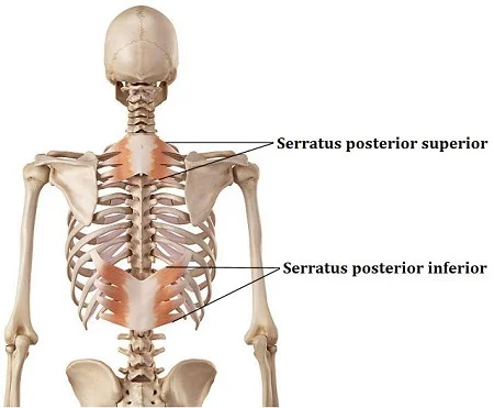
Serratus posterior muscles are two sets of muscles found in the upper and lower backs. These include:
- The serratus posterior superior muscle
- The serratus posterior inferior muscle
These muscles work together to form the back’s intermediate layer of extrinsic musculature.
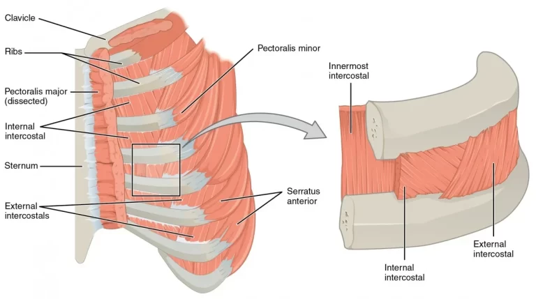
These muscles’ principal function is to facilitate breathing; the serratus posterior superior muscle lifts the ribs, while the serratus posterior inferior muscle lowers them. In the case of forced breathing, these activities are very crucial.
Structure
Origin & insertion
The serratus posterior muscles, also called spinocostal muscles, link the vertebrae’s spinous processes to the ribs.
The nuchal ligament, spinous processes of vertebrae C7-T3, and related supraspinous ligaments give birth to the serratus posterior superior muscle as a thin tendinous sheet. The muscle fibres stretch inferolaterally into the lateral portion of the posterior thoracic cage. Each of the four finger-like projections that form the muscle’s insertion attaches to the posterosuperior portion of ribs 2-5, lateral to their angles.
Then, directly lateral to their angles, it ascends anterolaterally and penetrates the inferoposterior side of ribs 9-12.
Blood Supply
The posterior intercostal arteries provide circulatory support to both muscles.The subcostal and upper lumbar arteries provide more blood to the serratus posterior inferior.
Nerve Supply
- The serratus posterior superior is innervated by the ventral rami of the intercostal nerves T1-T5.
- The T9-T12 intercostal nerve’s ventral rami innervate the serratus posterior inferior muscle.
Relations
The serratus posterior inferior muscle is deeper than the latissimus dorsi but lies closer to the thoracolumbar fascia and erector spinae.
Function
Both muscles play supportive roles in respiration and are situated deep to the larger superficial back muscles.
The precise function of the serratus posterior muscles is still contested, however most people believe they play a part in breathing.
The serratus posterior superior aids inspiration (an auxiliary muscle of inspiration) by elevating the ribs.
The serratus posterior inferior, on the other hand, facilitates expiration by lowering the ribs, an expiratory auxiliary muscle.
Clinical Importance
Scapulocostal syndrome can be caused by an overuse of the serratus posterior superior and other back muscles (e.g., holding the phone between the ear and shoulder). Pain and paraesthesia along the scapula’s medial edge that radiates to the neck, chest, and upper extremities are its defining features. One of the most noticeable symptoms is discomfort, especially when the little finger muscle is palpated. Activities that strain the serratus posterior superior muscle from the scapula, such as lifting objects with extended hands, may make the pain worse.
Clinically, scapulocostal syndrome is sometimes confused with cervical radiculopathy, a lesion of the cervical vertebral column’s spinal nerves that usually manifests as numbness, muscular weakness, and further neurological impairments (such as absent or weak reflexes). Rotator cuff rupture or arthritis should also be taken into consideration as a differential diagnosis.
Treatment
Strengthening Exercises
Prone Arm Lifts (for SPS)
- With your arms outstretched, lie face down. Squeeze your shoulder blades together and raise your arms slightly above the ground.
- Targets the SPS and other upper-back muscles.
Resisted Diaphragmatic Breathing (for SPS/SPI)
- Wrap a towel or resistance band around your lower ribcage. Take a deep breath when facing the adversary.
- activates both SPS and SPI, aiding in the training of rib movement.
Quadruped Thoracic Extensions (for SPI)
- While on all fours, focus on thoracic mobility by rounding your back up (cat) and then extending your upper spine (cow).
- Strengthens the SPI and other spinal muscles, especially when done carefully and gradually.
Stretching Exercises
Child’s Pose with Side Stretch
- To feel the stretch in your ribs and lower back, return to the child’s position and shift your hands to one side.
- SPI and the torso side.
Seated Forward Fold with Rib Expansion
- Fold forward with your legs outstretched, concentrating on softly breathing to lengthen your back.
- SPI and the thoracolumbar fascia are stretched.
Wall Angels (Active Stretch)
- “goalpost” position. Slowly lift and drop the arms while maintaining wall contact.
- extends the upper back and opens up the thoracic spine (SPS).
FAQs
What causes serratus posterior pain?
SPI muscle discomfort can be caused by a variety of factors, including resting on an excessively soft mattress, bending sideways, overstretching the body while reaching aloft, twisting the back when moving something heavy, and activating the back muscles rather than the legs.
How do you fix serratus pain?
How is serratus anterior pain treated?
Get some rest. Try to relax the muscle as much as you can and take it easy with your everyday tasks.
Ice. Apply an ice pack wrapped in a towel to the sore muscle for 20 minutes at a time, many times each day.
Compression. …
Elevation.
How to strengthen the serratus posterior?
Serratus Posterior
On the floor, place two heavy kettlebells shoulder-width apart.
As though performing a Romanian deadlift or bent-over row, stand behind the kettlebells with your feet hip-width apart and flex at the hips to drive your butt back.
Grab the grips and maintain the posture once you can reach the kettlebells.
What are the symptoms of serratus weakness?
When you reach or hold your arm overhead, you may have neck or shoulder pain as you rotate upward. weakness or shakiness when performing overhead pressing exercises or bench exercises at the gym. When you extend forward and above, your shoulder joint feels clunky or clicky.
References
- Serratus posterior muscles – 2023https://www.kenhub.com/en/library/anatomy/serratus-posterior-muscles
- Serratus posterior muscles: Anatomy, clinical relevance, and function – 2001 – Vilensky, Baltes, Weikel, Fortin, Fourie