Phalanges of Foot
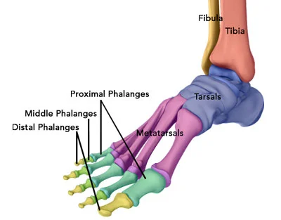
Phalanges of the foot are tiny fingerbones located in the feet. Each foot consists of 14-foot phalanges. The large toes or hallux have two phalanges the proximal and distal, whereas the others have three: proximal, middle, and distal.
For individual toes, the metatarsal-phalangeal articulation binds the metatarsals to the proximal phalanx. These joints are what make up the football. The big toe and the first metatarsal phalangeal joint are parallel to each other. This location frequently causes foot pain and other issues.
Types of Phalanges of Foot
The lengthy bones in the foot situated distal to the metatarsals are called phalanges. Comparable to the hand, every toe carries three phalanges i.e. proximal, middle, and distal phalanges.
Proximal phalanges
Proximal phalanges (Latin: phalanges proximales) are the proximal bones of the fingers of the hand and foot. Every foot has five proximal phalanges, which correspond to five toes. Out of the three, the proximal phalange is the biggest.
Middle phalanges
The middle phalanges, also known as intermediate phalanges (Latin: phalanges mediae), are essentially detached from the proximal and distal phalanges. Since the big toe lacks middle phalanges, each foot has four in total. In terms of their shape and position, the middle phalanges fall within the proximal and distal phalanges.
Distal Phalanges
The rounded portions of the bones at every single toe’s tilt are known as the distal phalanges (Latin: phalanges distales). Each foot bears five distal phalanges that are curved in form distally. The distal phalanges have flattened heads with rough, raised patches called tuberosities.
Anatomy of Phalanges of Foot
Interphalangeal joints are formed when the phalanges of each toe articulate with one another. Metatarsophalangeal joints develop when the proximal phalanges articulate with the metatarsals of the foot.
The bottom is positioned at the proximal end of every phalanx, followed by the head at the distal end and the body in the core of the bone.
- Base: The base or proximal extremity’s proximal section is enlarged. At the base every single proximal phalanx are circular and concave articular surfaces. However, a median ridge divides the two identical concave articular surfaces at the bases of the middle and distal phalanges.
- Body: The body, also occasionally referred to as the shaft, is the extending prolongation coming from the base. Every phalanx’s body is convex on both flanks, with the dorsal surface being more concave than the plantar surface. Flexor tendons can adhere to rough spots on the sides of the body.
- Head: The heads or distal extremities have rounded distal ends and are enlarged. Phalanges’ heads are smaller than their bases. Except for the distal phalanges, which have distal tuberosities and are flattened, each head has an articular surface.
Articulation
- Through the metatarsophalangeal joint, the proximal phalanges keep their connection to the metatarsals.
- The association between the proximal and middle phalanges occurs at the proximal interphalangeal joint.
- A distal interphalangeal joint is used to connect the middle and distal phalanges.
- An individual interphalangeal joint had been identified on the great toe.
Muscles Attachment
- The rear of the foot is where the extensor tendons are fastened. Extensor hallucis longus is received by the great toe’s distal phalanx, whereas extensor hallucis brevis is received by the proximal phalanx.
- The lumbrical, interossei, and digitorum extensors make up the extensor development. The tendons known as extensor digitorum brevis are connected to the second, third, and fourth toes’ extensor expansions.
- While flexor hallucis brevis enters the proximal phalanx through the two sesamoids, flexor hallucis longus joins to the great toe’s distal phalanx on the plantar surface.
- The flexor digitorum longus connects the distal phalanx, and the flexor digitorum brevis connects all of the additional toes to the sides of the middle phalanx.
- The pointed end of the fifth proximal phalanx can be utilized for attaching the flexor digiti minimi brevis and abductor digiti minimi brevis.
Ligaments Attachment
- Each metatarsophalangeal joint on the plantar surface is strengthened by the plantar ligament, a substantial fibrocartilage pad.
- The plantar ligaments that encircle the metatarsophalangeal joints are joined by the deep transverse metatarsal ligament, which is made up of four tiny bands of fibrous tissue.
- Collateral ligaments (similar to the hand) strengthen the sides of the metatarsophalangeal joint and interphalangeal joints.
Blood Supply
- Arterial Supply: The external plantar arch of the foot has been established by the lateral plantar artery. Across the bases of the second, third, and fourth metatarsals, this curvature is convex. The four webs and digits are nourished by plantar metatarsal arteries, which emerge from this arch. The hallux receives its flow of blood right from the medial plantar artery. Dorsal metatarsal arteries receive their distribution from the foot arch’s starting and metatarsal arteries.
- Venous drainage: The perforating veins, which accompany the perforating arteries, convey blood from the dorsal venous arch. The “sole pump” function of the plantar muscles facilitates the passage of blood back into the veins.
Nerve Supply
- Both the dorsal and plantar digital nerves innervate the complete phalangeal bone assembly.
Anatomical variation
- The plantar portion of the great toe’s metatarsophalangeal joint typically has two sesamoid bones, with the medial being the bigger one (refer to multipartite hallux sesamoid). Sesamoid bones can be discovered in the metatarsophalangeal joint joints of the second to fifth toes and the interphalangeal joint of the great toe.
Function of the Phalanges of Foot
- The proximal phalanges are important for proper toe bending.
- As such, they play an essential function in everything from walking to jumping.
- Additionally, these bones play a crucial role in lateral motions, which aid in maintaining your balance and navigating uneven surfaces.
Relevant Condition to Phalanges of Foot
The lateral four toes are nearly usually affected by toe deformities, which can result in incapacitating discomfort. The metacarpophalangeal, proximal interphalangeal, and distal interphalangeal joints dorsiflex due to claw toe. Women and older people are more likely to have this malformation. Certain neuromuscular conditions, including multiple sclerosis and cerebral palsy, can coexist with claw toe. Additionally, it can be observed in inflammatory conditions like rheumatoid arthritis and metabolic disorders like diabetes mellitus.
Hammertoe deformity
Hammertoe deformity is the most prevalent abnormality observed in the distal four toes. This deformity is typically caused by poorly fitting shoes, which leads to hyperextension of the metatarsophalangeal and distal interphalangeal joints as well as a flexion deformity of the proximal interphalangeal joint. Wearing the proper footwear and taping the toe are effective therapy options for this issue. After using these techniques, surgical correction might be necessary if the incapacitating discomfort persists.
Mallet toe
The term “mallet toe” refers to a distal interphalangeal joint flexion abnormality. This disorder, which may be caused by trauma or improper footwear, may result in calluses and nail abnormalities. Orthotics and toe guards are examples of non-operative therapy options.
Fractures
The fifth toe is the most frequently fractured; toe dislocation may also occur. These fractures are typically caused by trauma, such as a falling object or a toe stub, and they frequently happen distal to the metacarpophalangeal joint.
FAQs
What are the 5 phalanges?
Five proximal phalanges, one associated with every digit, are present in each finger. In terms of the distal and middle phalanges, they are the largest. The exception is the thumb’s proximal phalanx, which is broader and shorter than the others.
The fourteen phalanges are what?
The toes of both feet and the fingertips of both hands contain these 14 bones. The phalanges of each finger are proximal, middle, and distal. Only two are present in the thumb.
Which formula applies to phalanges?
A “phalangeal formula” that shows the number of phalanges in digits is frequently used to express the number of phalanges in animals. The thumb has two phalanges, whereas the remaining fingers each have three, according to the 2-3-3-3-3 formula seen in the hands and feet of the majority of land mammals, including humans.
What function do the toes’ phalanges serve?
The proximal phalanges are crucial for proper toe bending. They are therefore crucial for actions like walking and jumping. Additionally, these bones play a crucial role in lateral motions, which aid in maintaining your balance and navigating uneven surfaces.
References
- Phalanges of the foot. (2022, November 24). Kenhub. https://www.kenhub.com/en/library/anatomy/phalanges-of-the-foot
- Hacking, C. (2015). Phalanges of the feet. Radiopaedia.org. https://doi.org/10.53347/rid-41552
- Next, A. (n.d.-b). %s | Anatomy.app | Learn anatomy | 3D models, articles, and quizzes. https://anatomy.app/encyclopedia/phalanges-of-foot
- Foot phalanges. (n.d.). https://surgeryreference.aofoundation.org/orthopedic-trauma/adult-trauma/foot-phalanges
- Wikipedia contributors. (2024c, November 30). Phalanx bone. Wikipedia. https://en.wikipedia.org/wiki/Phalanx_bone

