4 Stages Of Disc Herniation
Introduction
Herniated disc-related back pain is so prevalent that it is now regarded as a global health issue! This disorder affects between 2% and 3% of adults in the United States. Furthermore, it is not an age-related issue.
In actuality, the risk of developing a herniated disc is higher between the ages of 30 and 50 than after the age of 70. In addition, males are twice as likely as women to get a herniated disc, making them more affected than women.
However, disc herniation does not always cause back and neck discomfort, nor does it usually cause pain to start right away. To determine whether your back discomfort is due to a herniated disc, keep reading.
Firstly, what is an intervertebral disc exactly?
Discs and vertebrae make up your vertebral column, which is the skeletal framework that encircles the spinal cord. The discs are between the vertebrae and function as shock absorbers. These little pillows:
- Avoid impact and friction injuries.
- Give the spine support.
- Give the spine the capacity to support its weight.
- The weight should be distributed throughout the spine.
- To put it simply, 23 of these little, half-inch guards put out constant effort to maintain the safety and health of your whole spine. The 12 discs in the thoracic area (behind your chest) usually won’t cause you any issues. The discs in the lumbar region—five in the lower back—are the most vulnerable to herniation. The second-highest risk rate is seen in those in the cervical region (6 discs in the neck).
Relevant Anatomy for Clinical Practice
Intervertebral discs: It connects two vertebral bodies. They make up the “functional unit of Junghans” together with the matching facet joints.
The disk consists of an annulus fibrosus, a nucleus pulposus, and two cartilaginous endplates. The annulus and nucleus can only be distinguished in youth since the disc’s homogeneity becomes more constant with age. Nuclear disk protrusions are hence rare after age 70. From a therapeutic standpoint, it is essential to view the disc as a single, cohesive unit, with each component’s integrity being critical to the disc’s normal function. This suggests that the other components will react adversely if one is destroyed.
The disc has an endplate, nucleus pulposus, and annulus fibrosus.
Etiology/Causes
The annulus fibrosus, a thick collagenous ring that encircles the nucleus pulposus, makes up an intervertebral disc.
- When all or a portion of the nucleus pulposus pokes through the fibrous annulus, disc herniation takes place. Failure in the innermost annulus rings initiates this herniation process, which then spreads outward radially.
- There appears to be a connection between disc annulus injury and frequently or prolongedly fully extending the spine.
- A herniation may appear all at once or gradually over several weeks or months.
- The degenerative process is the most frequent cause of disc herniation; as people age, their nucleus pulposus weakens and loses moisture, which can cause progressive disc herniation.
- Trauma is the second most frequent cause of disc herniation.
- Congenital conditions, including short pedicles and connective tissue abnormalities, are other reasons.
- The most prevalent location for disc herniation is the lumbar spine.
- The cervical spine comes next. Understanding the biomechanical pressures in the flexible portion of the spine can help explain the high rate of disc herniation in the lumbar and cervical spine.
- In the thoracic spine, disc herniation is less common.
- Disc injury can result from repetitive mechanical tasks like twisting and bending without pauses.
- Disc prolapse can also be brought on by a sedentary lifestyle, bad posture, obesity, and tobacco use.
Pathophysiology
- The nucleus pulposus, a gelatinous core made up of collagen, elastin, and a hydrated gel, and the annulus fibrosus, a complicated arrangement of fibrous rings, make up the disc. The anterior wall’s vertebral bodies, intervertebral discs, and ligaments, as well as the lateral wall’s vertebral arches and ligaments, combine to form the vertebral canal. This vertebral canal contains the spinal cord.
- Herniated discs are thought to be caused by a combination of the bulging nucleus pulposus mechanically compressing the nerve and the localized rise in inflammatory chemokines.
- The annulus fibrosus can rip. To impact neural anatomy, the nucleus pulposus material may pass through this rupture and enter the intervertebral or vertebral foramen.
- The changes consist of nuclear degeneration, nuclear displacement, and stage of fibrosis.
Types Of Herniations
- Posterolateral Disc Herniation – Protrusion is commonly posterolateral into the spinal canal. As the nerve passes over the level of the disc on its way to the foramen, the next lower nerve is often compressed by the protruded disc. (L5 protrusion, for example, usually affects S1)..
- Less often, central (posterior) herniation occurs. Cauda Equina Syndrome or spinal cord compression might result from a protruding disc above the second vertebra.
- Nerve root compression occurs above the level of lateral disc herniation. Most frequently, the L4 nerve root is affected.
A herniated disc: What is it?
The strong, rubber-like outer shell (Annulus fibrosus) of these intervertebral discs envelops a jelly-like nucleus. The idea is that, regardless of the location of the herniation, the nucleus presses against the annulus and causes it to protrude into the spinal canal.
The general idea is that, regardless of the location of the herniation, the nucleus pushes against the annulus and causes it to protrude into the spinal canal. Since the spinal canal is small, the bulging annulus eventually presses against the nerve root, resulting in back pain. If the problem is not addressed promptly, the nucleus may tear through the annulus and leak the jelly-like substance into the spinal canal.
The most prevalent symptoms of lumbar disc disease are as follows:
The location of the disk’s herniation and the nerve root it is pressing on determine the symptoms of lumbar disc disease. The most typical signs of lumbar disc disease are as follows:
Backache that comes and goes. Standing for extended lengths of time, coughing, sneezing, or moving about might worsen this.
Muscle spasm in the back
Sciatica. This type of discomfort begins in the area of the buttocks or back and moves down the leg to the calf or foot.
- Leg muscle weakness
- Soreness in the foot or leg
- Diminished knee or ankle reflexes
- Modifications to bowel or bladder function
Lumbar disc disease symptoms might mimic those of other illnesses. For a diagnosis, always consult your healthcare provider.
What separates a herniated disc from a disc bulge?
An air leak from a car tire is analogous to a bulging disc. The disc appears to be expanding outward as it sags. This is usually regarded as the first of a herniated disc’s four phases. A herniated disc may eventually develop a hole or tear in its outer layer.
As a result, the nucleus pulposus—the disc’s jelly-like core—leaks into the spinal canal and may press against the nerve, resulting in symptoms that extend from the lower back to the toes.
Disc herniation’s causes
A single excessive injury: While a single, severe shock has the potential to rupture the disc, this is uncommon unless the injury is the consequence of a severe impact, such as that which occurs in a vehicle accident or a terrible fall.
Ageing: The annulus becomes fragile, brittle, and thin once the spinal discs begin to dry up beyond the age of thirty. The hard outer ring may ultimately break as a result of this.
Obesity: Gaining too much weight can lead to several health problems, including problems with the spinal disc. Obesity increases the risk of disc injury and rupture by placing extra strain on the discs.
Repetitive motion: The discs may gradually deteriorate as a result of any activity (baseball, golf, etc.) that repeatedly stresses or traumatizes a specific region of the spine.
Straining the spine with lifting: When lifting incorrectly, you put a great deal of pressure on the discs and the vertebral column.
Genetics: Disc herniation is more common in people with a family history of the disorder.
How do you detect a herniated disc?
A doctor will use a three-step procedure to assess if a herniated disc is the cause of your back pain when you visit them.
Medical history and symptoms: This is done to determine whether disc herniation was caused by a hereditary component or an underlying or previous ailment.
Physical examination: The goal is to find out if you have muscular weakness or lack of feeling.
You could have one or more of the following tests in addition to a thorough medical history and physical examination:
X-ray: This test creates pictures of inside organs, bones, and tissues on film using invisible electromagnetic energy beams.
MRI: This process creates fine-grained pictures of the body’s organs and structures without the need for X-rays by combining radio frequencies, big magnets, and a computer.
Myelogram: This procedure uses dye injected into the spinal canal to make the structure visible on X-rays.
CT scan: This imaging technique creates fine-grained pictures of any area of the body, including the bones, muscles, fat, and organs, using X-rays and computer technology.
EMG stands for electromyography: This test gauges how the muscles react electrically when they are stimulated by a nerve. This examination looks for any related nerve injury.
Discogram: In this process, a needle is guided by X-rays and inserted into the disc.
Assessing the healing process of a herniated disc requires both routine monitoring and diagnostic instruments. These resources offer insightful information that enhances patients’ subjective accounts of their experiences.
Imaging Outcomes:
Healing is evident when MRI or X-ray pictures show positive improvements. Healthcare professionals can see the structural improvements in the damaged disc and surrounding tissues by comparing photographs over time.
Medical Examination: Insights into the healing process are obtained from feedback given by healthcare professionals during medical examinations. An all-encompassing awareness of the recovery state is facilitated by neurological evaluations, physical tests, and conversations regarding symptoms.
Follow-up Consultations:
Making follow-up visits with the medical staff on time is essential for monitoring progress. These consultations enable the treatment plan to be modified, any new issues may be addressed, and a cooperative attitude to the continuous healing process is ensured.
Study of neurophysiology
Some persons may benefit from additional diagnostic instruments or monitoring techniques, such as nerve conduction tests and electromyography (EMG).
Physical Assessment
The degree of compression can be localized with the use of a thorough neurological examination. The following describes the various levels of sensory loss, weakness, pain location, and reflex loss.
Typical results of a single nerve lesion in the lumbar spine caused by a herniated disc
- The inguinal area frequently experiences L1 Nerve discomfort and sensory loss. No stretch reflex is impacted, and hip flexion weakness is uncommon.
- Back discomfort that radiates into the anterior thigh and medial lower leg; sensory loss in the anterior thigh and occasionally the medial lower leg; weakness in hip flexion and adduction; weakness in knee extension; and a diminished patellar reflex are all symptoms of the L2-L3-L4 nerves.
- The L5 nerve is located in the back and radiates into the buttock, lateral thigh, lateral calf, dorsum of the foot, and great toe. It causes weakness in hip abduction, knee flexion, foot dorsiflexion, toe extension and flexion, foot inversion and eversion, and the semitendinosus/semimembranosus reflex.
- Sensory loss on the posterior calf, lateral or plantar aspect of the foot; weakness on hip extension, knee flexion, and plantar flexion of the foot; S1 Nerve-back, radiating into buttock, lateral or posterior thigh, posterior calf, lateral or plantar foot; Achilles tendon; medial buttock, perineal, and perianal area; sexual dysfunction, fecal and urine incontinence, and maybe mild weakness.
- S2–S4 Nerves: absence of bulbocavernosus, anal wink, sensory deficiency on the medial buttock, perineal, and perianal area; sacral or buttock discomfort extending into the posterior portion of the leg or the perineum
The Four Herniated Disc Levels
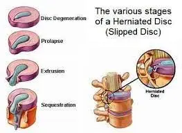
Level 1: Disc Bulging
The mildest type of herniation is a bulging disc, which happens when a disc’s outer layer weakens, and the jelly-like center pushes out against it. Lower back discomfort that extends outward is one of the common, moderate symptoms of a bulging disc. Medication, physical therapy, and rest are all possible treatments for a bulging disc to reduce inflammation and pain.
Level 2 – Protruding Disc
A bulging disc puts pressure on the spinal nerves when the disc’s jelly-like core pushes out past the outer covering. It can result in tingling or numbness that extends outward, along with moderate to severe lower back discomfort. Rest, physical therapy, and pain and inflammation-reducing drugs are often used to treat a bulging disc. In more difficult situations, surgery can be necessary.
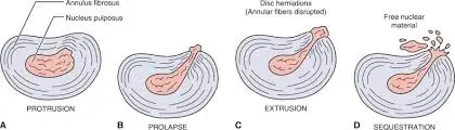
Level 3: Disc Extrusion
When a disk’s jelly-like core is driven out past its outer layer and no longer contained inside it, it is said to be extruded. In addition to numbness and tingling that travel outward, this might result in severe lower back discomfort. Rest, physical therapy, and pain and inflammation-reducing drugs are usually used to treat an extruded disc. To alleviate pressure on the spinal nerves and remove the extruded disc, surgery can be required in certain situations.
At this point, the outer shell is penetrated by the inner disc material. If the affected disc is in the lumbar region, as is typical with disc herniation, you may have lower back pain that radiates downhill after the inner material ruptures. Patients can frequently benefit from nonsurgical therapy at this stage, such as:
- Exercises for correcting posture;
- steroid injections and other drugs;
- physical treatment;
- hot and cold therapy; and activity modification
With the aid of the aforementioned therapies, the associated symptoms may now be entirely resolved or controlled. Nonetheless, some motions may still cause pain flare-ups.
Surgery may eventually be advised in some situations. Depending on which nerve is injured and how the bulging disc material is irritating or compressing it, you may or may not be affected. The good news is that at this point, surgery is only required around 10% of the time.
Level 4: Confined Disc
The most severe type of herniation is called a sequestered disc, in which the disc’s jelly-like core has detached from its outer covering. It may result in widespread tingling and numbness as well as severe lower back pain. Even loss of feeling and muscular weakening are possible. To relieve pressure on the spinal nerves, surgical excision is typically the best course of action for a sequestered disc. In certain instances, if the disc has seriously damaged the vertebrae, a lumbar fusion could also be required.
When the inner disc material completely separates from the shell and the spinal disc, disc sequestration takes place. This is a significant condition that frequently necessitates a discectomy. Additionally, it can cause some equally serious problems, particularly if it becomes stuck in the spinal canal. You could get extreme nerve discomfort in this situation. Patients are frequently bedridden at this stage because of how uncomfortable they are.
A discectomy or a less invasive microdiscectomy could be suggested if conservative therapy for your herniated disc is not working. Even though this is typically one of the most effective back surgery procedures, individuals who have a substantial hole in the disc’s outer ring are far more likely to experience herniation after the treatment. A huge hole in the disc’s outer ring more than doubles the chance of requiring another operation, and the surgeon frequently won’t know the hole’s size until the procedure starts.
Particular Examinations
The cervical
- Spurling test.
- Test of distraction.
- Test of upper limb tension.
- Test of shoulder abduction.
- The sign of Tinel
- Lumbar
The test of the straight leg raise:
- The straight leg lift test that is contralateral (crossing)
- Lasègue’s test.
- Bowstring examination
- Bending the knees prone
- Paresis reflexes or muscle weakness
- Test of hyperextension: While keeping the knees extended, the patient must passively move the trunk through its whole range of extension. If the radiating pain worsens, the test shows that disc herniation is the cause.
- Sensory and manual testing: Check for tingling, numbness, hypoaesthesia, or hypoalgesia.
Differential Diagnosis
From a clinical and imaging perspective, other diseases might mimic a herniated disc and should be taken into account.
These lesions include those originating from the:
Osteophytes and metastases in the vertebral body; a discal cyst in the intervertebral disc; neuromas in the intervertebral foramina; a synovial cyst in the interapophyseal joints; and hematoma and epidural abscess in the epidural space.
Additional differential diagnoses consist of:
- Spondylolysis
- Spondylolisthesis
- Causes of Cauda Equina Syndrome
- Spasms in the muscles
- Pain from a machine
- Pain in the myofascial region (causes sensory abnormalities, local and/or referred discomfort)
Among the spinal reasons are:
- Osteomyelitis is an infection from trauma
- Arachnoiditis and ankylosing spondylitis are examples of inflammation.
- Nerve roots are compressed by multiple myeloma, extradural tumors, and benign or malignant neoplasms.
Among the extraspinal reasons are:
- Peripheral vascular disease
- gynaecological state
- Sacroiliac joint disease or hip OA
- Lesions in peripheral nerves
Healing signs in herniated disc
Early Signs of Progress
After a herniated disc, there may be visible improvements in several physical well-being elements in the early phases of recovery. Since these indicators indicate progress in healing and recovery, both patients and medical practitioners need to recognize them.
Decreased Pain
Less discomfort is the first and most obvious indication. Your overall back pain will start to improve as the inflammation goes down and the strain on your nerves goes down. A decrease in the severity of sciatica or leg pain or its spread down the leg is a good indication of whether the herniation damaged a nerve that caused it.
Enhanced Mobility
A good sign of healing is when you can bend or move more easily without experiencing excruciating pain. You’ll probably notice an increase in your mobility and capacity to carry out everyday tasks without experiencing agony as your pain becomes more bearable.
Reduced Nerve Pain
If your arms or legs were tingly or numb from the herniation, a return to normal feeling is a sign that your nerves are mending. Generally speaking, a reduction in pins and needles is a positive indication that nerve irritation is abating.
Reduced Spasms of Muscles
Muscle spasms surrounding the affected area frequently relax and lessen with less discomfort, improving mobility.
Improved sleep quality
Sleep is frequently disturbed by pain. A positive indication of success may be better sleep patterns brought on by less discomfort at night.
Advancing through the phases of healing
Additional information on the total recovery from a herniated disc may be gained from the passage through several phases of healing. You may evaluate your progress and modify your expectations by being aware of these stages.
Changes in Pain
Sharp, continuous pain may give way to more or less tolerable pain. This shift denotes improvements in the underlying structures and less strain on the impacted nerves.
Function Recuperated
One of the main indicators of functional recovery is the return of the capacity to carry out everyday activities with greater ease. As the healing process progresses, patients may see improvements in function, strength, and coordination.
Reduced Drug Use
Progress in controlling pain through natural healing processes is indicated by a decrease in the need for painkillers. The need for medicine may lessen as symptoms become better, resulting in a more self-sufficient and long-lasting recovery.
Better Sleep:
For general well-being, better sleep quality as a result of less pain at night is essential. Patients frequently report better sleep patterns as they recuperate, which helps them recover more quickly and perform better during the day.
Others:
Other encouraging signs, such as a better mood, more energy, or a return to formerly favored hobbies, may result from individual variances.
Herniated disc treatment
- SPONTANEOUS RECOVERY: Over three to four months, a disc herniation may occasionally improve on its own.
NON-SURGICAL MEDICATION: But as you heal, you might want assistance with pain management. Usually, the following medications are recommended to treat pain:
- Taking a vacation from the activities that triggered the issue, resting, and slowing down.
- Intentional efforts to reduce activities that could worsen the problem, such as lifting and bending.
- To manage pain, doctors use nonsteroidal anti-inflammatory drugs (NSAIDs), most often ibuprofen or naproxen.
- Workouts that are specifically designed to assist tone and strengthening the muscles in the belly and lower back.
- If your severe pain keeps you from participating in physical therapy, epidural steroid injections could be advised.
- SURGICAL INTERVENTIONS: Disc herniation surgery is only contemplated after all other treatment options have been exhausted and the condition is still getting worse. Options for surgical therapy include:
DISCECTOMY: The least invasive procedure involves removing the disc portion that is pushing against the nerve.
LAMINECTOMY: To reach the disc from behind, the vertebral roof is removed.
FUSION AND INSTRUMENTATION: Only required in cases of severe spinal instability and disc injury.
LASER SPINE SURGERY: This minimally invasive procedure releases pressure by evaporating a portion of the disc using heat from a tiny laser.
Herniated disc healing process
A herniated disc often heals gradually and goes through several phases, each with unique traits and difficulties.
Acute Inflammation, Stage 1 (1-2 days to 2 weeks)
This first phase is frequently the most excruciating. You could encounter:
Severe discomfort in the affected location, such as your neck or lower back.
If a nerve in your lower back is affected by the herniation, you may get sciatica, or pain radiating down your leg.
- Contractions in the surrounding muscles.
- Tingling or numbness in the affected region.
- Having trouble bending or moving.
Treatment: At this point, the main goals are to control discomfort and stop more harm. This might include:
- To prevent aggravating the disc, rest and change your activities.
- Heat and ice treatment can help lower inflammation.
- Over-the-counter muscle relaxants or painkillers.
- Drugs that reduce inflammation.
You may learn safe stretches and exercises in physical therapy to aid with your recovery and avoid more issues.
Phase 2: Progressive Enhancement (two to six weeks)
During this phase, you should begin to see a slow improvement in your symptoms if the original therapy is successful. You could encounter:
- Reduced frequency and severity of discomfort.
- Less jerking of the muscles.
- Increased range of motion.
- Reduced tingling or numbness.
You can carry on with the stage 1 therapies, making any necessary adjustments in light of your development. To increase your mobility and strengthen your core muscles, you could also be able to start more difficult physical therapy activities.
Stage 3: Stabilization and Plateau (6–12 weeks)
Your discomfort may have somewhat lessened by this point, but it may still be there, particularly when engaging in particular activities. Additionally, you could encounter:
Occasional pain flare-ups following physically demanding activities.
There is still some tightness or stiffness in the affected region.
At this point, preserving your development and averting future issues are the major priorities. As you progressively resume your regular activities, you can resume your physical therapy exercises, being careful to pay attention to your body and prevent re-injury.
Stage 4: Extended-Duration Management (Over 12 Weeks)
By this point, the majority of patients with herniated discs have significantly improved and can return to their regular activities. However, if you don’t maintain proper posture, core, and flexibility, you can still experience some of the pain.
At this point, preventing such episodes and preserving your general health and well-being are the major priorities. This might include:
- Maintaining core strength and flexibility requires frequent physical therapy exercises.
- Use proper body mechanics and posture in your day-to-day tasks.
- Keeping a healthy weight in mind.
- Avoiding excessive alcohol use and smoking.
Remember that each person recovers at a unique pace. These phases are only meant to serve as basic guidelines; your exact healing period may differ. Age, general health, herniation severity, and degree of exercise all affect recovery.
Additionally, keep in mind that not all ruptured discs recover completely. The disc annulus tear frequently persists even though the bulge may decrease and the discomfort may subside. If conservative measures fail to relieve pain and impairment from a herniated disc, surgery may be necessary.
What lumbar disc disease complications could arise?
Back and leg discomfort that interferes with everyday activities might be a symptom of lumbar disc disease. It may result in numbness or paralysis in the legs as well as issues controlling the bowels and bladder.
How can lumbar disc disease be avoided?
Lumbar disc disease can be prevented by maintaining a healthy weight, exercising often, and adopting proper posture. Smoking should be avoided as it raises the risk of disc herniation.
Having lumbar disc disease and surviving
Patience is necessary for conservative treatment. However, your treatment plan might lessen the likelihood of disc degeneration or chronic back pain. Both surgery and conservative methods may not show results right away.
Summary
The degree of herniation determines the kind of therapy required for a mild to severe herniated disc. Treatments for a herniated disc may include medication, surgery, physical therapy, and rest. A spinal decompression device, a non-surgical therapy option that aids in pain relief and recovery, is another alternative you could consider.
Finding the herniation stage as soon as feasible is crucial for putting the right therapy in place. To make sure your patients get the treatment they require, pay attention to what they have to say about their problems.
When a disc in the lower back region of the spine protrudes or herniates between the bone segments of the spine, lumbar disc disease may result. Movement and exercise worsen lower back pain, leg discomfort, and weakness associated with lumbar disc degeneration.
Reducing pain and the chance of more spine injuries is the first step in therapy. Surgery may be required if the medical and physical therapy fails.
FAQs
Is an L4-L5 herniated disc serious?
Impotence and problems with reproduction are only two of the serious health problems that can result from an L4–L5 disc bulge, also known as a slipped disc. Infertility, loss of bladder or bowel control, paralysis in one or both legs, and even death are some possible outcomes.
What signs of a Stage 4 herniated disc are present?
Disc extrusion and sequestration in stages three and four
In stages three and four, tingling, numbness, and weakening in the surrounding tissues are frequently present along with pain.
What is Stage 4 disc degeneration?
Stage 4: Collapsing: Severe Degeneration and Loss of Height
The vertebrae fall at this advanced stage due to severe degradation of the intervertebral discs. The spine loses height as a result, and significant chronic pain and impairment may follow.
What is considered a severe herniated disc?
According to the so-called “6 mm rule,” researchers and medical professionals defined a big herniated disc as any spinal disc that protrudes 6 millimeters or more beyond its normal structure. Although there is no proof to support this classification, several internet sources claim that a big herniated disc is 3 mm or greater.
Do herniated discs fully heal?
Although your herniated disc may heal on its own, there is no assurance that it will fully recover to its pre-injury state. Rather, the disc will either shrink or dry up when your herniation enters remission. The majority of your symptoms will disappear with the correct care.
References
- (n.d.)https://www.physio-pedia.com/Disc_Herniation
- Team, B. (n.d.). Which four phases make up a disc herniation?https://blog.barricaid.com/blog/herniated-disc-stages
- What exactly are degenerative disc disease’s four stages? – Spine surgery. (2024, April 2). Spine Surgery. https://www.spine.md/insights/stages-of-degenerative-disc-disease
- (n.d.). Herniation of the lumbar disc, spine, and orthobullets.
- https://www.orthobullets.com/spine/2035/lumbar-disc-herniation
- The distinction between a herniated disc and a bulging disc. (2020, October 23). Miami Neuroscience Center. https://miamineurosciencecenter.com/en/conditions/herniated-disc/
- Spine MD’s Timeline & Recovery: Indications a Herniated Disc Is Healing. (2024, April 1). Spine Surgery. https://www.spine.md/insights/signs-herniated-disc-is-healing
- Allspineadmin. (2021, February 3). Herniated disc Treatment & symptoms. AllSpine | Centre for Laser and Surgery. Treatment symptoms for herniated discs: https://allspinesurgerycenter.com/
- Guelph Women’s Health Associates. (2024, July 4). Disc herniation or bulge – Guelph Women’s Health Associates. Guelph Women’s Health Associates – Guelph Women’s Health Associates Provides Patient-centred, Evidence-based Care for Women. GWHA Is a Place Where Women Are There for Other Women and Are Health Leaders That Can Use Their Knowledge and Passion to Support Women on a Path to Optimal Health. https://gwha.ca/disc-herniation-or-bulge/
- The Four Disc Herniation Stages | Disc Herniation Grades. | Levels of disc herniation. Antalgic Trak. https://www.antalgictrak.com/blog/the-4-stages-of-a-herniated-disc-what-you-need-to-know/