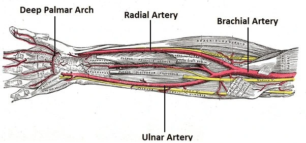
While experiencing stomach pain is never comfortable, it is more uncomfortable immediately following pasta consumption.
Contrary to popular belief, food allergies in adults are less frequent than you may think. It may take some time to pinpoint the specific foods that are causing your pain, so it’s important to talk to a doctor about your eating habits and any food-related symptoms. Speak with your doctor or a certified dietitian to determine the best course of action for you.
You guessed it: one of the most frequent reasons for post-meal stomach pain is dyspepsia, which is basically simply another word for indigestion. Bloating, post-meal feelings of fullness, and abdominal pain are symptoms of dyspepsia.
Your stomach may pain after eating because of an underlying problem, even though indigestion normally goes away on its own.
After a healthy meal, do you frequently experience stomachaches? If any of these health issues are the cause, it might be worth looking into.
Pasta has some elements that can exacerbate some medical issues, such as stomach ache. If you have a known food allergy, eating pasta may cause an allergic reaction. Pasta that contains soy, wheat, or eggs may aggravate intestinal inflammation and cause stomach pain. After eating pasta, stomach pain may potentially be a symptom of celiac disease or gluten sensitivity. The majority of pasta contains gluten, a wheat protein. People who are gluten intolerant have a range of digestive issues when they eat pasta.
Causes of Stomach Pain after Eating Pasta?
Food Allergy
If you have a food allergy, pasta may trigger an allergic reaction. Allergy-causing wheat, soy, eggs, and dairy items are commonly found in pasta dishes. Read the ingredients on the box before consuming any pasta to which you may have a professionally documented allergy. Pasta manufacturers are required by law to disclose the usage of any substances that the government considers to be common allergies. If the reaction is related to a food allergy, you may also have additional symptoms, such as asthma, hives, or congestion of the nose, in addition to stomach pain.
Gluten Intolerance
Pasta contains gluten unless it is labeled as “gluten-free.” Even a tiny quantity of pasta can cause excruciating stomach discomfort and other symptoms if you have celiac disease.
This digestive illness is caused by your immune system attacking the lining of your small intestine when you consume gluten-containing foods.
The cause of this immune system abnormality is unknown to doctors. For the benefit of your long-term digestive health, you must stay away from gluten if you have a gluten sensitivity.
Pasta Sauce
Sauces are frequently served with pasta. Pasta sauces contain two components that can cause
Dairy and tomatoes hurt. Tomatoes, the ingredient in red sauces, can trigger an allergic reaction in public.
Sauces that contain milk or cream may cause nausea if you have lactose intolerance or an allergic reaction.
A digestive disorder known as lactose intolerance results in gas, bloats, cramping in the stomach, pain, and diarrhea when dairy products are consumed.
Gastroenteritis is an infection of the stomach and intestines that manifests as nausea, vomiting, and diarrhea. Another common cause of food-related gastroenteritis, such as stomach pain after consuming tomato sauce, is food poisoning.
Although food allergies can mimic the symptoms of gastroenteritis, they do not really cause infections. Due to the presence of milk, fish, and tomatoes, pasta sauces might trigger allergic responses. If you have stomach pain after eating pasta sauce, you should speak with your doctor to determine the cause of your symptoms.
Gastroesophageal reflux disease (GERD)
Your problem is gastroesophageal reflux disease (GERD), which causes heartburn and stomach pain by irritating the lining of your oesophagus with stomach acid.
Your chance of having gastric reflux disease (GERD) is increased if you overeat or have a taste for spicy meals. This is because acid reflux may be very unpleasant and causes stomach acid to run up into your oesophagus.
Limit your consumption of alcohol, caffeine, and spicy foods if you think you could have gastric reflux disease (GERD). To help control your symptoms, you can also take over-the-counter antacids. If that doesn’t help, get in touch with your doctor.
Irritable bowel syndrome (IBS)
Stomach ache after eating pasta is undoubtedly one of the many symptoms of the intestinal problem, which can also include gas, diarrhea, constipation, and stomach pain. If you experience constipation or diarrhea, as well as chronic stomach pain after eating, it’s a good idea to visit your doctor and get tested for IBS.
Celiac disease
Celiac disease is an immunological reaction to gluten ingestion, and those who have it may have a range of symptoms. One is the ache in the stomach after eating gluten. For your information, celiac disease is not the same as mild gluten sensitivity, which is a condition in which your body makes it difficult for you to digest gluten.
When someone has celiac disease, ingesting gluten damages their small intestine; when someone has gluten intolerance, they may merely have physical symptoms like gas or diarrhea. To find possible reasons of the problem, you might collaborate with your physician.
Ulcer
In addition to other symptoms including weight loss, anemia, vomiting, difficulty swallowing, or blood in your stool, an ulcer may be the source of your ongoing pain after eating. Since ulcers, which are sores that develop in the lining of your stomach, small intestine, or oesophagus, are usually treated with drugs that lower acid production and, in some cases, antibiotics, you should definitely speak with a doctor about this.
Gastroparesis
It is also known as a “slow stomach,” which hinders proper digestion by partially paralyzing the stomach muscles. This prolongs the time that food remains in your stomach. Consequently, stomach pain and/or spasms result from your stomach’s incapacity to digest and absorb more meals. Additionally, nausea or vomiting may occur.
Most occurrences occur on their own and usually follow a stomach infection caused by a virus or bacteria. However, gastroparesis has also been linked to a recent COVID-19 infection.
This condition makes it easier for food to pass from your stomach, therefore you should eat smaller, more frequent meals. You may also want to avoid meals high in fiber, such as celery, beets, broccoli, and oranges, and choose cooked fruits and vegetables instead of raw ones. Your doctor may also suggest medications to reduce the ensuing nausea and tighten the muscles in your stomach.
Small intestinal bacterial overgrowth (SIBO)
occurs when there are too many bacteria in your small intestine. When there are too many harmful bacteria, the beneficial bacteria needed for digestion are overpowered. After meals, bloating, diarrhea, and abdominal pain might be caused by poor digestion.
SIBO risk factors include advanced age, prior stomach surgery, autoimmune disorders, and chronic constipation.
A gastroenterologist should be consulted if you suspect SIBO. Treatment entails dietary changes and the use of medicines to eradicate the bacterial overgrowth.
Gallbladder disease
Gallbladder disease, which encompasses a number of disorders that produce pain in the upper-right quadrant of your abdomen as well as around your back, is more prevalent among women in their 40s.
Greasy meals (think fried foods, cheese, sausage, potato chips, and butter) can cause mild to severe abdominal pain because fat activates the gallbladder. Gallstone-induced inflammation that blocks the ducts that lead to your small intestine can also cause excruciating pain. When you wake up in the middle of the night with severe stomach discomfort, it’s usually a sign that something’s wrong with your gallbladder.
Gallbladder issues typically don’t go away on their own, so if you have frequent or severe stomach pain after eating or any other concerning symptoms, you should see your doctor immediately. In more extreme situations, they may prescribe medicine to ease the pain and suggest surgery to remove the gallbladder.
Crohn’s disease
Any part of the GI tract, from the mouth to the anus, may be affected by this type of inflammatory bowel disease.
Inflammation associated with Crohn’s disease can cause mild to severe symptoms, including cramping, nausea, vomiting, diarrhea, fatigue, and blood in the stool. Although the exact origin of Crohn’s disease is unknown, food and heredity are typically implicated. Your doctor may recommend surgery, dietary changes, and medication to alleviate symptoms and provide long-term comfort, despite the fact that it is a chronic condition that requires constant monitoring and management.
Ulcerative colitis
Another type of inflammatory bowel illness that usually affects the colon and causes little ulcers all over the colon or rectum is ulcerative colitis. The symptoms usually develop gradually and include abdominal or rectal pain, bloody diarrhea, rectal bleeding, urgency to use the restroom, fatigue, and weight loss. Foods like cake, butter, coconut oil, and bacon that are heavy in sugar or saturated fats make stomach pain worse.
The exact reason is unknown, but potential contributing factors include immune system dysfunction, stress, nutrition, and inheritance. If you have severe stomach pain after eating or if you see blood in your stool, consult a physician. Surgery may be necessary in more severe situations, however anti-inflammatory medications are usually utilized as treatment.
Pancreatitis
Upper abdomen pain that may also radiate to the back is a symptom of pancreatitis, an inflammation of the pancreas. When the pancreas releases its digesting enzymes too soon, it attacks the organ rather than the food in the stomach, resulting in pancreatitis.
Alcoholism, gallstones, or pancreatic disorders can cause pain that develops gradually or that occurs quickly. Severe cases may require surgery, while minor cases of acute pancreatitis can be treated with rest and pain medication and resolve in a few days. If you have severe upper abdominal tenderness, fever, nausea, vomiting, or sudden onset pain, contact your doctor immediately.
Again, overeating might be the culprit, but if you frequently get stomach pain after meals, it’s wise to see a doctor to find out what’s causing it.
Symptoms of Stomach Pain after Eating Pasta?
There are many different types of stomach pain and distress. You’ve probably already come across a lot of them.
Typical symptoms and indicators include of:
Pain
Nausea
abdominal cramps
Acid reflux and bloating
feeling very full during or after eating;
abdominal tightness or bloating;
Gas Burning in the lower abdomen and arms or chest that ranges from mild to severe
partial regurgitation of the contents of the stomach If you know someone who is in excruciating pain from a stabbing.
Diagnosis of Stomach Pain after Eating Pasta?
Your doctor might be able to identify the reason of your stomach ache if you describe your symptoms to them. However, there may be times when more invasive testing is necessary. This could include:
Endoscopy and colonoscopy
CT scan
X-ray, and pH monitoring
Blood tests
MRI
Stool collection
The best way to find out whether you have a food intolerance is often to try different foods. Maintaining a meal journal may help you keep an eye on your symptoms. Your doctor may also recommend an elimination diet.
Treatment of Stomach Pain after Eating Pasta?
Maintaining adequate hydration is essential for managing gastroenteritis. The body may lose essential fluids as a result of excessive vomiting and diarrhea caused by gastroenteritis. To promote the development of bulky stool, increase your consumption of clear liquids and avoid bland foods such white rice, bananas, white bread, plain yoghurt, and apple sauce. As soon as you experience any symptoms of dehydration, such as dry lips, dry skin, or a sluggish feeling, give your doctor a call.
Here are some examples of over-the-counter remedies:
Simethicone, another name for Gas-X, helps painful bloating.
To reduce burning feelings, antacids like Tums, Alka-Seltzer, and Rolaids neutralize stomach acid.
Acid-reducers such as Pepcid reduce stomach acid output for up to 12 hours.
Beano helps avoid gas. Diarrhea and associated symptoms are stopped by imodium and other antidiarrheals.
Lansoprazole and esomeprazole (Prevacid, Prilosec) prevent the generation of acid and promote oesophageal healing when taken on a regular basis.
Pepto-Bismol soothes nausea and diarrhea and relieves burning by coating the lining of the oesophagus.
Benadryl, also known as diphenhydramine, is used to treat nausea, vomiting, and symptoms associated with an adverse immunological response.
Periodic constipation and the accompanying bloating are lessened with stool softeners and laxatives. Acetaminophen (Tylenol) does not irritate the stomach, although aspirin, ibuprofen, and naproxen can.
Probiotics generally enhance digestion.
Prognosis:
With dietary changes, the majority of patients who have stomach pain after eating pasta have a good prognosis. However, for an accurate diagnosis and treatment, a medical assessment is advised if symptoms are severe, ongoing, or linked to exhaustion, weight loss, or other digestive problems.
Summary
There are several causes of stomach aches after eating. There is probably nothing major wrong with it, and it is easy to treat. It’s critical to consider portion control and your food choices. However, if the problems persist, it may be worth visiting your physician.
Indigestion, acid reflux, and food intolerances (such gluten sensitivity or celiac disease) are the most frequent causes of stomach pain that only appears after eating pasta. Pasta additives (such as dairy or specific sauces) can occasionally cause pain.
It is best to see a doctor for a proper evaluation and diagnosis if symptoms intensify, last longer, or are accompanied by other problems (such diarrhea, vomiting, or weight loss). Making dietary changes, like avoiding thick sauces or trying gluten-free pasta, may help determine the source and reduce symptoms.
FAQs:
What causes stomach ache when eating pasta?
Wheat-based foods Cereal, grains, pasta, bread, baked products, crackers, and granola are examples of this. Despite the possibility that you have a gluten sensitivity or intolerance, Harris-Pincus argues that fructan in wheat products may be the cause of some IBS symptoms.
Does GERD get aggravated by pasta?
Even while pasta is safe to eat if you have acid reflux, your favorite sauces may not be! As previously stated, tomatoes may exacerbate symptoms of acid reflux; therefore, it is advisable to substitute low-fat milk sauce or broth for tomato-based sauces. In many of our diets, milk can be an essential ingredient.
For what length of time does pasta remain in your stomach?
For instance, simple carbs like pasta and rice often remain in your stomach for 30 to 60 minutes. On the other hand, meals like avocado and peanut butter that are higher in fat and protein can take two to four hours to pass through your stomach.
Which meals make you feel sick to your stomach?
Foods high in fructose include high-fructose fruits such as apples, pears, cherries, and mangoes, as well as any item that has added sugars or high-fructose corn syrup. Foods that contain a lot of specific oligosaccharides: Consider lentils, chickpeas, artichokes, wheat, onions, and garlic.
Why is it so difficult to digest pasta?
However, because Italian-style pasta is prepared from a unique hard wheat called durum wheat, it digests more slowly than most refined grain items. At the height of the low-carb mania in North America, in February 2004, Jenkins made a very public point about this issue at a pasta conference in Rome.
Why does eating pasta make me feel sick?
If you have gluten intolerance, ingesting gluten may make you feel ill. You may have gassiness, nausea, or bloating. Although gluten intolerance and celiac disease are not the same, they have many symptoms. An autoimmune condition called celiac disease damages the digestive system.
What is the duration of pasta bloat?
After eating, how long does bloating last? After eating, occasional bloating is usually just transient, lasting anywhere from a few hours to a day or two.
Why does eating spaghetti make my stomach hurt?
After consuming gluten, people with gluten sensitivity may develop gastrointestinal symptoms or other symptoms like exhaustion, joint discomfort, and headaches. When a person stops eating gluten, these symptoms usually go away.
Is the stomach irritated by pasta?
Even if they do not have celiac disease, people with IBS may suffer bloating, constipation, diarrhea, and other gastrointestinal issues when they eat wheat-based foods. Cereal, grains, pasta, bread, baked products, crackers, and granola are examples of this.
Why does pasta make me so sensitive?
You may have a wheat allergy, which affects millions of Americans, if you experience specific symptoms after eating cereal, bread, or pasta. For example, you may get a stomachache, hives, or a rash, or your nose may feel stuffy or run.
Why does eating pasta cause pain in my body?
Pasta, Inflammation, and the Glycaemic Index Rapid blood sugar rises from high GI foods can trigger an inflammatory response because they produce pro-inflammatory chemicals and create oxidative damage.
Why does using the muscles in my stomach hurt?
The most frequent reasons, such gas, indigestion, or a torn muscle, are typically not life-threatening. While the location and pattern of stomach discomfort can be helpful indicators, the duration of the pain is particularly helpful in determining its etiology.
What causes my stomach to swell when I eat pasta?
Wheat. Some people may experience bloating, gas, stomach pain, and diarrhea due to the gluten protein found in wheat. Gluten can be found in bread, spaghetti, and a variety of baked foods. A disorder known as celiac disease, which affects roughly 1% of Americans, can cause sensitivity to gluten.
When you eat spaghetti and your stomach aches, what does that mean?
Consuming gluten-containing meals can cause a variety of digestive issues, including diarrhea, which can have an especially disagreeable odor. stomach pains. Farting (flatulence) and bloating.
References
Mojidra, D. (2023, November 23). Stomach pain after eating pasta. Mobility Physiotherapy Clinic. https://mobilephysiotherapyclinic.net/stomach-pain-after-eating-pasta/
Osborn, C. O. (2024, May 7). Why does my stomach hurt after eating? Healthline. https://www.healthline.com/health/stomachache-after-eating
Clinic, C. (2024, December 9). 5 ways to get rid of a stomachache. Cleveland Clinic. https://health.clevelandclinic.org/how-to-get-rid-of-a-stomach-ache
Triceps tendonitis, also known as tricep tendinitis, is characterized by inflammation and irritation of the triceps tendon. The triceps tendon is a thick band of connective tissue that connects the triceps muscle on the back of the upper arm to the olecranon, which is the bony prominence at the tip of the elbow.
When the triceps muscles are used excessively or repetitively, tendonitis develops, which can cause minor rips or damage to the tendon. This can result from repetitive arm movements, such as weightlifting, throwing, or sports like tennis and golf. It is also common among individuals who engage in activities that require frequent overhead arm movements.
The most typical signs of triceps tendonitis are elbow, shoulder, or triceps pain and weakness. Pain frequently worsens when using the triceps, such as when pressing with the arm. There may also be elbow, triceps, or shoulder aches.
Injuries to the triceps tendon are an uncommon clinical issue rarely discussed in the literature.
Rest, ice, immobilization, nonsteroidal anti-inflammatory drugs, and physical therapy are conservative treatment options for partial tendon tears and triceps tendinopathies with retained strength.
If conservative treatment is unsuccessful for six months or if an examination shows weakness deficits, surgery should be considered.
Anatomy of Triceps Muscle:
The triceps brachii, also known as just the triceps, is the name of the big muscle on the rear of the upper arm. It is in charge of straightening the arm by extending the elbow joint. In the muscles of the triceps muscle, there are three heads:
Long head: The scapula’s (shoulder blade) infraglenoid tubercle is the source of this head. The only head that crosses the elbow and shoulder joints is this one.
The lateral head is located above the radial groove on the posterior aspect of the humerus, or upper arm bone.
Medial head: This head, which lies beneath the radial groove, likewise comes from the humerus’s posterior surface.
The triceps muscle’s three heads unite to form a single tendon that attaches to the ulna’s (one of the forearm bones) olecranon process.
Functions:
In contrast to the biceps brachii muscle, the triceps muscle is principally in charge of extending the forearm at the elbow joint. Additionally, it aids in elbow joint stability when performing delicate tasks like writing.
The radial nerve innervates the triceps muscle.
Causes of Triceps Tendonitis:
Tendonitis in the triceps is common in people who lift weights or play particular sports regularly. People who start a sport or fitness program and start exercising excessively too soon are also often affected by this illness. Your body is telling you that a muscle has been overworked. Rather than occurring suddenly, tricep tendonitis develops gradually.
Tricep tendinitis is frequently caused by the following factors:
Tricep tendon extension or overuse regularly.
Forceful motions, such as pounding or bench pressing.
Actions that aren’t typical of the body, such as excessive arm extension.
Inadequate technique or form when engaging in physical exercise.
Quick, sharp motions.
Rheumatoid arthritis and diabetes are two other illnesses that can cause tendinitis.
Signs and Symptoms of Triceps Tendonitis:
Pain and soreness in the rear of the elbow, close to the tip, which may spread down the back of the arm, are the main signs of triceps tendonitis. Moving your arms or applying pressure to the affected area may make the discomfort worse. There may occasionally be edema or a warm feeling around the elbow.
Some indicators that you may develop triceps tendonitis include the following:
Soreness in the triceps, shoulder, or elbow muscles is what you’re experiencing.
The discomfort you feel when you work on your triceps.
Your arm’s range of motion is limited.
A lump or swollen area directly above the elbow on the back of your upper arm.
Weakness in your triceps, elbow, or shoulder.
A popping noise or feeling right after being hurt.
Diagnosis:
Your doctor will ask you about your symptoms and the onset of your discomfort before making the diagnosis of tricep tendonitis. They may assess your range of motion and apply pressure to specific parts of your arm to see whether they hurt.
They may prescribe tests to examine the condition within your arm and look for infections or other problems if they believe you may have tricep tendonitis. These examinations could consist of:
Ultrasounds
X-rays and magnetic resonance imaging (MRI) to examine the nearby bones
Aspirations of the joints to check for infection
To help with the discomfort and swelling, your doctor may suggest a nonsteroidal anti-inflammatory medicine (NSAID), such as naproxen or ibuprofen. NSAID creams, which are applied topically to the area, can also help with pain.
Following these therapies, your doctor may suggest a steroid injection if your symptoms don’t get better. In rare circumstances, you might need surgery.
Treatment of Triceps Tendonitis:
To promote healing and stop the problem from getting worse, early diagnosis and suitable treatment are essential. In the early stages of triceps tendonitis, RICE (rest, ice, compression, and elevation) is frequently advised to relieve pain and reduce inflammation. Avoiding activities that make the discomfort worse and taking anti-inflammatory drugs may also help.
R: Rest: Stay Away from activities or movements that could further injure or exacerbate your triceps tendon.
I- Ice: Apply ice to the affected area for 20 minutes or so, several times a day, to lessen pain and swelling.
C: Compression: Use bandages or wraps to compress and support the area until edema has subsided.
E-Elevate: Maintain the affected area above the level of your heart to minimize swelling.
Drugs:
CorticosteroidsInjections:
Corticosteroid injections can help reduce pain and edema. The drug will be injected by your physician into the region surrounding your triceps tendon.
For tendonitis that has lasted more than three months, this treatment is not recommended since repeated steroid injections may weaken the tendon and increase the risk of further damage.
Your physician may suggest a platelet-rich plasma (PRP) injection for your tendonitis. The platelets and other blood components that aid in healing are isolated from a sample of your blood during PRP.
This solution is then injected into the area around your triceps tendon. The injection may help by providing nutrients to hasten the healing process because tendons have a poor blood supply.
Physical therapy:
Modalities:
Laser therapy and electric shock (IFT, TENS)
Physical therapy may also be used to treat your triceps tendonitis. It focuses on strengthening and increasing the flexibility of your triceps tendon through a regimen of targeted workouts.
Here are some simple exercises you could attempt. Before starting any of these workouts, don’t forget to consult your doctor because performing certain exercises too soon after an accident could make your condition worse.
Bending and straightening the elbow:
Elbow Joint Range of Motion Exercise
Keep your hands by your sides in a loose fist.
Your hands should be at shoulder height or higher.
Drop your hands slowly while keeping your elbows straight after you have them back at your sides.
Stop after ten to twenty times.
The French stretch
French stretch
Standing, clasp your hands together and raise them above your head.
Keeping your hands clasped and your elbows close to your ears, drop your hands behind your head as you attempt to touch your upper back.
For fifteen to twenty seconds, maintain the posture decreased.
Three to six times.
Static triceps stretch:
Triceps stretching
At the elbow, your injured arm should be 90 degrees bent. In this position, your hand should be in a fist with the palm facing inward.
Press down with the fist of your bent arm on the open palm of your other hand to tighten the triceps muscles at the back of your injured arm.
Hold for five seconds.
Tighten your triceps 10 times as much as you can without feeling uncomfortable.
Towel resistance:
shoulder press with a towel
Hold one end of a towel in each of your hands.
Place the damaged arm behind your back and the uninjured arm over your head while you are standing.
With the other hand, gently pull the towel down while you raise your injured arm towards the ceiling.
Hold the position for 10 seconds.
Ten times overall.
Triceps kickback
Leaning forward and supporting oneself with a table or chair under the hand of one arm.
Place the right foot on a step or platform, place the right forearm on the thigh, or let the arm drop just below the shoulder to support the back.
Using your left hand to hold a weight, raise your elbow to body level.
Focus on strengthening your triceps muscle as you extend your arm behind you while maintaining that elbow position.
Maintain this posture for a short while.
Reduce the forearm’s angle to around 90 degrees.
Then return to your neutral position.
Then unwind.
Do this exercise five to ten times.
Maintain the immobility of your upper arms against your body throughout the exercise.
Bench Tricep Dips
bench tricep dips
Place two flat benches parallel to each other and three to four feet apart.
Sitting on one bench and facing the other, place your hands on the side of the bench.
Using your hands to support your weight, raise your feet to the top of the other bench, leaving the rest of your body hanging.
The two benches are on the other side.
Stretch one foot over the other.
To get your forearms and upper arms at a straight angle, progressively bend your elbows towards the floor.
Avoid bending your shoulders below a 90-degree angle since this can cause tension.
Then return to your neutral position.
Then unwind.
Do this five to ten times.
Supine Triceps Extension
Triceps-extension
Lay flat on a table or bed to start.
With your feet firmly planted on the floor, rest your head on the bed.
Raise a dumbbell over your shoulders at arm’s length in each hand.
Lower the dumbbells until you are in a comfortable position while bending your elbows.
Slowly return to the beginning position.
Hold this position for a short time.
Going back to the starting position.
Then relax.
Do this exercise five to ten times.
Overhead Tricep Extension
Begin by standing in a comfortable position.
Keep your back straight.
Hold a weight in the air with both hands.
Lower the weight until your elbows are at a 90-degree angle behind your head while maintaining your biceps near your ears.
Tighten your triceps and straighten your arms.
Then return to your neutral position.
Then unwind.
Do this exercise five to ten times.
Keep your core tight and refrain from arching your back during the workout.
Surgery:
It is advised to treat triceps tendonitis with more conservative measures such as physical therapy, medicine, and rest.
However, if the damage is severe or if other therapies have not worked, surgery might be required to repair your torn triceps tendon. This is often recommended in cases where the tendon is partially or completely torn.
Repairing tendons:
The purpose of triceps tendon repair is to reattach the damaged tendon to the elbow’s olecranon. The olecranon is located in the ulna, one of the long bones of your forearm. Usually, the entire treatment is performed under general anesthesia, which puts you to sleep.
The wounded arm is immobilized while an incision is made. Following appropriate exposure of the injured tendon, sutures are used to connect it to the olecranon by inserting bone anchors or suture anchors into the bone.
For a precise diagnosis and a customized treatment plan depending on the severity of the triceps tendonitis and the demands of the individual, it is imperative to speak with a healthcare provider. Targeted rehabilitation exercises, adequate rest, and early intervention can greatly enhance the outcome and enable people to resume their normal activities pain-free.
Tips for Avoiding Tricep Tendonitis:
There are precautions you may take to reduce your risk of tendinitis, even if there is no surefire method to avoid it.
Get warm-up. Your risk of injury is reduced and blood flow is increased as you warm up your muscles.
Execute workouts properly. Spend some time learning how to perform exercises correctly. This lowers your chance of getting hurt and improves your performance.
Avoid overstretching yourself. Excessive exercise on fatigued muscles might result in tendinitis and larger muscular tears.
When to Consult a Physician:
It could be necessary to see a doctor if your symptoms don’t go away after three days of rest. Additional indicators that you should consult a physician include:
An ongoing redness and swelling of the tendon
Severe discomfort and pain
Limitations in movement and symptoms that worsen even after rest
Home Treatments:
Resting the affected tendon for up to three days can help treat some types of tendonitis at home. You can attempt the following while your muscles are at rest:
Prevent from moving. For two to three days, avoid moving your arm or tricep. As much as possible, rest it and avoid moving it.
Ice the area. Place a bag of frozen peas or a bag of ice wrapped in a towel on the tendon. Every three hours, leave it there for twenty minutes.
Get ready. To support the muscle, try applying a supportive bandage or brace. The support should fit comfortably without being too tight. Before you go to bed, take it off.
Until the tendon has completely healed and the discomfort has subsided, avoid making any sharp or violent motions again.
Summary:
Triceps tendonitis most commonly manifests as elbow, shoulder, or triceps pain and weakness. Pain usually worsens when using the triceps, like when pressing or using the arm. There may also be elbow, triceps, or shoulder aches.
Injuries to the triceps tendon are an uncommon clinical issue rarely discussed in the literature. Treatment for triceps tendinopathies depends on the mechanism of damage and the patient’s motor evaluation.
Rest, ice, immobilization, nonsteroidal anti-inflammatory drugs, and physical therapy are conservative treatment options for partial tendon tears and triceps tendinopathies with retained strength.
If conservative treatment is unsuccessful for six months or if an examination shows weakness deficits, surgery should be considered. Based on the data that is now available, the “best” surgical technique is still unclear.
FAQs
What is the treatment for tricep tendonitis?
Rest, ice, immobilization, nonsteroidal anti-inflammatory drugs, and physical therapy are conservative treatment options for partial tendon tears and triceps tendinopathies with retained strength. If conservative treatment is unsuccessful for six months or if an examination shows weakness deficits, surgery should be considered.
How long does it take to heal from tricep tendonitis?
Since most occurrences of triceps tendonitis are treated non-operatively, the tendon usually heals in four to six weeks. Athletes can also assist prevent further injuries by warming up correctly before play and applying ice to the damaged elbow if it hurts after exercise.
Is it possible to reverse tricep tendonitis?
In many cases, triceps tendonitis can be resolved with rest alone. Sometimes medication is required to treat the pain.
Could you work out if you have tendinitis in your back arm muscles?
First and foremost, the guilty movement should be avoided, changed, or lessened. We advise weightlifters, for example, to use a lesser weight when performing shoulder and bench presses, which are popular pressing exercises done in the gym. Occasionally, switching up shoulder and chest exercises can be beneficial.
Is heat a good treatment for tendinitis in the back of the arm?
For persistent tendon discomfort, sometimes referred to as tendinopathy or tendinosis, heat may be more helpful. Because heat can enhance blood flow, it may help mend tendons. Additionally, heat relaxes muscles, which helps ease pain.
Which workouts are best for treating triceps tendonitis?
Exercises for flexibility, stretching, and joint strength. These exercises include push-ups, plank triceps kickbacks, alternating dumbbell floor presses, dumbbell overhead shoulder presses, and elbow bending and straightening.
How long does tendinitis last?
While persistent tendinitis takes about six weeks to recover, the most common type of tendonitis usually goes away in one to three weeks.
Does tendinitis benefit from exercise?
Start working out to strengthen the muscles surrounding the aching joint within a day or two. A lengthy warm-up should be performed first to lessen tissue shock. Next, try lifting little weights or using an elastic training band.
References
What to know about tricep tendonitis. (n.d.). WebMD. https://www.webmd.com/fitness-exercise/what-to-know-about-tricep-tendonitis
Patel, D. (2023d, December 13). Triceps tendonitis – Cause, symptoms, treatment, exercise. Mobile Physiotherapy Clinic. https://mobilephysiotherapyclinic.in/triceps-tendonitis-treatment/
Bariya, D. (2023, December 13). 26 Best exercise for triceps tendonitis – Mobile Physio. Mobile Physiotherapy Clinic. https://mobilephysiotherapyclinic.in/26-best-exercise-for-triceps-tendonitis/
Seladi-Schulman, J., PhD. (2019, April 22). How to treat triceps tendonitis. Healthline. https://www.healthline.com/health/sports-injuries/tricep-tendonitis
Tietze syndrome is a rare, benign condition characterized by inflammation of the costal cartilage, typically where the upper ribs meet the sternum (breastbone). It causes localized chest pain, tenderness, and swelling, often mimicking heart-related pain.
Where your sternum (breastbone) joins your upper vertebrae (the first 10) is where the cartilage points (costal cartilage) are located. Cartilage is also present at the costochondral joints, which are the connections where these points join.
Although Tietze syndrome is more specific. Additionally, it is known for causing significant edema. The cause of Tietze syndrome is also known as costochondral junction syndrome. It doesn’t appear to be serious. With rest and painkillers, you can treat it at home.
Relevant anatomy
The main anatomical feature of Tietze syndrome is the inflammation of the cartilage at the costosternal junction, which results in severe chest pain and swelling. Tietze syndrome is characterized by localized pain, tenderness, and occasionally visible swelling at the costochondral joints, specifically the cartilage where the upper ribs (usually the second and third) connect to the sternum (breastbone).
Affected area: While additional upper ribs may be impacted, the costochondral junction of the second and third ribs is the most frequently affected area.
Inflammation: Inflammation of the costal cartilage at its attachment to the sternum is the main pathophysiology.
Unilateral presentation: Only one side of the chest is often impacted by Tietze syndrome.
Pain characteristics: Tietze syndrome pain can spread to the arms, shoulders, and neck and is frequently described as acute, hurting, or clutching.
Epidemiology
It is unknown how common Tietze syndrome is and how often it occurs. Geographical location, occupation, race, or biological sex don’t reveal anything about incidence rates. Males and females seem to be affected by this syndrome in roughly equal amounts. The syndrome affects people under 40 more often than it does people over 40. Although there have been documented examples, cases involving people 40 and older are rare.
Pathophysiology
The hallmark of Tietze syndrome is a small, painful, swollen lump that typically lacks erythema or a pustulation rub. About 70% of patients have the disease limited to one side, with the most common relationship being with the cartilage of either rib 2 or rib 3. However, the xiphisterna and sternoclavicular joints have also been found to be affected by this Tietze syndrome.
The idea that this syndrome process is inflammatory and could be a component of a more extensive seronegative pathology is still being discussed. Recurrences of Tietze syndrome are conceivable, but the symptoms usually go away on their own.
Sign and Symptoms
The most noticeable symptom of Tietze syndrome is chest pain, which may develop gradually or suddenly.
Some claim that when you’re motionless, it seems dull, but as you move or twist your upper body, it becomes acute and gripping. Exercise, sneezing, and coughing might exacerbate it. Usually on one side, you will feel it in the front and upper part of your chest. Additionally, it could radiate to your neck, shoulder, and arm.
The other main sign of costochondral junction syndrome is localized edema. Somewhere in your upper sternum, usually at your second or third rib, you can see a little, swollen lump. In addition, it could appear red or discolored and feel warm. All of these are signs of inflammation. One characteristic of Tietze’s disease is swelling, which frequently goes away last.
Causes
Tietze syndrome is characterized by inflammation and irritation of the cartilage at that uncomfortable location in your rib cage. However, medical professionals are unsure of the reason for this and why it occurs in the specific manner that it does. Tietze syndrome differs from ordinary costochondritis in these ways.
Traumatic injury. Tietze’s syndrome may be caused by an automobile accident, a fall, or an athletic injury that affects your upper rib cage. Perhaps this region’s soft tissues just respond to trauma differently.
Microtrauma. Over time, minor but recurrent stress on your rib cage may cause a particular inflammatory response, such as intense coughing or repeated chest hits.
Risk Factor
If you have a persistent condition that weakens or irritates the cartilage in your rib cage, or if you are recuperating from another syndrome, you may be at a higher risk of developing Tietze’s disease. Among the examples are:
Autoimmune diseases.
Hereditary connective tissue diseases.
Degenerative or inflammatory arthritis.
Chronic chest infections.
Chronic coughing or vomiting.
Recent surgery in the area.
Diagnosis
When you present your chest pain to your doctor, they will first make sure it isn’t a heart attack. They will carefully go over your symptoms and check your chest after ruling that out. Imaging studies will be performed later to check for signs of injury and inflammation inside your chest. Once other potential causes of your symptoms have been checked out, medical professionals diagnose Tietze’s disease.
In addition to heart attacks, Tietze syndrome can resemble other heart and lung disorders. To rule out any further problems that might be more serious or require different treatment, you must see your doctor.
Tietze syndrome cannot be diagnosed with a single test. Your doctor will ask you to describe your symptoms and perform a physical examination to determine what’s causing them. To look for areas of pain and edema, they will most likely feel or apply pressure on your chest and ribs.
Investigation
Tests might include:
Chest X-ray
CT scan
MRI
Ultrasound
Electrocardiogram (EKG)
Biopsy
Differential Diagnosis
When acute chest pain first appears, it is important to think about a wide differential diagnosis because Tietze syndrome is identified by ruling out other possible causes. Because anterior chest wall pain that palpates at the osteochondral connections is a common symptom of both disorders, clinicians sometimes misdiagnose this condition as costochondritis. Multiple ribs, usually ribs two through five, are linked to costochondritis. Furthermore, there is no correlation between costochondritis and localized swelling over the affected joints.
Costochondritis will not show any notable results on ultrasound. Ultrasonography, on the other hand, actively visualizes edema and inflammation to diagnose Tietze syndrome. Performing a physical examination to check for obvious edema might also help make this diagnosis.
Exclude further conditions including acute coronary syndrome, hypertensive crisis, pleural infections or inflammatory processes, cancers, fractures related to chest trauma, rheumatoid or pyogenic arthritis, gastroesophageal reflux syndrome, or psychogenic disorders. Rib fractures, lupus, and fibromyalgia are among the other syndromes on the differential diagnostic list.
Seronegative spondyloarthropathy
Spondyloarthropathy(SA)
Rheumatoid Arthritis(RA)
Slipping Rib Syndrome
Myelomalacia
Early forms of soft tissue and bone cancers
Chondrosarcoma of chondrocostal joints
Costochondritis
Chest or pulmonary tumors
Metastases of breast, kidney, and prostate neoplasms
Rib Fracture
Treatment
Your soft tissues require relaxation and time to heal from inflammation. Typically, medical professionals recommend just that. Pain and swelling can be reduced using over-the-counter pain medications. You shouldn’t have to miss work or school, but while you’re recovering, take it easy and stay away from strenuous activity. As you begin to feel better, you can progressively increase your level of activity.
After ten days, if you still feel that you need to take NSAIDs, speak with your doctor. Long-term use of these may result in adverse effects. At the site of irritation, they may administer a cortisone injection or advise you to switch to acetaminophen. Before their chests heal, some people may require treatment to halt coughing, sneezing, or vomiting. Discuss your recuperation with your provider.
Medical treatment
Conservative therapy is the cornerstone of treating Tietze syndrome, and patients are reassured that the syndrome will typically go away in a matter of weeks with no lasting effects. Months or even a year may pass while this condition persists. Rest and topical or oral anti-inflammatory and analgesic medications are the first-line treatments.
Given that inflammation is the primary cause, nonsteroidal drugs are usually more effective than acetaminophen or opioids. Clinically advise taking an anti-inflammatory dose of scheduled nonsteroidal medication for up to 10 days, if there are no contraindications. If there are no contraindications, a brief course of oral steroids like prednisone or methylprednisolone may also be taken into consideration.
If rest and nonsteroidal drugs are unable to significantly reduce swelling, medical professionals may inject a local anesthetic, steroid, or a combination of the two at the region of greatest swelling and use ultrasonography to monitor the area. Warming pads applied to the affected area have been beneficial to some patients. Clinicians have suggested resecting cartilage as a therapy option in certain severe, ongoing instances. In general, clinicians advise against using this treatment and advise just evaluating it in specific cases.
Nonsteroidal anti-inflammatory medications(NSAIDs). This covers medications like naproxen, ibuprofen, and aspirin. Before taking any over-the-counter drug, consult your physician or pharmacist. They will inform you if certain medications can worsen pre-existing diseases or interfere with other medications you are taking.
Steroids. Your doctor may prescribe corticosteroid pills or provide an injection directly into the affected area if your pain is severe and doesn’t go away with previous treatments. When alternative therapies fail, this can rapidly lessen joint pain and swelling.
Injection of lidocaine. To relieve pain, your doctor may administer an anesthetic or numbing shot. This is something you might receive when over-the-counter painkillers don’t work, much like the steroid shot.
Physical Therapy treatment
Tietze’s condition is not yet perfectly treated. Whether the sickness is dealt with definitively or not is unknown. Therefore, Tietze syndrome is primarily treated symptomatically. There are currently insufficient reliable clinical trials on the treatment of Tietze’s condition.
Range-of-motion exercises
Advise on how to perform exercises and demonstrate proper body alignment. Patients must strike a healthy balance between rest and exercise. The physiotherapist must provide accurate information regarding the patient’s posture when sitting and performing regular tasks. Additionally, the patient must refrain from repetitive motions or activities.
As soon as feasible, range-of-motion exercises should be introduced. While performing the activities, the patient might not experience any pain. You should cease and stay away from vigorous exercises if they make your symptoms worse.
Exercises for range of motion to increase flexibility, and gradually increase chest mobility.
Strength training: After the pain goes away, specific shoulder and chest strengthening activities can be added.
Stretching of pectoralis major
Stretching can be helpful.
Standing in a corner for 10 seconds with both hands against the wall (like in a push-up) is a good way to stretch the pectoralis major.
You must repeat it for one or two minutes several times a day.
Stretching exercises to release tension in the chest region, concentrate on the pectoralis major muscle.
Mobilization of soft tissues
A light massage to increase blood flow and relieve tense muscles.
To prevent thoracic rigidity and to lessen symptoms, mobilize the spine and ribs.
Instruct the patient on breathing techniques.
modalities to reduce pain
To apply TENS. Shoulder and back exercises can be provocative, thus they should only be used once the problems have subsided. They can apply electroacupuncture and transcutaneous electrical stimulation to the painful spot. The solid filiform needle, another name for the acupuncture needle, is inserted into the affected spinal region. After that, the implanted needle is subjected to low-frequency electrical currents.
Dry Needling: Assessing and treating musculoskeletal chest wall pain has historically been challenging. It has previously been reported that costochondral-related chest wall pain can be treated with injection therapy using local anesthetics or corticosteroids. According to the findings of an earlier study, dry needling may be just as successful for several syndrome as injection therapy.
Breathing exercises, electrotherapy (such as ultrasound), and cryotherapy (ice) are examples of physiotherapy for Tietze’s condition. You might also receive a soft tissue massage and additional stretching and strengthening exercises. Hydrotherapy is another crucial physiotherapy treatment. To help the patient manage their pain, introduce them to cognitive behavioral therapy and pain neuroscience education.
Home care advise
Get some rest. Exercise and other forms of physical activity should be taken occasionally to avoid straining your chest and ribs.
Ice or heat. For a specific period, your doctor could advise you to use hot or cold compresses intermittently. Using cold or heat packs to reduce pain and inflammation in the affected area
Postural correction: Teaching the patient how to stand correctly to reduce rib strain.
Heat and cold therapy. To assist prevent muscle overload and reduce pain, use massage and heat/cold pads. Both cold and heat pads work just as well. For the patient to select his favorite. The patient may also apply Vanpooling spray to the affected regions in place of cold pads. This spray can help with chest pain.
Breathing techniques: Deep breathing exercises to ease pain and increase chest mobility
Complications
Complications Injections, drugs, or surgery may provide a danger, however, complications directly linked to Tietze syndrome are generally uncommon.
Severe drug reactions
Infection
Pneumothorax
Prognosis
You should anticipate fully recovering from costochondral junction syndrome, which is a transient ailment. While some bodies take longer to heal, most people do so in a few weeks. After the pain subsides, the swelling may occasionally persist for several months. Sometimes Tietze’s syndrome appears to disappear and then returns. In certain circumstances, Tietze syndrome may be more persistent than in others due to varying causes and risk factors.
Tietze syndrome often has a good prognosis and resolves on its own. Because the cause or causes of Tietze syndrome are not fully understood, there is uncertainty about how to prevent this disorder. With conservative treatment, the majority of patients report complete symptom relief in 1–2 weeks; however, some patients report that the typical pain and swelling persist for up to a year. Moreover, recurrences are probable. The majority of individuals recover completely from Tietze syndrome.
Having Tietze Syndrome
Tietze syndrome usually resolves on its own without medical intervention. However, there are things you may do to improve your mood while you’re recovering.
You could:
Use painkillers that are available over the counter.
If your pain is severe, ask your doctor for a stronger medication.
Where it hurts, apply ice or heat.
Discuss with your physician what activities you should refrain from during your recuperation.
Find out when you can resume chest exercises without risk.
If your symptoms don’t improve in a few weeks or if you get any new chest pain or swelling, consult your physician.
Which information about Tietze syndrome is the most important to understand?
Tietze syndrome is an uncommon ailment that often affects the second or third rib and is characterized by chest pain and swelling of a single costal cartilage. It is distinguished from costochondritis by the swelling. Young people under 40 are usually the ones who exhibit it. The results of the clinical history and physical examination, along with the elimination of potentially fatal causes of chest pain, are used to make the diagnosis of Tietze syndrome. Painkillers and reducing physical activity until the inflammation subsides are the mainstays of treatment.
What should I avoid if I have Tietze’s condition?
You shouldn’t disregard your symptoms if you think you might have Tietze’s syndrome. Try to utilize a supported cough if you can, and stay away from any activity that can make your symptoms worse. This and other pain-reduction techniques will be taught to you by your physiotherapist.
Is Tietze syndrome dangerous?
However, you should still discuss it with your physician. They will want to rule out other medical disorders like lupus, broken ribs, acid reflux, and dangerous conditions like heart attacks or tumors of the chest wall that may exhibit the same symptoms but need different therapies.
Conclusion
Chest pain and edema are symptoms of the uncommon inflammatory disease Tietze syndrome. It is rarely severe and typically resolves on its own in a few weeks. However, if you believe you have it or if your symptoms persist, consult your physician. You can recover more quickly with rest, medicine, or other therapies.
FAQs
What is the duration of Tietze?
Tietze syndrome is thought to be a self-limiting syndrome that often goes away with rest in a few months. Analgesics and nonsteroidal anti-inflammatory medications (NSAIDs), such as ibuprofen, aspirin, acetaminophen (paracetamol), and naproxen, are typically used to treat Tietze syndrome.
Which medications address Tietze syndrome?
Your soft tissues require relaxation and time to heal from inflammation. Typically, medical professionals recommend just that. Pain and swelling can be reduced using over-the-counter pain medications. Nonsteroidal anti-inflammatory medicines, or NSAIDs, are frequently suggested by providers.
What distinguishes costochondritis from Tietze syndrome?
There is typically no accompanying redness or swelling in costochondritis. 70% of cases involve the second or third ribs, and Tietze syndrome is typically unilateral, affecting only one joint. A history of recent syndrome with coughing or recent intense exertion is frequently present in cases of Tietze syndrome.
What is the Tietze syndrome injection?
Tietze syndrome is typically treated with basic analgesics, such as nonsteroid anti-inflammatory medications, or rest. A local corticosteroid injection is known to be helpful in situations where the patient may remain immobilized for several months.
How much pain does Tietze syndrome cause?
The localized pain may be minor or even severe. Only the upper ribs, specifically T1 through T4, are usually affected by the syndrome; the most frequently impacted levels are the second and third ribs. When it comes to Tietze syndrome, cardiac, pulmonary, and neurological examinations are usually benign.
What might be confused for the syndrome of Tietze?
Though rarer and more significant, Tietze syndrome is sometimes mistaken for costochondritis and is characterized by acute chest pain and localized swelling at the breastbone-rib junction.
What adverse effects might Tietze syndrome cause?
Chest swelling and pain are the most typical signs of Tietze syndrome. It may be bland or acute, or it may be minor or severe. Redness or a warm sensation where it hurts are further symptoms of Tietze syndrome.
Is there a cure for Tietze syndrome?
Physical therapy and stretching or strengthening exercises may be helpful for an early recovery once movement is pain-free. The majority of Tietze syndrome cases go away on their own in a few weeks, however, they can sometimes recur or continue longer.
Is it possible to cure Tietze’s disease?
If I have Tietze syndrome, what can I anticipate? You should anticipate fully recovering from costochondral junction syndrome, which is a transient ailment. While some bodies take longer to heal, most people do so in a few weeks. After the pain subsides, the swelling may occasionally persist for several months.
What is the duration of Tietze syndrome?
Symptoms usually disappear in a few weeks or months. However, some people have swelling for months or more. If you find it difficult to control your symptoms on your own, let your doctor know. To reduce your pain and inflammation, you might need to take further measures.
Reference
Tietze Syndrome. (2025, January 24). Cleveland Clinic. https://my.clevelandclinic.org/health/diseases/23565-tietze-syndrome
Wiginton, K. (2024, February 11). Tietze Syndrome. WebMD. https://www.webmd.com/pain-management/tietze-syndrome
Wikipedia contributors. (2024, August 17). Tietze syndrome. Wikipedia. https://en.wikipedia.org/wiki/Tietze_syndrome
RIB pain | Aurora Health Care. (n.d.). https://www.aurorahealthcare.org/services/heart-vascular/conditions/rib-pain
Pt, S. R. B. (2024, September 23). Can dehydration make costochondritis worse? CARESPACE Health+Wellness. https://carespace.health/post/can-dehydration-make-costochondritis-worse/
Arthritis is a broad term for conditions causing inflammation and pain in the joints. It affects people of all ages, with the most common types being osteoarthritis, which results from wear and tear, and rheumatoid arthritis, an autoimmune disorder.
Symptoms often include joint pain, stiffness, swelling, and reduced mobility. Management strategies vary depending on the type but may include medication, physical therapy, exercise, and lifestyle changes to reduce pain and improve joint function.
The hard, slick substance that surrounds the ends of vertebrae where they unite to create a joint, called cartilage, deteriorates due to osteoarthritis. The disease known as rheumatoid arthritis occurs when the immune system targets the joints starting with the joint lining.
A high blood level of uric acid can result in uric acid crystals, which can lead to gout. Infections or underlying conditions like lupus or psoriasis can induce different kinds of arthritis. Depending on the type of arthritis, different treatments are used. Reducing symptoms and enhancing quality of life are the primary objectives of arthritis therapy.
What is Arthritis?
One condition that damages your joints is arthritis. Where two bones unite in your body is called a joint. As you age, some joints deteriorate naturally. Following that typical, lifelong wear and tear, many people acquire arthritis. Certain forms of arthritis develop as a result of joint damage from injuries. Arthritis can also be caused by certain medical disorders.
Although arthritis can occur in any joint, it most frequently affects people:
Hands and wrists.
Knees.
Hips.
Feet and ankles.
Shoulders.
Lower back (lumbar spine).
A medical professional will assist you in managing pain and stiffness. Eventually, some patients with severe arthritis require surgery to replace the damaged joints.
If you feel like you can’t move or utilize your joints as well as normal, or if your joint pain is severe enough to interfere with your everyday activities, you should see a healthcare professional.
What is the function of a joint?
Where two or more bones touch, like in the fingers, knees, and shoulders, is called a joint. Bones can move freely within certain bounds and are held in place by joints.
The majority of our body’s joints are encased in a robust capsule. A viscous substance that aids in joint lubrication fills the capsule. Our bones are held in place by these capsules. Ligaments assist them in doing this. They resemble extremely durable elastic bands.
Cartilage lines the ends of the bones that make up a joint. As you move, the bones can slide over each other because of this resistant yet smooth layer of tissue. The muscle pulls a tendon that is connected to the bone when our brain signals the muscle to move the bone. As a result, muscles are crucial for maintaining a joint.
Types of arthritis
Arthritis comes in over a hundred varieties. Among the most prevalent kinds are:
Osteoarthritis
Osteoarthritis
Osteoarthritis, the most prevalent kind of arthritis, is caused by wear and tear on the cartilage, which is the firm, slippery layer that covers the ends of bones where they unite to form a joint. The ends of the bones are cushioned by cartilage, which permits almost frictionless joint motion. However, if the cartilage is sufficiently damaged, the bone may grind against the bone, causing pain and limited mobility. A joint injury or infection may accelerate this wear and tear, or it may develop over many years.
The connective tissues that hold the joint together and connect muscle to bone deteriorate and the bones change as a result of osteoarthritis. The lining of a joint may swell and become inflamed if the cartilage is seriously injured. Osteoarthritis is the most prevalent kind of arthritis. In the UK, an estimated 8.75 million people have visited a doctor for osteoarthritis. Cartilage roughening is the first sign of osteoarthritis.
If this occurs, the body may attempt to compensate for the loss of this vital component by implementing a “repair” mechanism. Then, the following may occur:
Osteophytes are tiny pieces of additional bone that can grow at the ends of bones in joints.
The quantity of viscous fluid within the joint may rise.
The joint may become deformed due to the stretching of the joint capsule.
Osteoarthritis can occasionally develop in its early stages with little pain or difficulty. Nevertheless, it may result in pain, stiffness, and internal joint injury. Osteoarthritis often affects adults over 45 and is more common in women. The knees, hands, hips, and back are the body parts most frequently impacted.
Maintaining an active lifestyle will help you keep your weight in check, which will ease the strain on your joints. Regular exercise will maintain the strength of the muscles surrounding a joint, which will support and stabilize an osteoarthritis-affected joint. Being overweight can exacerbate osteoarthritis and increase your risk of developing it.
You can keep active and lessen your symptoms by using non-steroidal anti-inflammatory medicines (NSAIDs) like ibuprofen and painkillers like paracetamol. Additionally, being active will lessen edema, stiffness, and pain. You might apply NSAID creams topically to the affected area. Trying these first would be a good idea.
Numerous options are available for pain alleviation. A doctor or physiotherapist can offer you particular assistance if you’re having trouble managing the pain associated with osteoarthritis.
Your doctor may talk to you about having surgery if your osteoarthritis worsens, especially in your knees and hips. This is typically only taken into consideration after every other therapeutic option has been exhausted. These days, joint replacements are highly effective and advanced.
Rheumatoid arthritis.
Rheumatoid arthritis
The lining of the joint capsule, a strong membrane that surrounds every joint component, is attacked by the body’s immune system in rheumatoid arthritis. The synovial membrane, the lining, swells and becomes inflammatory. Eventually, the disease process may cause the joint’s bone and cartilage to be destroyed.
One kind of inflammatory arthritis is rheumatoid arthritis. It’s referred to as an auto-immune disease. The body’s natural defense mechanism, the immune system, keeps us safe from diseases and illnesses. In an autoimmune disease, the body’s immune system unintentionally targets healthy tissues, like the joints, resulting in inflammation.
Normally, inflammation is a useful immune system tool. It happens when the body fights an infection by directing more blood and fluid to that location. This is what happens, for instance, when a cut becomes infected and the surrounding skin swells and changes color.
But with rheumatoid arthritis, the excess fluid and inflammation in a joint might lead to the following issues:
It may cause pain and difficulty when moving the joint.
The fluid’s chemicals have the potential to harm joints and bones.
The joint capsule may get stretched by the excess fluid. A joint capsule never fully returns to its initial position after being extended.
The fluid’s chemicals have the potential to cause pain by irritating nerve endings.
Inflammation can harm a joint permanently in addition to producing pain and stiffness. Early initiation of successful treatment can help minimize damage.
Rheumatoid arthritis symptoms can include:
Swollen joints.
Joint edema and stiffness in the morning that persists for more than 30 minutes; extreme exhaustion, sometimes known as fatigue; and a general sense of being ill.
In addition to affecting the same joints on both sides of the body simultaneously, rheumatoid arthritis frequently begins in the little joints of the hands and feet. It may begin more forcefully or more slowly and then progressively worsen.
Adults of any age can develop rheumatoid arthritis. People between the ages of 40 and 60 are most likely to experience its onset. Women are more likely than men to have it.
Some medications can lessen joint pain and swelling by slowing down an overactive immune system. These include biological therapy and are referred to as disease-modifying anti-rheumatic medications (DMARDs). Conventional DMARDs are the first line of treatment for rheumatoid arthritis. When your immune system is overactive and harming your body, these can have the overall effect of lowering its activity.
Doctors will consider considering the more recent biological therapies if these medications have been tried and have failed. The immune system is more specifically affected by biological treatments.
Gout
Gout
Gout arthritis causes sharp uric acid crystals to form in your joints. One form of inflammatory arthritis that can cause excruciating joint swelling is gout. Although the big toe is usually affected, other joints in the body may also be impacted.
Gout-affected joints may turn red and heated. Additionally, the skin may peel and seem glossy. It is caused by an excess of urate, also referred to as uric acid, in the body. Everybody has some urate in their bodies.
However, some people may have higher levels of urate in their bodies due to being overweight or consuming excessive amounts of certain foods and alcoholic beverages. Your inherited genes may increase your risk of developing gout.
Urate can crystallize into substances that stay in and around the joint if it reaches a high enough quantity. They can remain there for some time without posing any issues or even letting the person know they’re there. The crystals may fall into the soft area of the joint as a result of a knock on one part of the body or a fever. Swelling and pain will result from this.
Gout attacks can be avoided by taking medications that lower the body’s urate levels. Febuxostat and allopurinol are two examples. Also, you will want temporary pain treatment if you are experiencing a gout attack. Paracetamol and non-steroidal anti-inflammatory medicines (NSAIDs) can be good first-line medications.
Gout is more frequent in women after menopause, but it can strike men as early as their mid-20s. Gout risk may be increased by taking water tablets. Calcium crystals can also form in and around joints as a result of certain situations.
Calcium is necessary for healthy bones and teeth in all of us. On the other hand, excessive calcium levels in some persons might result in the formation of crystals around joints. This may result in excruciating edema.
Diseases caused by calcium crystals usually resolve on their own. The pain and swelling can be reduced by using an ice pack covered with a moist cloth and using NSAIDs and medications.
Spondylarthritis
The term “spondylarthritis” refers to a group of disorders that mostly affect the joints of the spine and produce pain and swelling.
Entheses, which are tiny fragments of connective tissue, become inflamed in these situations. These are strong, tiny cords that connect tendons or ligaments to bones.
Ankylosing spondylitis
Arthritis that affects joints near your lower back. Ankylosing spondylitis is a form of spondylarthritis that mostly affects the joints of the spine and produces pain and swelling.
The body may produce extra calcium in this condition as a result of inflammation surrounding the spine. The body often uses this mineral to strengthen bones. Nevertheless, in ankylosing spondylitis, the excess calcium may induce new bone fragments to form in the spine, resulting in stiffness and pain.
Usually, this problem results in back pain in the second half of the night and swelling that lasts longer than 30 minutes in the morning.
This process can be slowed down by certain medications. Being active can assist in reducing spinal stiffness, and having proper posture will help to prevent the spine from becoming more curved. Typically, ankylosing spondylitis strikes people in their 20s to 30s. Men are more likely to have it.
Psoriatic arthritis
Psoriatic arthritis
Arthritis affects people who have psoriasis. An autoimmune disease is psoriatic arthritis. It is a form of spondylarthritis as well.
The immune system of the body might result in psoriasis, a red, scaly skin rash, as well as excruciating joint swelling and stiffness. The elbows, knees, back, buttocks, and scalp are among the body parts that may be impacted by the rash. Fatigue, another name for extreme exhaustion, is also frequent.
Biological treatments and disease-modifying anti-rheumatic medications (DMARDs) can address the underlying cause of joint inflammation. Psoriasis can also be treated with a range of products, including pills and lotions.
People who already have psoriasis are typically affected by psoriatic arthritis. Some people, nevertheless, get arthritis before psoriasis. One may have arthritis without any psoriasis at all. Although it tends to affect adults, this illness can affect persons of any age.
Juvenile arthritis
Juvenile arthritis
Arthritis in kids and teens younger than 16. Juvenile idiopathic arthritis is the term used to describe inflammatory arthritis diagnosed in a person before the age of sixteen.
Different kinds of JIA exist. These are autoimmune diseases, and joint pain and swelling can be caused by the immune system. The sooner a person receives a JIA diagnosis, the better. This is to minimize any harm to the body and begin an effective treatment.
Non-steroidal anti-inflammatory medicines (NSAIDs) and painkillers are two medications that can help with the symptoms. Biological treatments and disease-modifying anti-rheumatic medications (DMARDs) can reduce or even eliminate the arthritis that is causing the body to swell.
Depending on the kind of arthritis you have, it may result in inflammation (swelling) or degeneration (breakdown) of the natural tissue in your joint. Degeneration results from inflammation caused by certain kinds.
Other illnesses with symptoms resembling those of arthritis. Different disorders can cause joint pain and even edema.
Lupus
Lupus An auto-immune disease is lupus. The body’s healthy tissues are mistakenly attacked by the immune system.
Lupus can have a wide range of symptoms. It may have an impact on the heart, lungs, and other body organs.
Lupus frequently causes joint pain and swelling, especially in the little joints of the hands and feet. In lupus, joint pain may shift from one joint to another.
Since lupus can present a wide range of symptoms that frequently mimic those of other conditions, it can be challenging to diagnose.
Fibromyalgia
A chronic illness that can produce pain and soreness throughout the body is fibromyalgia.
The symptoms may resemble those of arthritis. But rather than the joints, the muscles are where the symptoms are most noticeable.
The following are the most typical signs of fibromyalgia:
Widespread pain
Sleep can be unrefreshing when it does occur.
Fatigue
Headaches
Amnesia
You’re most likely extremely sensitive to pressure or pain if you have fibromyalgia.
Polymyalgia rheumatic (PMR)
The disorder known as polymyalgia rheumatica (PMR) causes stiff and aching muscles. Thighs, shoulders, and hips are frequently impacted.
Having both arms raised above your head can be challenging and painful. Usually, the stiffness and soreness are greatest in the morning. Fatigue and a general sense of being ill are additional symptoms.
The majority of those affected are above 70.
A disorder known as giant cell arteritis (GCA) can strike some individuals with polymyalgia rheumatica. This might cause pain and tenderness around the side of the head because it affects the blood vessels in the head.
In rare instances, giant cell arteritis can result in visual issues or even blindness. It can also cause pain in the jaw or tongue during chewing. You should see a doctor immediately if you encounter any of these symptoms. Giant cell arteritis can cause irreversible vision loss, including blindness if treatment is not received.
Steroids, usually in the form of pills, are an effective treatment for both giant cell arteritis and polymyalgia rheumatica.
Back pain
Many of us suffer from back pain, which is a typical issue. It is typically not the result of a significant issue, but rather of a simple strain of a muscle or ligament. Perhaps there isn’t even a cause.
In most cases, back pain goes gone in a few weeks.
The greatest course of action when taking painkillers is frequently to stay active.
Tendinopathy
Tendons, the robust cords that connect muscles to bones, can become painful when a person has tendinopathy.
The affected area may be red, swollen, and heated. This may make it challenging to move that bodily part. Additionally, you may experience a grating feeling.
Overuse of that bodily part may be the cause of this. The first step to rehabilitation can be to stop or change the activity that created the issue.
Taking pain medication and staying generally active can also be beneficial.
Pain and swelling can also be decreased by applying an ice pack, such as a tea towel wrapped around a bag of frozen peas.
Signs and symptoms
The joints are the site of the most prevalent arthritic symptoms. The following signs and symptoms may be present, depending on the type of arthritis:
Stiffness or decreased range of motion (the amount of movement a joint can produce).
Swelling (inflammation).
Skin discoloration.
Sensitivity or tenderness to touch in the surroundings of a joint.
A sensation of warmth or heat close to your joints.
Swelling and pain in any number of joints are symptoms of arthritis. Joint stiffness and pain are the primary signs of arthritis, and they usually get worse with age. The two most prevalent forms of arthritis are rheumatoid arthritis and osteoarthritis.
The type of arthritis you have and the joints it affects will determine where your symptoms appear. Flares or flare-ups are the sporadic waves of symptoms associated with some kinds of arthritis. Others cause constant pain or stiffness in your joints, especially after physical activity.
Causes
The causes of arthritis differ based on the type you have:
As you age, osteoarthritis develops naturally because continuous use of your joints can wear down the cartilage that cushions them.
If you have hyperuricemia or too much uric acid in your blood, you may have gout.
When your immune system accidentally destroys your joints, it can result in arthritis, particularly rheumatoid arthritis.
COVID-19 is one of the viral diseases that can cause viral arthritis.
Arthritis can occasionally occur without a known cause or trigger. Physicians refer to the condition as idiopathic arthritis.
Rheumatoid arthritis and osteoarthritis, the two primary forms of arthritis, cause various kinds of joint damage.
Risk Factor
Among the risk factors for arthritis are:
Family background. Since some forms of arthritis are inherited, having parents or siblings with the condition may increase your risk of getting it yourself.
Age. As people age, their risk of developing various forms of arthritis, such as gout, rheumatoid arthritis, and osteoarthritis, rises.
Sex. Rheumatoid arthritis is more common in women than in men, whereas gout, another form of arthritis, is more common in males.
Prior joint damage. Individuals who have had a joint injury, possibly during athletic activities, are at an increased risk of developing arthritis in that joint in the future.
Overweight. Being overweight strains your joints, especially your spine, hips, and knees. Arthritis is more likely to occur in obese people.
Use of tobacco: Using tobacco products, including smoking, raises your risk.
Activity level: If you don’t engage in regular physical activity, you may be at a higher risk of developing arthritis.
Additional medical conditions: Your risk of developing arthritis is increased if you have autoimmune illnesses, obesity, or any other condition that affects your joints.
Some individuals are more likely to get arthritis, such as:
Individuals above 50.
Women.
Sportsmen, particularly those who participate in contact sports.
Those who work in physically demanding occupations or perform tasks that strain their joints, such as standing, squatting, spending a lot of time on their hands and knees, etc.
Diagnosis
A medical professional will use a physical examination to diagnose arthritis. They will interview you about your symptoms and inspect the affected joints. Inform your healthcare physician about the onset of symptoms such as pain and stiffness and whether certain activities or times of day exacerbate them.
Your range of motion, or how far a joint can move, will most likely be assessed by your healthcare professional. The range of motion of one joint might be compared to that of other, comparable joints (your other knee, ankle, or fingers, for instance).
Investigation
Your doctor may capture images of your joints using imaging tests, such as:
X-ray.
Ultrasound.
Magnetic resonance imaging (MRI).
A computed tomography (CT) scan.
Your doctor can detect joint deterioration with the use of these tests. They can also assist your healthcare professional in ruling out other conditions or traumas, such as bone fractures, that may be causing similar symptoms.
If your doctor believes you have gout, they may perform blood tests to measure your uric acid levels. Additionally, blood testing may reveal indications of autoimmune disorders or infections.
Treatment for arthritis
What is the treatment for arthritis?
Although there isn’t a cure for arthritis, your doctor can help you find ways to manage your symptoms. The cause of your arthritis, its type, and the joints it affects will determine the treatments you require.
The most popular therapies for arthritis include:
Anti-inflammatory medications, such as acetaminophen or NSAIDs. Prescription anti-inflammatory drugs, such as cortisone injections, are known as corticosteroids.
Medications are known as disease-modifying antirheumatics (DMARDs) if you suffer from psoriatic or rheumatoid arthritis. You can increase your strength, range of motion, and self-assurance when moving with the use of physical therapy or occupational therapy.
Exercise for arthritis
Low-impact exercise is typically the most effective for people. Exercises like yoga, Pilates, Tai Chi, swimming, cycling, and brisk walking have all been beneficial for those with arthritis. Finding something you enjoy will help you stick with it.
When you work out, you could experience some pain. This sensation is common and should subside a few minutes after you’re done. It doesn’t mean you’re harming yourself. Exercise can help you better manage your arthritis and lessen pain.
You can exert yourself and engage in intense exercise, but it’s crucial to avoid going beyond. If you experience unbearable pain during or after your activity, you will have to Another crucial component of keeping a healthy weight is regular exercise. Relieving joint pressure will alleviate your pain. Inflammation is more prone to occur in those who are overweight.
Eating a balanced, healthy diet that is low in fat and sugar is the greatest method to reduce weight. Make sure you consume a lot of fresh produce, stay hydrated, and get regular exercise. You will lose weight if your daily caloric expenditure exceeds your daily caloric intake.
See a doctor or physiotherapist if you are ever having trouble staying active and need encouragement or support.
Another choice, if you have the funds, would be to regularly work out with a personal fitness trainer at a gym. Someone who is certified as a level two or higher personal trainer will be able to track your progress and offer you advice on the exercises that will work best for you. Be sure to explain your problem to them.
Surgery (typically only if your symptoms are not relieved by nonsurgical therapy).
Surgery for arthritis
If various therapies are ineffective for your severe arthritis, surgery may be necessary. Joint replacement and joint fusion are the two most popular forms of arthritis surgery. The process of surgically connecting bones is known as joint fusion. It most frequently affects the bones in your ankle (ankle fusion) or spine (spinal fusion).
You may require an arthroplasty (joint replacement) if your joints are damaged or you have lost bone. Your damaged natural joint will be removed by your surgeon and replaced with an artificial joint, or prosthesis. A partial or complete joint replacement may be necessary.
You will learn what to anticipate and what kind of surgery you will require from your physician or surgeon.
There is not always a way to prevent arthritis because some types develop naturally or as a result of uncontrollable medical disorders. Nonetheless, you can reduce your risk of arthritis.
Complication
It might be challenging to perform daily duties if you have severe arthritis, especially if it affects your hands or arms. Weight-bearing joint arthritis might make it difficult to sit up straight or walk comfortably. Joints may occasionally progressively lose their form and alignment.
Prevalence
Arthritis is very prevalent. According to experts, about one-third of Americans suffer from some form of joint arthritis. The most prevalent kind is osteoarthritis. According to studies, approximately 50% of adults will eventually get osteoarthritis.
Staying away from tobacco products.
Maintaining a healthy diet and exercise regimen.
Engaging in low-impact workouts.
When engaging in any activity that could harm your joints, always wear the appropriate protective gear.
Prognosis
You will likely need to manage your arthritis symptoms for the remainder of your life. Your doctor will assist you in locating therapies that lessen the frequency and severity of arthritis’s effects on your day-to-day activities.
As they get older, some arthritis sufferers have worsening symptoms. Find out from your doctor how frequently you should schedule follow-up appointments to monitor any changes in your joints.
What age does arthritis usually start to manifest?
Any age can be affected by arthritis. The type you have and the cause will determine when it begins. Adults over 50 are typically affected by osteoarthritis. Adults between the ages of 30 and 60 are typically affected with rheumatoid arthritis.
Near that particular trigger is typically where other sorts that have an improved direct cause begin. Gout doesn’t appear until after high uric acid levels have been present for at least a few months, and patients with post-traumatic arthritis don’t get it until after their joints have been harmed.
Discuss your risk for arthritis and when to begin looking for symptoms or changes in your joints with your healthcare professional.
How can arthritic pain be relieved the quickest?
No one solution works for everyone because every person’s body reacts differently to various therapies. To alleviate your arthritis pain, your healthcare practitioner will assist you in identifying a mix of treatments. When you notice that your arthritis symptoms are getting worse, you may be able to start taking prescription or over-the-counter medication. To prevent pain before it becomes severe enough to interfere with daily activities, some people take arthritis medication regularly.
One of the best strategies to reduce the symptoms of arthritis is to continue being active. Stretching and exercising your body can help ease pain and stiffness and keep them from getting worse, but avoid forcing yourself to do anything that hurts a lot.
Being an Olympic weightlifter or ultramarathon runner is not necessary. Stretching or yoga, swimming, biking, and walking are all excellent ways to develop your muscles and support your joints.
Consult your healthcare physician or physical therapist about healthy exercise options. They will provide ways for you to be active in a safe manner.
FAQs
What is arthritis’s primary cause?
Overuse or gradual joint wear and tear are the two main causes of osteoarthritis. The body’s immune system attacking its tissues is the cause of rheumatoid arthritis, lupus, and scleroderma. Gout is caused by crystal buildup in the joints. Genes may play a role in some types of arthritis.
How can arthritis be treated?
Self-care: Reduce weight if necessary; substitute low-impact exercises like swimming or walking for high-impact ones like running; and stay away from exercises that could exacerbate the problem, such as squats and lunges. For pain, use heat or ice, and discuss using NSAIDs with a physician.
Can someone with arthritis lead a normal life?
Your arthritis won’t worsen as long as you exercise at the appropriate level and kind for your condition. Regular exercise will help you lose weight and ease the strain on your joints when combined with a nutritious, well-balanced diet. The kind and intensity of exercise that is best for you can be suggested by your doctor.
Why does arthritis hurt?
The ends of the bones are cushioned by cartilage, which permits almost frictionless joint motion. However, if the cartilage is sufficiently damaged, bone may grind against bone, causing pain and limited mobility. A joint injury or infection may accelerate this wear and tear, or it may develop over many years.
How does arthritis begin?
Arthritis comes in more than 150 varieties. Some types of arthritis have no known origin, while others might be caused by illness, infection, genetic flaw, trauma, or overuse. Over 50 million persons in the US suffer from arthritis, which is the leading cause of disability in the country.
Can we completely cure arthritis?
Even while there isn’t a cure for arthritis, recent years have seen significant advancements in treatment, and early intervention is beneficial for many types of arthritis, especially inflammatory arthritis. It could be hard to pinpoint the source of your arthritis.
Which five vegetables should people with arthritis avoid?
Some arthritis sufferers claim that nightshade vegetables, like peppers, eggplants, tomatoes, and potatoes, trigger flare-ups in their condition. Tomatoes might be an exception to the general rule that there is no evidence linking arthritis pain to nightshades. That’s because they cause uric acid levels to rise.
Is arthritis a serious condition?
Certain forms of arthritis need to be treated right away. Receiving treatment as soon as possible can assist in maintaining joint function and avert other major health issues if you have a form of arthritis that can result in irreversible joint damage.
Reference
Arthritis – Symptoms and causes. (n.d.). Mayo Clinic. https://www.mayoclinic.org/diseases-conditions/arthritis/symptoms-causes/syc-20350772
Arthritis. (2025, February 7). Cleveland Clinic. https://my.clevelandclinic.org/health/diseases/12061-arthritis
Branch, N. S. C. a. O. (2025, January 8). NIAMS health information on arthritis. National Institute of Arthritis and Musculoskeletal and Skin Diseases. https://www.niams.nih.gov/health-topics/arthritis
Arthritis. (n.d.). Versus Arthritis. https://versusarthritis.org/about-arthritis/conditions/arthritis/
Deep Vein Thrombosis (DVT) is a condition where a blood clot forms in a deep vein, mainly in the legs. It can cause pain, swelling, and redness, though some cases are asymptomatic. If a clot breaks loose, it can travel to the lungs, causing a life-threatening pulmonary embolism (PE).
Long-term immobilization, surgery, specific medical problems, and lifestyle choices are risk factors. Prevention involves movement, hydration, and, in some cases, medication.
Your vein’s ability to carry blood may be entirely or partially blocked by the blood clots. The majority of DVTs occur in the lower leg, thigh, or pelvis, although they can also develop in the arm, brain, intestines, liver, or kidney.
How dangerous is DVT?
The blood clots may break free and move through your bloodstream, even though DVT is not a life-threatening condition in and of itself. When moving blood clots, or emboli, become stuck in your lung’s blood veins, it can cause a pulmonary embolism (PE). You need a prompt diagnosis and treatment because this ailment might be fatal.
Up to 50% of people who suffer a DVT in the legs experience sporadic limb pain and swelling, which can linger for months or even years. These symptoms, known as post-thrombotic syndrome, can occur when your veins’ inner lining and valves are damaged, causing blood to “pool” more than it should. In addition to causing discomfort and swelling, this raises the amount of pressure inside your veins.
This condition’s features include:
A collection of blood.
Persistent edema in the legs.
Elevated blood vessel pressure.
Increased skin discolouration or pigmentation.
Venous stasis ulcers are leg ulcers.
What is the prevalence of deep vein thrombosis?
In the US, between 1 and 3 out of every 1,000 adults get a deep vein or pulmonary embolism annually, and up to 300,000 individuals lose their lives to DVT/PE. After heart attacks and strokes, it is the third most prevalent vascular disease. Although acute DVT/PE can happen at any age, it is more common in people over 60 and less frequent in children and adolescents. More than half of all DVTs occur after surgery or when a patient is in the hospital due to a medical condition. After a hospital stay, DVTs are more likely since you spend most of your time in bed rather than going around like you normally would.
Blood clots that form in a vein near the skin’s surface are known as superficial venous thrombosis, phlebitis, or superficial thrombophlebitis. Unless they first pass from the peripheral system into the deeper venous system, these blood clots hardly ever make it to your lungs. A doctor can use a physical examination to identify superficial vein clots, but only an ultrasound can identify DVT.
Deep vein thrombosis (DVT) types
Chronic DVT
Acute DVT
Acute DVT:
A blood clot that develops in a deep vein, typically in the thigh or lower leg, is known as acute deep vein thrombosis (DVT). It may happen unexpectedly and necessitate prompt medical care.
Restoring blood flow is the aim of treatment for acute DVT. Pain and swelling usually go away after the clot is eliminated or disintegrated.
To eliminate the blood clot, Stanford offers a range of methods, medications, and equipment. A catheter, which resembles an IV, is inserted into the occluded vein during the surgery. The type of device we utilize is determined by the size and age of the thrombi.
Chronic DVT:
Any clot that is more than a month or two old is referred to as “chronic.” The vein is scarred and the clot gets harder. This process causes the vein to shrink significantly, making it difficult for blood to pass through.
Signs of persistent DVT
Leg swelling, discomfort, and frequently skin darkening below the knee are symptoms of persistent DVT.
In order to alleviate these symptoms, compression stockings are usually administered to these patients.
These symptoms are caused by a blocked vein that prevents blood from leaving the leg.
Causes and Symptoms
What signs of deep vein thrombosis are present?
Usually, a DVT develops in the veins in your arms or legs. Up to 30% of DVT patients have no symptoms, though occasionally there are extremely minor ones that are not cause for alarm. Acute DVT symptoms include the following:
Swelling in your arm or leg, which can occasionally occur all at once.
You could only experience pain or discomfort in your arm or leg as you stand or walk..
It’s possible that the swollen or painful part of your arm or leg is warmer than normal.
Skin that is stained or red.
It’s possible that the veins close to the surface of your skin are bigger than usual.
When blood clots damage the veins deep within your belly, you may have flank or abdominal pain.
Severe headaches, which typically appear suddenly, and/or seizures, which occur when blood clots damage the brain’s veins.
Deep vein thrombosis: what causes it?
Your chance of developing deep vein thrombosis may be elevated by certain conditions:
The chance of blood clots is increased if you have a genetic or hereditary disorder.
Undergoing chemotherapy and other cancer therapies.
Having a family or individual history of deep vein thrombosis.
Having restricted deep vein blood flow as a result of an injury, surgery, or immobilization.
Long-term immobility following surgery or a catastrophic injury, such as sitting for extended Periods of time while traveling by car, truck, bus, rail, or airplane.
Having recently given birth or being pregnant.
Being over 40, even though DVTs can strike anyone at any age.
Being obese or overweight.
Suffering from an autoimmune condition such as inflammatory bowel disease, vasculitis, or lupus.
Consuming tobacco products.
Having veins that are varicose.
Using hormone therapy or birth control tablets.
Having a pacemaker or central venous catheter.
Having COVID-19.
Diagnosis
How is a diagnosis of DVT made?
In addition to reviewing your medical history, your healthcare professional will perform a physical examination. Imaging tests are also required.
Diagnostic tests for DVT
The Homan’s Sign Test
A physical examination technique called the Homan’s sign test is performed to check for DVT. When combined with other clinical indicators, a positive Homan’s sign could be a rapid indicator of DVT.
John Homan established Homan’s sign test, frequently referred to as the dorsiflexion sign test, in 1941. A physical examination technique called the Homan’s sign test is used to check detect Deep Vein Thrombosis (DVT). For patient treatment, the clinical examination by itself is not sufficient. However, when done correctly, it can still be helpful in identifying whether more tests are required (such as pulmonary angiography, multidetector helical computerized axial tomography (CT), ultrasonography, and the D-dimer test).
Homan sign
Method
The patient must voluntarily extend his knee in order to complete this exam.
The examiner lifts the patient’s straight leg to ten degrees after the knee is extended, then suddenly and passively dorsiflexes the foot then squeezes the calf using the other hand.
Tenderness and deep calf pain could be signs of DVT.
The technique’s mechanism has been described as follows: knee flexion combined with passive and sudden ankle dorsiflexion results in mechanical pressure on the anterior tibial vein, which in turn activates lower limb pain-sensitive tissues.
Take Precaution
This examination may be risky because surgeons utilize forceful dorsiflexion on the foot to remove clots from the veins.
Typical Mistakes
The examiner frequently makes the mistake of not dorsiflexing the patient’s foot sufficiently to obtain an accurate evaluation. Additionally, the patient’s knee needs to be fully extended.
Venous duplex ultrasound: Because it is readily accessible and non-invasive, this test is the most frequently used to diagnose DVT. This test shows blood flow and clots that occur in your veins using ultrasonic waves. As they scan your arm or leg, a vascular ultrasonography technologist applies pressure. A blood clot may be present if the force of blood does not cause your vein to collapse. Your healthcare professional may employ an additional imaging test if the duplex ultrasound results are unclear.
Venography: In order to determine whether any blood clots are totally or partially obstructing blood flow within your veins, your doctor will numb the area around your neck or groin and then use a tube called a catheter to inject a particular dye (opposed material) into your veins. Nowadays, venography is rarely utilized, although occasionally it is required.
MRI stands for magnetic resonance imaging, or magnetic resonance venography: An MRI shows pictures of the inside organs and structures of your body. Images of the veins in specific body sections are displayed by MRV. More information can frequently be obtained via MRI and MRV than from a CT scan or duplex ultrasonography.
An X-ray called a computed tomography (CT) scan can reveal internal body structures: A CT scan may be used by your doctor to detect a blood clot in your lungs (pulmonary embolism) and a DVT in your brain, pelvis, or abdomen.
You could require certain blood tests if your doctor believes you might have an acquired or genetic clotting issue. This could be significant if:
Your doctor is unable to identify another reason for your history of blood clots. You had a clot of blood in an uncommon place, like a vein in your brain, liver, kidney, or intestines. Your family has an extensive record of blood clots. There is a particular genetic clotting disease in your family.
Treatment
You may initially find it more difficult to move around due to limb pain and swelling if you have a DVT. However, you will be able to gradually resume your regular activities.
Place your heels 5 to 6 inches higher in bed if your limbs feel heavy or bloated.
This reduces edema and enhances circulation.
Furthermore:
If you spend a lot of time sitting still, work out your calf muscles. Every hour while awake, and particularly during a lengthy flight or road journey, get up and take a little walk.
Medical Treatment
As directed by your healthcare professional, take the drugs exactly as prescribed.
Get the blood tests your doctor prescribes, and attend all of your lab appointments on time.
Ask your doctor before beginning or stopping any medicine, including over-the-counter medications and vitamins.
Discuss your diet with your healthcare physician. According to the medication you take, you might need to adjust.
Anticoagulants, which thin the blood. This kind of drug makes blood clotting more difficult. Anticoagulants also inhibit blood clots from migrating and from growing larger. Blood clots are not destroyed or “melted” by anticoagulants. Sometimes a clot doesn’t entirely go away, but your body may dissolve it on its own. If not, they typically contract and turn into tiny “scars” inside your veins. These “old” clots frequently don’t cause any symptoms, but occasionally they might cause swelling in the legs.
Oral Xa inhibitors, heparin, and warfarin are among the various kinds of anticoagulants. The optimum kind of medicine for you will be discussed with you by your doctor.
You may be required to take an anticoagulant for a limited period of time (often three to six months) or for an indefinite period of time. Your treatment duration may vary based on each person’s unique circumstances, such as if:
Clots are nothing new to you.
You’re undergoing treatment for another disease, such as cancer or an autoimmune condition (anytime your risk of a clot is elevated, you might require the use of an anticoagulant). The most frequent adverse effect of anticoagulants is bleeding. If you discover that you break or bleed readily while taking this drug, you should contact your doctor immediately.
Put on compression stockings that reach your knees. If used on a daily basis, these reduce leg discomfort and edema by at least 50%.
Avoid behaviors that could seriously hurt you.
Always drink plenty of water, but especially when traveling.
Deep vein thrombosis (DVT) physical therapy aims to increase blood flow and circulation.
Additionally, it can lessen pain and swelling and stop additional clots.
Physical Therapy Treatment
Compression treatment: One way to lessen swelling and avoid blood clots is to use intermittent pneumatic compression devices or wear compression stockings.
Massage therapy can lessen muscle tension and increase circulation.
Aerobic exercisescan help alleviate symptoms, such as jogging, swimming, dancing, walking, and trekking.
Exercises for range of motion: Leg stretches, ankle circles, and foot pumps can all improve blood flow and muscular strength.
Patients receive education regarding the prevention, risk factors, symptoms, and repercussions of DVT.
Additional DVT control
Earlier mobilization
Easy workouts
Medicines with mechanical compression
Changes in lifestyle, include eating a healthy weight, drinking plenty of water, and avoiding prolonged bouts of inactivity
Complication
A pulmonary embolism (PE) occurs when a fragment of the clot separates and moves to the lungs, obstructing blood flow and possibly leading to death. Chest pain, breathing difficulties, and blood in the cough are among the symptoms.
Chronic venous insufficiency
A chronic illness that causes blood to collect in the veins, resulting in leg pain and swelling
post-thrombotic syndrome
A chronic illness that causes leg pain, edema, redness, ulcers, and sores
Prevention
How do I lower my risk?
You must lower your risk of developing another DVT or PE clot after having one by:
following your doctor’s instructions to the letter when taking your meds.
Maintaining your subsequent appointments with the lab and your physician. These inform your doctor about the effectiveness of your treatment.
modifying one’s lifestyle to include things like quitting smoking, eating better, and exercising more.
If you are at a higher risk of getting a DVT but have never had one, make sure to:
Exercise your calf muscles if you have to sit still for extended periods of time.. If you’re on a long flight, get up and take a stroll at least every 30 minutes. Alternatively, if you’re on a lengthy road trip, get out of your car every hour.
After being ill or having surgery, get out of the bed and walk about as soon as you can. You have a lower likelihood of getting a DVT the earlier you move around.
To lower your chance of a clot following surgery, take medicine or, if recommended by your doctor, wear compression stockings.
To lower your chance of a clot, follow the instructions of your provider as instructed and heed their advice.
Prognosis and Outlook
If I have a deep vein thrombosis, what can I anticipate?
Because a DVT can take weeks to a year to heal, you will need to continue taking blood thinners as prescribed and wearing stockings with compression until your doctor instructs you to stop. To ensure you’re taking blood thinners at the proper dosage, you might require blood testing. To determine whether your blood clot continues to be there, getting better, or getting bigger, your doctor might want to perform additional ultrasounds in the future.
FAQs
What is the primary cause of thrombosis in the deep vein?
One or more of the following risk factors increases your chance of developing DVT: becoming sedentary as a result of bed rest or prolonged periods of sitting still, like when traveling. A family history of thrombosis. containing a tube on a blood vessel, such as an indwelling catheter.
How can a DVT test be performed at home?
This test, called Homan’s Test, involves extending the knee of the suspicious leg while lying flat on your back. Ask a friend or relative to pinch the calf after raising the extended leg to a 10-degree angle. The presence of significant calf pain could be a sign of DVT.
Is it possible to fully heal DVT?
After beginning blood thinner medication, the majority of persons with DVT and PE will experience improvement somewhat rapidly (within days to weeks), and by three months, their symptoms will be completely resolved. As shown below, longer-term symptoms can occur in certain circumstances.
What is DVT’s last stage?
If DVT is not treated, the blood clot may rupture and travel to the lungs through the bloodstream, obstructing circulation. We refer to this as a pulmonary embolism (PE). Venous thromboembolism (VTE) is a potentially lethal and life-threatening illness that includes both PE and DVT.
For DVT, what is the first line of treatment?
Direct oral anticoagulants (DOACs) are advised as the initial line of treatment for acute PE or DVT. For the majority of patients without moderate-to-severe liver disease, antiphospholipid antibody syndrome, or severe renal insufficiency (creatinine clearance <30 ml/min), DOAC therapy is recommended over vitamin K antagonists (VKAs).
Does DVT benefit from exercise?
Exercise can also help with DVT symptoms like redness, pain, and edema. Engaging in exercise can also boost your energy levels. Being moving is especially crucial for your legs if you have DVT. Blood clots typically form there.
References
Deep Vein Thrombosis (DVT). (2024, May 1). Cleveland Clinic. https://my.clevelandclinic.org/health/diseases/16911-deep-vein-thrombosis-dvt
Acute DVT. (2019, September 26). Stanford Health Care. https://stanfordhealthcare.org/medical-conditions/blood-heart-circulation/deep-vein-thrombosis/types/acute-dvt.html#about
Edoema, sometimes written edema, is the buildup of fluid in soft tissues. Other names for it include swelling, hydropsy, dropsy, and fluid retention. Most often affected are the arms or legs. Among the symptoms are stiff joints, heavy spots, and the feeling of tight skin. Any other symptoms will depend on the underlying cause.
Some of the potential causes include angioedema, certain medications, liver disorders, low protein levels, heart failure, deep vein thrombosis, and lymphedema. It can also occur to those who are immobile because of a stroke, spinal cord injury, aging, or temporary immobility caused by prolonged standing or sitting, menstruation, or pregnancy. It is more harmful if the illness worsens quickly or if there is pain or dyspnea.
The fundamental reason determines the course of treatment. A diuretic and less salt may be ingested if sodium retention is the underlying cause. Support stockings and leg elevation may be beneficial for leg edema. More elderly people are frequently affected. The word “da edema,” which means “swelling” in Greek, is the source of the name.
Pathophysiology:
Edema is caused by anything that increases endothelial permeability, decreases oncotic pressure, raises capillary pressures, or hinders lymphatic drainage. A common cause of edema, including congestive heart failure, left ventricular failure resulting from pulmonary edema, and right ventricular failure, is elevated capillary pressure. The arterial pressure that is pressed onto the capillary is determined by the autoregulatory capacity of capillary hydraulic pressure, which permits variations in resistance at the precapillary sphincter.
On the other hand, the capillary’s venous end is poorly regulated, which causes variations in venous pressure to cause parallel variations in capillary hydraulic pressure. There are two ways to raise venous pressure. First, when the volume of blood is increased; second, when the venous end is blocked.Cirrhosis or right heart failure causes venous blockage, whereas heart failure and renal illness cause volume expansion, both of which eventually contribute to edema. In addition to causing deep vein thrombosis, external compression, and superior vena cava blockage, local venous obstruction can also raise capillary pressure.
Common causes of reduced oncotic pressure include diabetic nephropathy, lupus nephropathy, amyloidosis, minimal change disease, membranous glomerulonephritis, HIV-associated nephropathy, focal segmental glomerulosclerosis, IgA nephropathy, light chain associated renal disorders, chronic glomerulonephritis, and radiation nephropathy. Reduced oncotic pressure is usually caused by hypoalbuminemia and occurs in a number of diseases, including renal disease, where albumin is lost across the glomerulus (nephrotic syndrome). Reduced oncotic pressure and eventually edema can also result from hepatic diseases including cirrhosis and chronic liver disease caused by insufficient albumin synthesis, as well as malabsorption or malnutrition caused by insufficient albumin intake and synthesis, such as kwashiorkor.
Edema is caused by increased capillary permeability, usually as a result of vascular damage, for a number of reasons. Damage to vessels increases the capillary walls’ porosity, which in turn increases net filtration. Additionally, the difference between the oncotic pressure of the capillary and the oncotic pressure underneath the endothelium glycocalyx narrows when the coefficient of proteins across the capillary wall diminishes. Edema develops when the oncotic pressure gradient decreases. When histamine and oxygen free radicals cause microvascular damage and direct physical harm, capillary permeability usually rises in burn patients.
Capillary permeability is also increased by vascular endothelial growth factor or recombinant human interleukin 2 treatment (PMID:3495213, PMID:10836914). Edema, particularly pulmonary edema, is caused by an increase in pulmonary capillary permeability that happens in any situation when cytokines like interleukin 1 or tumor necrosis factor are released, as in respiratory distress syndrome. Some even contend that increased capillary permeability may contribute to edema in cases of diabetes mellitus or kwashiorkor.
Edema is caused by eating after three or more days of fasting. This is thought to be because re-feeding with carbs raises insulin levels, which improves sodium reabsorption.
Furthermore, one well-known cause of edema is lymphatic blockage; other common causes include tumors, fibrosis, inflammation, infection (e.g., Filariasis from Wuchereria bancrofti), surgery, congenital anomalies, and lymphedema. Interstitial albumin and other proteins build up in myxedema, which is usually caused by thyroid disorders. This results in an excess of interstitial fluid and protein without an increase in lymphatic movement. According to some theories, this occurs because filtering proteins attach to interstitial mucopolysaccharides and block the lymphatics’ ability to remove them. Although there are numerous causes of edema, the underlying cause determines the unique physiology.
Mechanism of Edema:
Edema can develop as a result of six factors:
increased hydrostatic pressure,
decreased colloidal or oncotic pressure in blood vessels,
elevated tissue colloidal or oncotic pressure, increased permeability of blood vessel walls (including inflammation),
blockage of fluid clearance in the lymphatic system, and modifications to the tissues’ ability to retain water.
Increased hydrostatic pressure frequently indicates that the kidneys are retaining water and salt.
The Starling equation’s forces govern the generation of interstitial fluid. Water tends to seep out of blood vessels into the surrounding tissue due to hydrostatic pressure. As a result, the protein concentrations in tissue and blood plasma differ. Consequently, the greater protein content in the plasma tends to suck water back into the blood vessels from the tissue due to the colloidal or oncotic pressure. The difference between the two forces and the vessel wall’s permeability to water, which establishes the flow rate for a particular force imbalance, determines the fluid leakage rate, according to Starling’s equation.
Capillaries and post-capillary venules, which have semi-permeable membrane walls that let water flow more easily than protein, are where the majority of water leaks occur. (The protein is said to be reflected, and a reflection constant of up to one indicates how effective the reflection is.) Permeability to water increases first if the spaces between the vessel wall’s cells widen, but as the spaces get bigger, permeability to proteins also rises while the reflection coefficient decreases.
By increasing the hydrostatic pressure inside the blood vessel, decreasing the oncotic pressure inside the blood vessel, or increasing the permeability of the vessel wall, changes in the variables in Starling’s equation can cause edemas to occur. The latter has two outcomes. By making it easier for protein to exit the vessel, it lowers the colloidal or oncotic pressure differential and permits water to flow more freely.
The lymphatic system, another group of vessels, functions as a “overflow” and has the ability to replenish the bloodstream with a significant amount of extra fluid. However, even the lymphatic system can get overloaded. If the lymphatic system is clogged or there is just too much fluid, the fluid will stay in the tissues and cause swelling in the legs, ankles, feet, belly, or any other portion of the body.
An increase in tissue colloidal or oncotic pressure; a decrease in blood vessel colloidal or oncotic pressure; or a rise in hydrostatic pressure. an obstruction of the removal of lymphatic fluid; an increase in the permeability of blood vessel walls (as in inflammation). Additionally, changes in the tissues’ own capacity to retain water. An rise in hydrostatic pressure usually indicates that the kidneys are retaining salt and water.
The Starling equation’s forces govern how much interstitial fluid is produced. The hydrostatic pressure in blood arteries causes water to tend to seep into the tissue. Consequently, the protein composition of tissue and blood plasma varies.
Consequently, the higher protein content in plasma tends to draw water from the tissue back into the blood vessels by oncotic or colloidal pressure. Starling’s equation states that the difference between the two forces and the vessel wall’s permeability to water determine the rate of fluid leakage for a given force imbalance.
Most water leakage happens in capillaries or post-capillary venules, which have a semi-permeable membrane wall that allows water to enter more easily than protein. (The protein is said to be reflected; the efficacy of reflection is indicated by a reflection constant up to 1.) As the gaps between the cells of the vessel wall expand wider, the permeability to water first increases. However, as the gaps get larger, the permeability to protein also increases, and the reflection coefficient decreases.
There are two basic processes in the production of edema. A change in capillary hemodynamics that promotes fluid flow from the vascular space into the interstitium is the first. Edema can also result from the kidneys retaining water and sodium that has been given intravenously or through diet. Initially, fluid enters the interstitium from the arterial space, which lowers tissue perfusion and plasma volume. The kidney keeps water and salt in response to these changes. The plasma volume recovers to normal, and some fluid remains in the vascular space. Edema is the outcome of retained fluid entering the interstitium due to this alteration in capillary hemodynamics.
Higher capillary hydraulic pressures or greater capillary permeability, endothelial glycocalyx rupture, decreased interstitial compliance, lower plasma oncotic pressure, or a combination of these can also cause edema. Because filtered fluids do not normally return to the systemic circulation, lymphatic blockage can also result in fluid accumulation. The lower extremities are especially vulnerable to fluid collection because edema can be localized or widespread and gravity is a major factor in fluid accumulation.
Signs and symptoms:
Specific area
Edema may arise in some organs due to tissue-specific mechanisms. Several instances of edema in various organs:
Peripheral edema
Peripheral edema, sometimes referred to as “dependent” edema of the legs, is a buildup of extracellular fluid in the lower extremities caused by gravity. It occurs when fluid accumulates in the hands, feet, or legs. This often occurs in immobile patients, such as paraplegics or quadriplegics, pregnant women, or otherwise healthy individuals, due to hypervolemia or extended standing or sitting.
Cerebral edema is the accumulation of extracellular fluid in the brain; it may occur under toxic or abnormal metabolic conditions, such as systemic lupus or low oxygen levels at high altitudes; it causes somnolence or unconsciousness, which leads to brain herniation and death; it can also occur in people with elevated hydrostatic venous pressure or decreased oncotic venous pressure; it can be caused by congestion in the lymphatic or venous veins draining the lower leg; it can also occur in people with elevated hydrostatic venous pressure or decreased oncotic venous pressure.
Pulmonary edema
When the pressure in the lung’s blood vessels rises, pulmonary edema occurs because of a blockage in the pulmonary veins. This is frequently caused by the left ventricle of the heart failing. Altitude sickness or the inhalation of toxic chemicals can also cause it. One sign of pulmonary edema is dyspnea. Pleural effusions may result from an accumulation of fluid in the pleural cavity.
Edema in the cornea of the eye can also result from keratitis, acute conjunctivitis, glaucoma, and following surgery. Bright lights could be surrounded by colored haloes for those who are impacted.
Periorbital edema, often known as swollen eyes, is the term used to describe an edema that encircles the eyes. The fluid’s gravitational redistribution in the horizontal posture may be the cause of the periorbital tissues’ most noticeable swelling just after awakening.
Common causes of cutaneous edema, sometimes called contact dermatitis, include mosquito bites, spider bites, bee stings (both wheal and flare), and skin contact with certain plants, like western poison oak or poison ivy.
Myxedema
Another type of cutaneous edema caused by an increased deposit of connective tissue is called myxedema. The tissue’s increased inclination to hold water in its extracellular space causes edema in myxedema and several other rare illnesses. This is caused by an increase in hydrophilic, carbohydrate-rich molecules, most likely hyaluronic, that are deposited in the tissue matrix in myxedema.
The reason why edema in dependent areas is more common in older adults who sit on chairs a lot at home or on airlines is unknown. Estrogens change the water content of tissues, which has an impact on body weight. The transfer of water from tissue matrix to lymphatics may be hindered in a number of poorly understood situations due to changes in the hydrophilicity of the tissue.
Myoedema
Percussion, such as flicking a relaxed muscle with the thumb and fingers, can cause myoedema, a localized swelling of muscle tissue. A noticeable, hard, and non-tender mound is formed 1-2 seconds after the tactile stimulation, and it returns to normal in 5-10 seconds. It is a sign of a hypothyroidism-related myopathy, such Hoffmann syndrome.
Lymphedema
The failure of the lymphatic system leads to inappropriate clearance of interstitial fluid in lymphedema, which can be caused by blockage (e.g., pressure from enlarged lymph nodes or cancer), damage to the lymphatic channels from radiotherapy, or infection (e.g., elephantiasis) infiltration of the lymphatics, but it is most obviously caused by muscle weakness resulting from immobility in conditions such as multiple sclerosis or paraplegia.
It has been suggested that the edema that some people experience after taking cyclo-oxygenase inhibitors that function similarly to aspirin, such as ibuprofen or indomethacin, may be caused by the suppression of lymph heart activity.
Generalized edema
Hydrostatic pressure rises with heart failure. Osmotic pressure decreases in both liver failure and nephrotic syndrome.
Edema-causing factors that are generalized throughout the body may cause edema in different organs as well as peripherally. For example, severe heart failure can cause pulmonary edema, pleural effusions, ascites, and peripheral edema; the medical term for such severe systemic edema is anasarca.
The majority of physicians note that although a low plasma oncotic pressure is often cited as the cause of the edema associated with nephrotic syndrome, the edema may appear before any detectable proteinuria or a decrease in plasma protein levels. The biochemical and structural alterations in the basement membrane of the capillaries in the kidney glomeruli that cause most types of nephrotic syndrome also affect, albeit to a lesser extent, the vasculature in most other body tissues; if the other capillaries are also more permeable, the resulting increase in permeability that causes protein in the urine may help to explain the edema.
In addition to the previously mentioned symptoms, some women often experience edema in the later stages of pregnancy; this is more common in those with a history of poor circulation or pulmonary problems, and it is worse in women who already have arthritis. Women with pre-existing arthritic conditions are more likely to need medical attention for discomfort caused by excessive swelling. Pregnancy-related edema is usually found in the lower leg, usually from the calf down. A child with hydrops fetalis will have fluid accumulation in at least two different body compartments.
Other:
Edema can have a variety of reasons, and the appearance will vary depending on the cause. Edema typically manifests as swelling in the ankles, but it can sometimes spread higher. Malabsorption, protein calorie malnutrition, obstructive sleep apnea, nephrotic syndromes, liver illness (cirrhosis), allergic reactions (urticaria or angioedema), congestive heart failure, constrictive pericarditis, pregnancy, or adverse drug reactions are common causes. Venous thrombosis is suspected when edema is unilateral or asymmetrical. When determining the precise site of edema in heart failure, the specific cause is crucial. For instance, pulmonary edema but not peripheral edema are common in conditions like coronary heart disease, hypertension, or left-sided valvular disease.
Cor pulmonale, on the other hand, is characterized by edema in the limbs and is initially pure right ventricular failure. Cardiomyopathies cause the left and right ventricles to be equally involved, and they frequently result in peripheral and pulmonary edema at the same time. Heart failure is also strongly suggested by an S3 heart sound, particularly when pulmonary or widespread edema is present. A chest x-ray displaying enlarged pulmonary vasculature, cardiomegaly, and haziness of vascular borders, which suggest fluid overload, are classic indicators of congestive heart failure. Additionally, patients may exhibit pitting edema and dyspnea.
Cellulitis, chronic venous insufficiency, deep vein thrombosis, lymphedema, or May-Thurner syndrome are the most common causes of localized edema. Infectious and/or thrombotic reasons should be suspected when the patient’s vitals are unstable (febrile, tachycardic, or tachypneic) and the edematous area is warm.
Antihypertensives (calcium channel blockers, minoxidil, or hydralazine), antidepressants (trazodone and MAO inhibitors), antivirals (acyclovir), chemotherapeutics (docetaxel, cyclophosphamide, and cyclosporine), fludrocortisone, pramipexole, hormones (estrogens, progesterones, and anabolic steroids), thiazolidinediones, and non-steroidal anti-inflammatory medications (celecoxib and ibuprofen) are the most common medications that cause edema.
Increased intracranial pressure can also result from brain edema. If treatment is not received, this is frequently fatal. Numerous factors, such as widespread hypoxia, trauma, abscesses, or tumors, can result in intracranial edema.
Edema can also be clinically caused by fluid in the bodily cavities. Ascites (caused by cirrhosis, heart failure, or tumors), pericardial effusion (caused by inflammation or tumors), and pleural effusion (caused by heart failure, inflammation, or tumors) are among the etiologies. Abdominal distention, fluctuating dullness, and a fluid wave upon abdominal percussion are the usual symptoms of ascites.
The etiology of widespread edema greatly influences the course of treatment. Treating the root cause is the first step in the therapy process. In some cases, such pulmonary edema, the illness can be fatal and needs to be treated very once. In some situations, it is possible to reduce interstitial fluids more gradually. Fluid removal with diuretics must be carefully controlled if retention results from compensatory factors, such as cirrhosis or heart failure, as treatment may impair arterial blood volume and, consequently, tissue perfusion. Edema fluid mobilization can happen quickly when edema is caused by heart failure, nephrotic syndrome, or sodium retention. In particular, two to three liters of fluid can be removed in a 24-hour period from a patient with anasarca without causing clinically noticeable changes in plasma volume.
Dietary changes can also assist minimize fluid overload. If hypoalbuminemia is present, consider increasing protein consumption to 1g/kg/dL and lowering sodium intake to 2 g/dL. Edema fluid can be decreased with diuretics, particularly loop diuretics like torsemide, bumetanide, and furosemide. Diuretics should be used with caution in individuals with liver cirrhosis and ascites who do not have peripheral edema or who have localized edema caused by lymphatic or venous blockage or cancer. After fluid levels drop in these situations, hypovolemia may result. The etiology determines the clinical profile of edema, and meticulous examination of the patient’s underlying illness maintains therapy.
Causes of edema:
Edema is common since it can be caused by a number of factors. Since minor cases of edema go away on their own, it is difficult to determine the exact rate of recurrence.
Heart
Healthy blood vessel pressure should be maintained in part by the heart’s ability to pump blood. However, if the heart begins to fail (a condition known as congestive heart failure), the pressure changes may cause very significant water retention. Although this sickness is most noticeable in the legs, feet, and ankles, water sometimes accumulates in the lungs, causing a chronic cough. This condition is usually treated with diuretics since, in the absence of them, water retention may make breathing difficult and increase cardiac strain.
Kidneys
Significant water retention can also be caused by renal failure, which occurs when the kidneys are unable to filter fluid from the bloodstream and convert it to urine. As with diseases like lupus or nephrotic syndrome, inflammation is often the initial indication of kidney disease. Ankle and leg swelling is a typical symptom of this type of water retention.
Liver
One common cause of the development of edema in the legs and abdomen is cirrhosis, or scarring, of the liver.
Veins
The most prevalent kind of edema (about 90%) is phlebetic lymphedema, also known as phlebolymphedema, which occurs in patients with untreated chronic venous insufficiency. Defective “leaky” veins that permit blood to backflow (venous reflux) and impede the return of blood to the heart (venous stasis) are the source of this combined venous/lymphatic illness. Standing causes a significant difference in the venous pressure in the legs when compared to lying down. The height of the individual determines the pressure; for an average adult, it is 8 mm Hg while they are laying down and 100 mm Hg when they are upright.
In order to evacuate the blood through the lymphatic system, venous stasis in venous insufficiency causes excessively high venous pressure (venous hypertension) and increased permeability of blood capillaries (capillary hyperpermeability). Since the lymphatic system is not as effective as an unimpaired circulatory system, swelling (edema) is evident, especially in the ankles and lower leg, even though it gradually drains extra fluid and proteins from the veins in the lower legs and transports them upward. An inflammatory reaction caused by the chronically elevated fluid in the lymphatic system and capillary hyperpermeability results in tissue fibrosis of the veins and lymphatic system as well as the opening of arteriovenous shunts, all of which exacerbate the condition in a vicious cycle.
Others:
Inactivity is another common cause of water retention in the legs. Exercise may improve the leg veins’ ability to defy gravity and pump blood back to the heart. If blood flows too slowly and starts to pool in the leg veins, too much fluid may be driven out of the capillaries in the legs and into the tissue spaces. Capillaries may burst, causing tiny blood spots to show beneath the surface. Veins themselves may be impacted by varicose veins, a condition that causes swelling, discomfort, and distortion. Muscle movement not only keeps the blood flowing through the veins but also aids the lymphatic system in carrying out its “overflow” function.
Long-term bed rest, lengthy travel, mobility-impairing conditions, etc., can all lead to water retention. Simple exercises like rotating your ankles and wriggling your toes will help reduce it.
Water retention is a common side effect of some drugs. These include beta-blockers, non-steroidal anti-inflammatory drugs, and estrogens, which also include hormone replacement therapy (HRT) drugs and oral contraceptives like the pill.
Pregnancy
Long-term bed rest, lengthy travel, mobility-impairing conditions, etc., can all lead to water retention. Simple exercises like rotating your ankles and wriggling your toes will help reduce it.
One of the most frequent adverse effects of several medications is water retention. Among these are estrogens, which also include oral contraceptives such as the pill or hormone replacement therapy. in addition to beta-blockers and non-steroidal anti-inflammatory drugs.
Because pregnant hormones encourage fluid retention, the body retains more water and salt than usual. The lower limbs, hands, feet, and face may swell.
Eclampsia, or elevated blood pressure that develops during pregnancy, can also cause edema. Bloating and breast discomfort are common symptoms of premenstrual water retention.
Conditions affecting the brain
Some causes of brain enlargement include the following:
Head injuries: Fluid accumulation inside the brain may result from a head injury.
Brain enlargement can result from severe strokes.
Brain tumors: A brain tumor will produce a buildup of water around it as it develops new blood vessels.
Allergies
Edema of the face or skin can occur in those who are allergic to or sensitive to particular foods and insect bites. Severe swelling is one of the signs of anaphylaxis.
Swelling of the throat can obstruct the airway, making breathing impossible. There is a medical emergency.
Issues involving the extremities
Some causes of edema in the extremities include the following:
Any obstruction, including a blood clot in a vein, can halt the flow of blood. When venous pressure increases, fluid leaks into the surrounding tissue, resulting in edema.
Broken valves are frequently the cause of varicose veins. As internal pressure increases, veins start to expand.
Furthermore, the pressure increases the possibility of fluids leaking into nearby tissue.
Any bulge that presses up against a lymphatic duct or vein, whether it be a tumor, growth, or cyst, can cause edema. As pressure rises, fluids may leak into the surrounding tissue.
When there is lymphedema, the lymphatic system helps to drain excess fluid from the tissues. Any disruption to this system, such from a tumor, an infection, or surgery, might result in edema.
Miscellaneous conditions
Other possible reasons for edema include:
Long-term inactivity: Skin edema can develop in those who are not active for an extended period of time. Both fluid buildup in gravity-sensitive areas and the pituitary’s release of the antidiuretic hormone may cause this.
High altitude: When combined with physical exertion, this may increase the risk of edema. Acute mountain illness can cause high-altitude brain edema or high-altitude pulmonary edema.
Burns and sunburns: Burns cause the skin to retain moisture. Localized edema is the effect of this.
In any tissue, swelling is a typical sign of inflammation or infection. This is usually most noticeable on the skin. The skin is typically where this is most obvious.
Menstruation: Throughout the menstrual cycle, hormone levels change. In the days preceding menstruation, progesterone levels are decreased, which may cause fluid retention.
Birth control pills: Any medication that contains estrogen has the potential to cause fluid retention. When people first start taking birth control tablets, they frequently gain weight.
Menopause: Fluid retention may be caused by hormonal changes that occur during this time. Additionally, hormone replacement therapy may cause edema.
Thyroid disease: Edema can result from hormonal abnormalities linked to thyroid issues.
Due to hydrostatic pressure
By increasing the hydrostatic pressure inside the blood artery, modifications to the variables in Starling’s equation can result in the development of edemas. enhancing the vessel wall’s permeability or reducing the oncotic pressure inside the blood vessel.
The latter yields two outcomes. By making it easier for proteins to exit the vessel, it increases water flow and reduces the colloidal or oncotic pressure differential.
Another blood vessel system called the lymphatic system acts as a “overflow” and has the capacity to return a sizable volume of excess fluid to the bloodstream. Even the lymphatic system, though, has its limitations. either the lymphatic system is blocked or there is simply too much fluid. Legs, ankles, feet, belly, or any other part of the body will enlarge as a result of the fluid remaining in the tissues.
Risk factors for edema:
Edema is primarily caused by infections, but there are other variables that can raise your risk of getting it.
Mild edema can result from a bad diet, particularly one that contains excessive amounts of salt. Additionally, a poor diet can exacerbate edema when paired with other illnesses.
Low protein consumption combined with malnutrition can also result in hypoalbuminemia, which can cause edema.
Edema can also result from prolonged standing and sitting, particularly during hot weather. Pregnancy and obesity are also linked to an increased incidence of edema.
You may be more susceptible to edema if you have other medical issues. For instance, damaged or varicose veins in your legs may cause edema. Any procedure that removes lymph nodes has the potential to cause edema, depending on the site. Lymphedema is the term for this type of edema.
Types of Edema:
Peripheral edema. Though it can also occur in the arms, this typically affects the legs, feet, and ankles. It can indicate issues with your kidneys, lymph nodes, or circulatory system.
Pedal edema. This occurs when fluid builds up in your lower legs and feet. Pregnant women and older people are more likely to experience it. You can have less sensation in your feet, which might make it more difficult to move around.
lymphedema. The most common cause of this swelling in the arms and legs is injury to the lymph nodes, which are tissues that aid in the removal of waste and pathogens from the body. Radiation and surgery used to treat cancer may be the cause of the harm. Additionally, the cancer itself may obstruct lymph nodes and cause fluid accumulation.
Pulmonary edema. Pulmonary edema occurs when fluid builds up in the lungs’ air sacs. Breathing becomes difficult as a result, and it gets worse when you lie down. You might cough up a foamy spittle, occasionally including blood, feel smothered, and have a rapid heartbeat. If it occurs unexpectedly, dial 911.
Cerebral edema. Fluid accumulates in the brain in this extremely dangerous illness. A severe blow to the head, a clogged or broken blood artery, a tumor, or an allergic reaction can all cause it.
Macular edema. This occurs when fluid accumulates in the macula, a region in the middle of the retina, the tissue in the back of the eye that is sensitive to light. It occurs when fluid seeps into the retina from damaged blood vessels.
Diagnosis of Edema:
Grading of edema:
Absent: Absent
Mild: Both feet/ankles
Moderate: Both feet, plus lower legs, hands or lower arms
Severe: Generalised bilateral pitting edema, including both feet, legs, arms, and face.
Pitting and non-pitting edema
Pitting edema and non-pitting edema are the two primary types of edema. When pressure is applied to a small area and then released, an indentation is left behind, which is known as pitting edema. Water retention causes the most common type of peripheral pitting edema, as shown in the figure. In addition to systemic ailments and pregnancy in some women, it can be caused directly or indirectly by heart failure. Local illnesses including varicose veins, thrombophlebitis, insect stings, and dermatitis can also cause it.
Non-pitting edema is observed when the indentation disappears. It is associated with conditions including myxedema, lipedema, and lymphedema.
Edema caused by malnutrition is known as kwashiorkor, an acute form of childhood protein-energy malnutrition that manifests as edema, irritability, anorexia, ulcerating dermatoses, and an enlarged liver with fatty infiltrates.
Pitting-edemanon-pitting edema
Assessment of Oedema:
History – Should include:
When did the edema start to occur? Acute compartment syndrome from trauma, cellulitis, ruptured popliteal cysts, deep vein thrombosis (DVT), or recent introduction of calcium channel blockers are more likely to cause acute swelling of a leg lasting less than 72 hours. The development or worsening of long-term systemic diseases such hepatic, renal, or congestive heart failure (CHF) is the cause of the persistent buildup of more widespread edema.
Edema variations with position
DVT, venous insufficiency, tumor-induced venous obstruction (such as iliac vein tumor obstruction), lymphatic obstruction (such as from a pelvic tumor or lymphoma), or lymphatic destruction (such as congenital vs. secondary from a tumor, radiation, or filariasis) can all cause unilateral edema. A systemic cause, such as CHF (particularly right-sided), pulmonary hypertension, chronic renal or hepatic illness (which results in hypoalbuminemia), protein-losing enteropathies, or severe malnutrition, may be the cause of bilateral or widespread swelling.
Evaluation of systemic illnesses and medication history.
Physical Examination:
Pitting, soreness, skin changes, and temperature are assessed during a physical examination.
Pitting: Pitting and non-pitting edema are the two forms of edema. An indentation that persists in the edematous area following pressure application is known as pitting edema. Treatment response determines its location, time, and extent. It is mostly evaluated on the dorsum of the foot, the bony part of the tibia, and the medial malleolus. Myxedema, lipedema, and lymphoedema all exhibit non-pitting edema.
Tenderness: Reflex sympathetic dystrophy, also known as complicated regional pain syndrome type 1, and DVT are linked to pain on palpation across the edematous area.On the other hand, palpating lymphoedema usually does not cause any pain.
Alteration in skin temperature, color, and texture: Acute DVT and cellulitis are linked to warmth in the edematous region. Notable symptoms include redness, shiny skin, and ulcers. Venous insufficiency is characterized by yellow-brown hemosiderin accumulation.
Methods of Assess Oedema:
The following instruments are most frequently used to measure edema:
Volume measurements (with a water volumeter)
Girth measurements (with a tape measure).
Pitting edema assessment.
Water Displacement ( Volume measurements)
The volumeter:
Glisson introduced it to medicine in 1622.
makes use of the same water displacement concept that was initially identified by the Greek mathematician Archimedes.
This principle states that the volume of water displaced is equal to the volume of the item submerged in the water.
Water is poured into a 13 x 5 x 9-inch clear acrylic rectangular box with a spout on one of the short sides until water shoots out of the spout.
The patient places one foot in the volumeter once the water level is steady, and the displaced water is measured in a graduated cylinder. The volume of the foot, ankle, or hand is equal to the amount of water displaced in milliliters.
The participant’s hand is inserted slowly into the volumeter with the forearm pronated, fingers adducted, and thumb facing the spout until the web of the middle and ring fingers rests on the stop dowel of the volumeter. The ankle can be tested using either sitting or standing, with the knees 90 degrees in the sitting position and the foot flat in the volumeter’s base.
Advantage: It is the most reliable instrument for determining edema.
Disadvantages: In a clinical context, these approaches have a number of drawbacks.
Because the water level must be steady, it must be set up several minutes prior to the test, which takes time.
Once filled with water, it becomes difficult to move.
It calls for specific equipment.
It is undesirable for some patient populations since it is untidy and requires the patients to submerge their hands in water.
Girth measurements (with a tape measure)
Circumferential Method
Among the methods for measuring girth is the circumferential method. Each lower or upper extremity is identified by a semi-permanent marking at a certain location in relation to the bony prominences for consistent measurements.
Figure-of-Eight method
It is among the methods for measuring girth as well. Because it covers a larger region, it is more dependable than the circumferential technique. When measuring edema, a tension-controlled measuring tape is better than regular tape since it may be wrapped around the hand, foot, or ankle. When it comes to hand and ankle swelling, the figure of eight approach is typically recommended. For consistency, it makes its own unique points.
Pitting edema
Evaluation: Firmly press each extremity with your thumb for at least two seconds.
above the foot’s dorsum
Behind the malleolus medialis
Above the medial malleolus in the lower calf
The depth of the pit and the recovery time the amount of time it takes for the skin to return to its pre-injury state are noted.
Pit depth (visually evaluated) and recovery time are used to grade edema from grade 0 to 4. The severity is rated using the scale, and the results are as follows:
Grade 0: No edema in the body
Grade 1: Minor pitting (2 mm depth) that instantly bounces back and shows no signs of deformation.
Grade 2: Rebounds in less than 15 seconds, with a slightly deeper pit (4 mm) and no obvious distortion.
Grade 3: The dependent extremity is full and bloated, with a noticeable deep pit (6 mm) that takes up to 30 seconds to recover.
Grade 4: The dependent extremity is severely deformed and takes more than 30 seconds to recover from a very deep pit (8 mm).
Grades of edema
Treatment of edema:
The face of a man was swollen.
edema of the face caused by venous obstruction during sleep.
The same person who has no facial edema
After being upright all day, the swelling subsides.
The underlying problem is typically addressed as part of treatment. Diuretics are commonly used to treat heart or kidney problems.
During treatment, the affected body parts may need to be positioned to improve drainage. For example, sitting with the feet raised on cushions or reclining down in bed may help reduce ankle or foot swelling. Intermittent pneumatic compression can be used to pressurize tissue in a limb, causing fluids such as lymph and blood to drain from the compressed area.
Complications of edema:
If edema is not addressed, it may cause:
Itchy, stretched skin infection at the area of edema and scarring between tissue layers with inadequate blood flow reduction in joint, artery, and vein suppleness uncomfortable edema with increasing stiffness, discomfort, and difficulty walking.
Ulcerations on the skin.
An underlying ailment needs to be treated to prevent it from getting worse.
Prevention:
Compression stockings help reduce swelling and pain associated with edema. A few self-care techniques can help reduce or prevent edema.
These consist of:
Reducing salt intake, losing weight if needed, exercising frequently, raising the legs to increase circulation, wearing support stockings bought online, avoiding extended standing or sitting, getting up and moving around a lot when traveling, and avoiding hot baths, showers, and saunas.
When it’s cold, wearing warm clothes
A physical therapist or massage therapist may help remove the fluid by using strong strokes toward the heart. The use of oxygen may be beneficial for certain types of edema. For example, a person with cardiogenic pulmonary edema may need extra oxygen if they have problems receiving enough of it.
According to an earlier 2004 study, oxygen delivered via the nose may improve vision impairment caused by diabetic macular edema. However, a particular study suggests that hyperbaric oxygen therapy may raise the risk of pulmonary edema.
Prognosis:
If you have swelling or edema in your body, it’s critical that you see your doctor. Your skin may become stretched by edema, which could worsen and lead to major health issues if left untreated.
Depending on what causes it, edema may be a temporary or permanent condition. You can make easy lifestyle adjustments to lessen swelling and fluid accumulation in your body, or you can get treatment to help manage any underlying illnesses that may be the cause of your edema.
When is edema an emergency?
Even though edema symptoms usually go away with rest and at-home care, they can indicate more serious health issues such renal or heart failure.
In more specific cases, breathing difficulties may be a sign of pulmonary edema, therefore you should get medical help immediately. Additionally, if you get abrupt edema during pregnancy, let your doctor know immediately since it may be a sign of problems.
Summary
Each person with edema has a different etiology, which affects how severe their condition is. If you are pregnant, it is normal to experience swelling as your due date draws near. You might have mild edema, which normally resolves on its own. There are options for therapy and medication if your disease is more severe.
Make an appointment for a check-up with your physician if you notice unexpected swelling in a particular area of your body and you are not pregnant. Early identification and treatment of any underlying medical condition that edema may be a sign of may lead to the best prognosis.
FAQs
What is the best oil for edema?
Extra virgin olive oil is applied topically to the skin of the foot and ankle to alleviate edema. The best benefits are obtained when 5–15 milliliters of extra virgin olive oil are applied topically.
What is the edema water pill?
Furosemide is a member of the class of medications known as loop diuretics, or water pills. Furosemide is used to treat swelling caused by congestive heart failure, liver disease, renal disease, and other illnesses, as well as fluid retention (edema).
What is the duration of edema?
Edema may be either transient or permanent, depending on the reason for your diagnosis. Usually, swelling subsides after a few days. Your swelling will be at its worst during the first two days and should begin to go down by the third day.
Can edema be lessened by bananas?
Potassium may assist reduce edema that is caused by an excess of salt. Therefore, eating bananas may help minimize foot swelling and the amount of extra fluid in your body.
What is edema’s first line of treatment?
Pregnancy, heart failure, liver failure, renal failure, or trauma can all cause the illness in younger people. Loop diuretics are typically the first drug prescribed by a physician when treating edema directly.
Does consuming water help those with edema?
Drink at least eight glasses of water each day to help eliminate edema if you are experiencing it. Avoiding alcohol is also a smart idea. This is due to the fact that alcohol tends to dehydrate the body, which makes your body eliminate fluids from the blood more quickly than it would otherwise.
What is the best fruit for edema?
To lessen edema, concentrate on including foods high in water, such as cucumbers and melons, as well as foods high in potassium, such as sweet potatoes and bananas. Remember the health advantages of fish’s omega-3 fatty acids and the anti-inflammatory qualities of spices like ginger and turmeric.
Is edema dangerous?
Edema can occur in one area of the body, be a mild and benign water retention issue that resolves on its own, be a sign of a serious illness that requires medical attention, or develop into a chronic and severe condition (such as lymphedema following cancer treatment or edema in one leg after deep vein thrombosis).
Does increasing water intake lessen edema?
The medical term for swelling is edema, and it may just affect one foot. It is possible to minimize edema by implementing effective techniques. Drinking plenty of water, elevating the feet frequently, and reducing salt intake can all help manage swollen feet.
Is it possible to cure edema?
The only way to treat edema is to address the underlying issue. Your doctor might prescribe a drug called a diuretic. Another name for this is a water tablet. These medications aid in the body’s urine-based removal of excess fluid and salt.
Are leg edemas permanent?
You can still take steps to reduce swelling, ease discomfort, and postpone some of the long-term damage that edema may cause, even if some types of it may be irreversible. Depending on the underlying cause, missing edema treatment can have different outcomes.
How is edema measured?
The most common techniques for measuring edema are as follows: Measurements of volume (with a water volumeter) (A tape measure is used.) Girth measurements. The length and depth of the depression are used to evaluate the pitting edema.
Is it possible to cure edema?
The only way to treat edema is to address the underlying issue. Your doctor might prescribe a drug called a diuretic. Another name for this is a water tablet. These medications aid in the body’s urine-based removal of excess fluid and salt.
Does walking help with edema?
Over the course of the day, edema and leg pain can be reduced with the use of walking and elastic stockings.
How can I naturally lessen edema?
Home treatments that are healthy Put pressure on: If the arm or leg is affected by edema, using gloves, sleeves, or compression stockings may help. transfer: It may be beneficial to transfer fluid back towards the heart by using the muscles in the swollen area of the body, especially the legs. Lift the leg. Protect, massage, and so on Reduce the amount of salt you eat.
Is edema a dangerous illness?
Receive medical help immediately if your edema suddenly gets worse, is unpleasant, or appears for the first time. You should also seek help if you have breathing problems or chest pain. The latter could be a sign of pulmonary edema, a serious condition in which the lung cavities fill with fluid.
References
Parmar, D. (2023, December 13). Edema – cause, symptoms, treatment. Mobile Physiotherapy Clinic. https://mobilephysiotherapyclinic.in/edema/
Edema. (2025, February 18). Cleveland Clinic. https://my.clevelandclinic.org/health/diseases/12564-edema
Wikipedia contributors. (2025, February 3). Edema. Wikipedia. https://en.wikipedia.org/wiki/Edema
Brazier, Y. (2023, November 13). Everything you need to know about edema. https://www.medicalnewstoday.com/articles/159111
Heitz, D. (2023, December 21). What you should know about Edema. Healthline. https://www.healthline.com/health/edema
Asthma, often known as bronchial asthma, is a lung condition. Excess mucus causes your airways to swell, narrow, and become obstructed. These symptoms can be treated with medication. Since it is a chronic (ongoing) disorder, it requires constant medical attention.
Currently, about 25 million Americans suffer from asthma. Over 5 million youngsters are included in this total. If you don’t receive treatment, asthma can become fatal.
When irritants, dust, pollen, or other particles are breathed into the lungs, the bronchioles constrict and generate mucus, which in turn limits the supply of oxygen to the alveoli. It is distinguished by reversible airflow restriction, easily induced bronchospasms, and fluctuating and recurrent symptoms. Shortness of breath, chest tightness, coughing, and wheezing bouts are among the symptoms. A couple of times a day and a few times a week, these might happen. Depending on the individual, exercise or the night may exacerbate asthma symptoms.
It is believed that both environmental and genetic factors contribute to asthma. Allergens and air pollution exposure are examples of environmental influences. Medications like beta blockers and aspirin are also possible factors. Spirometry lung function testing, the pattern of symptoms, and the patient’s reaction to treatment over time are typically used to make the diagnosis.
The frequency of volume of forced expiration in a single second, or FEV1, symptoms and the maximum expiratory flow rate are used to categorize asthma. It can also be categorized as either atopic or non-atopic, with atopy denoting a tendency to experience an allergy 1 hypersensitivity reaction.
Related conditions
In addition to asthma, patients with asthma are more likely to have rhinosinusitis, obstructive sleep apnea, and gastroesophageal reflux disease (GERD). Additionally, psychological illnesses are more prevalent , with mood disorders accounting for 14–41% and anxiety disorders for 16–52% of cases.
Whether psychological issues create asthma or asthma causes psychological issues is unknown. Increased mortality from heart disease, chronic lower respiratory tract disease, and all causes is linked to current asthma but not to past asthma. The occurrence of chronic obstructive pulmonary disease (COPD) is closely linked to asthma, especially severe asthma. People with asthma are more likely to experience radiocontrast responses, particularly if their condition is inadequately managed.
Pathophysiology
Over the last ten years, our knowledge of the pathogenesis of asthma has grown. The inflammatory component is crucial to the pathophysiology of symptoms in this bronchial hyperactivity disease.
Airway inflammation and an abnormal buildup of cells that are inflammatory in the bronchioles are the hallmarks of asthma. Increased tracheobronchial tree response to a wide range of stimuli, either alone or in combination, is linked to asthma. In addition to bronchospasm, inflammation of the airways, mucosa edema, and mucus plugging, there is an increase in expiratory resistance. Air trapping, more dead space, or hyperinflation result from this.
A personal or familial history of allergic conditions including urticaria, rhinitis, or eczema is often linked to extrinsic or allergic asthma. A personal or family history of allergies is not linked to nonallergic or intrinsic asthma. There is typically no external etiology identified for intrinsic asthma. Nonallergic asthma usually has normal serum immunoglobulin E type.
Various environmental allergens, dust mites, pollen, molds, animal dander, occupational substances, smoking, cold air, exercise, sinus diseases, emotional factors, widely upper respiratory infections, and medications like aspirin, nonsteroidal anti-inflammatory drugs, or beta blockers are some of the stimulation or triggers of asthma attacks.
Asthma has both a early and late response. In asthma, a trigger starts the inflammatory response in the airways. These triggers have the potential to activate local airway mast cells and induce immunoglobulin E cross-linking on a mast cell surface after inhalation. Histamine will be released as a result, and prostaglandins, leukotrienes, and other enzymes will be produced. At the same time, the lung will receive signals from other inflammatory cells and their mediators through cytokines produced by the mast cell.
Wheezing, bronchospasm, mucus secretion, increased vascular permeability, and airway inflammation are the outcomes. Because they happen in a matter of minutes, these occurrences are known called the early asthmatic reaction. Bronchospasm is a key element of the early reaction.
Hours pass before the late asthmatic reaction occurs. It is caused by numerous inflammatory cells that keep the inflammation going. T cells are a significant subset of inflammatory cells. Chronically engaged T helper cells may be exposed to a range of allergenic antigens from antigen-presenting cells. The local inflammatory reaction is then maintained and exacerbated by the numerous cytokines secreted by these cells. The cytokines produced by the T cells will cause a response from numerous other inflammatory cells, such as mast cells and eosinophils. Cytokines, which are produced by these inflammatory cells, intensify the inflammatory reaction and the cellular response.
Inflammatory cells are migrating into the circulation into the submucosa of the airways and the pulmonary vasculature. The arachidonic acid pathway, which produces leukotrienes, is essential to both the inflammatory process and its management.
In order to treat asthma, it is clinically important to comprehend the differences between early and late asthma. At different stages of the disease process, other treatments might work better. Beta-2 agonists are expected to be most beneficial in the early response to asthma because they will stabilize mast cells. In the late asthmatic response, corticosteroids and leukotriene antagonists work better because of their anti-inflammatory properties.
Increased airway resistance, reduced maximum expiratory flow, air trapping, elevated airway pressure, hypoxemia, hypercarbia, pulsus paradoxus, and respiratory exhaustion and failure are all symptoms of airway blockage.
Forms of asthma:
Asthma allergies
Asthma that is not allergic
Seasonal asthma
Asthma at work
Asthma induced by exercise
Having trouble breathing
Severe asthma
Asthma eosinophilic
Childhood asthma
Adult-onset asthma
Asthma allergies
Allergens such as dust mites, pollen, and pets can cause allergic asthma. Atopic asthma is another name for it. Allergies affect about four out of five asthmatics.
To determine whether you’re allergic to any common allergens, a skin prick or blood test may be performed if you have been diagnosed with asthma. You will gain a better understanding of what triggers your asthma.
In addition to having allergy triggers for their asthma, many people also have non-allergic causes, such as cigarette smoke and chilly temperatures.
Asthma that is not allergic
Asthma that isn’t caused by an allergen, such as dust mites or pollen, is referred to as non-allergic asthma or non-atopic asthma. Compared to allergic asthma, it is less prevalent. Non-allergic asthma affects about 1 in 5 asthmatics. Asthma that is not allergic frequently appears later in life.
You may have non-allergic asthma if allergens such as dust mites, pollen, or dogs do not appear to trigger your asthma. The following can cause non-allergic asthma:
colds,
flu,
chest infections,
stress,
recreational substances,
cigarette smoke, and
air pollution.
Asthma in season
Some people only have symptoms of asthma during specific seasons, such hay fever season or cold weather. This is sometimes called “seasonal” asthma.
It’s crucial to stick to your asthma action plan and use your preventer inhaler as directed, even if your symptoms only appear during specific seasons of the year.
Your doctor or nurse can recommend an AIR treatment plan if you only require assistance with managing your asthma during specific seasons of the year.
You can better control seasonal stressors like pollen and weather by following our suggestions.
Asthma at work
Asthma caused by exposures at work is known as occupational asthma. Occupational asthma affects roughly 10% of adults who acquire asthma.
You may have asthma related to your job if:
Your symptoms of asthma began when you were an adult, and they get better on the days when you’re not working. Numerous factors can contribute to occupational asthma. For instance, latex may be a trigger if you work in the medical field, while flour dust may cause symptoms if you work in a bakery.
Asthma caused by exercise
9 out of 10 asthmatics experience airway tightness as a result of exercise. We refer to this as bronchoconstriction. It can also happen to persons without asthma.
Bronchoconstriction symptoms may appear during or after physical activity. Among them are:
breathing difficulties, coughing, wheezing (a whistling sound made during breathing), and a constricted chest.
Having trouble breathing
Asthma that necessitates high dosages to manage symptoms is referred to as difficult asthma. Difficult asthma affects about 1 in 5 persons with asthma. It is sometimes referred to as difficult-to-treat or difficult-to-control asthma.
For the majority of patients, asthma symptoms can be reduced with proper management. This implies:
Using an asthma action plan and taking your preventer or MART inhaler as directed each day will help you know what to do in the event that you experience asthma symptoms. You should also have an asthma review at least once a year.
Severe asthma
Even with large dosages of medication, severe asthma is a form of problematic asthma whereby symptoms are challenging to manage. Severe asthma affects about 1 in 25 persons with asthma. A specialized asthma clinic is often where severe asthma is diagnosed and treated.
Asthma symptoms could be severe if:
You’ve had two or more asthma episodes in a year that required oral steroids, you’ve got one or more asthma attacks that required hospital treatment in a year, and your asthma symptoms are interfering with your ability to sleep, even with prescribed medicines.
Asthma eosinophilic
High concentrations of blood cells known as eosinophils within the airways are the cause of eosinophilic asthma. Inflammation is caused by these blood cells.
Eosinophilic asthma can coexist with other forms of asthma. Some people, for instance, suffer from severe eosinophilic asthma or allergic eosinophilic asthma.
Biologic therapy are frequently used to treat eosinophilic asthma when conventional asthma medications are ineffective. As a result, your airways contain less eosinophils.
Childhood asthma
The most prevalent chronic illness harming children in the United Kingdom is asthma. Asthma affects about 1 in 11 kids in the UK.
As they get older, some kids with asthma find that their condition gets better or goes away entirely. We call this childhood asthma. Later in life, asthma might occasionally resurface.
Read these tips to assist your child stay healthy if they have asthma.
Adult-onset asthma
Although asthma typically first manifests in children, some adults receive their first asthma diagnosis. We call this late-onset asthma or adult-onset asthma.
Adult-onset asthma has several causes, including:
occupational asthma, which is asthma caused by chemicals you are exposed to at work. smoking, secondhand smoke, female sex hormones, and obesity.
Classification
What are the different stages of asthma?
The National Institutes of Health established the following guidelines to help doctors assess the severity of your child’s asthma:
Step 1: Mild intermittent asthma is characterized by symptoms that occur less than twice a week, have short flare-ups lasting a few hours to a few days, and experience nighttime symptoms less than twice a month.
Step 2: mild, ongoing asthma More than twice a week, but not multiple times a day, symptoms The flare-ups may have an impact on his or her activity levels. Beyond twice a month, symptoms occur at night.
Step 3: Moderate persistent asthma in step three Daily symptoms take their life-saving medication every day. has flare-ups at least twice a week. The flare-ups can have an impact on his or her degree of activity. has symptoms at night on multiple occasions a week.
Step 4: severe, chronic asthma
Persistent symptoms has reduced their type of physical exercise has regular flare-ups and frequently encounters symptoms at night.
Causes
Allergies: Asthma risk might be increased by having allergies.
Environmental factors: After being exposed to items that irritate the airways, people may acquire asthma. These contaminants include second- or third-hand smoking, fumes, poisons, and allergies. Infants and young children, whose immune systems are still growing, may be particularly vulnerable to them.
Genetics: You are more likely to get asthma or other allergy disorders if your family has a history of them.
Respiratory infections: The developing lungs of young children may sustain harm from several respiratory infections, such as respiratory syncytial virus (RSV).
Although each person’s triggers are unique, some common ones are as follows:
Dust mites: Although invisible, dust mites are present in our homes and can trigger an asthma attack if you have an allergy to them;
Exercise: Exercise can trigger an asthma attack for some people; and air pollution: a variety of outdoor factors, such as factory emissions, vehicle exhaust, smoke from wildfires, and more, can trigger an asthma attack.
Mold: If you have asthma, mold might be problematic since it grows in damp areas. To have an attack, you don’t even need to have allergic to mold.
Pests: Asthma attacks can be caused by mice, cockroaches, and other household pests.
Pets: Your animals may trigger asthma episodes. Inhaling pet dander, which is made up of dried skin flakes, can cause irritation to your respiratory system if you have an allergy to it.
Tobacco smoke: You are more likely to get asthma if you smoke and someone in your household does. The best course of action is to give up smoking, though you should never smoke within enclosed spaces like your home or automobile. Your provider can assist you.
strong odors or chemicals. Some people may have attacks as a result of these factors.
specific exposures at work. At work, you may be exposed to a variety of substances, such as cleaning supplies, wood or wheat dust, and other chemicals. If you have asthma, any of these could be triggers.
Signs and Symptoms
pressure, pain, or tightness in the chest.
coughing, particularly in the evening.
breathlessness.
wheezing.
You might not experience every one of these symptoms during every flare-up of asthma. When you have chronic asthma, you may have various indications and symptoms at different periods. Additionally, symptoms may vary from one asthma attack to the next.
Who can get Asthma?
Asthma can strike anyone at any age. Asthma is more common in those who have allergies or have been around tobacco smoke. This covers secondhand smoke, which is when you are around someone else who is smoking, and thirdhand smoke, which is when you are around clothes or surfaces where someone has smoked.
Asthma is more common in women than in males. Furthermore, compared to other races, Black people are more likely to be impacted.
Diagnose
Physical examination
To rule out other potential illnesses, like a respiratory infection or chronic obstructive pulmonary disease (COPD), your doctor will do a physical examination. In addition, your doctor will question you about any other health issues as well as your symptoms.
Lung function tests
To find out how much air enters and exits your lungs during breathing, you could be subjected to lung function testing. These examinations could consist of:
Spirometry. By measuring how quickly and the amount of air you can exhale after taking a deep breath, this test calculates how small your bronchial tubes are. maximum flow. A basic tool that gauges how difficult it is to exhale is a peak flow meter. Peak flow readings that are lower than normal indicate that your asthma may be worsening and that your lungs may not be functioning as effectively. You will receive instructions from your physician on how to monitor and manage low peak flow values.
Incentive Spirometry
Imaging examinations. Any diseases (such an infection) or structural anomalies that may cause or worsen breathing issues can be found with the use of a chest X-ray. checking for allergies. A skin test or a blood test can be used for allergy testing. They identify any allergies you may have to dust, mold, pollen, or pets. Your doctor might suggest allergy injections if allergens are found.
Provocative testing for cold-induced asthma and exercise. Your doctor will measure your airway obstruction during these tests both before and after you engage in strenuous physical activity or inhale cold air multiple times.
Treatment
What asthma treatment choices are there?
You have alternatives to help manage your asthma. Your healthcare practitioner may prescribe drugs to control symptoms. These include:
Medical Treatment
These medications, known as bronchodilators, relax the muscles surrounding your airways. The airways can flow air because the muscles are loosened. Additionally, they facilitate the easier passage of mucus through the airways. These medications treat both chronic and intermittent asthma by reducing symptoms as they arise.
Anti-inflammatory medications: These medications lessen airway edema and mucus production. To manage or avoid your chronic asthma symptoms, your doctor could recommend taking these daily. When symptoms of severe asthma don’t go away with appropriate inhaler medication, biologic medicines are used.
Physical Therapy Treatment
The majority of patients suffering from asthma will seek physical therapy for dyspnea and hyperventilation. Physical therapist treat asthma in different ways to improve breathing techniques.
Physical therapy techniques for asthma are in addition to medication and should never be used as a replacement for prescribed medication, however may reduce the dosage required.
Techniques for Breathing Retraining
For mild-to-moderate asthma, breathing strategies might be more beneficial. By stabilizing respiratory rate and boosting expiratory airflow, breathing retraining aims to restore normal breathing patterns. The following elements are included in the physical therapist’s instructions on how to perform this technique:
Taking fewer breaths (lowering the respiratory rate)
Lowering Tidal Volume by Taking Smaller Breaths
Deep breathing (diaphragmatic breathing using the lower thoracic chest movement and abdominal muscles)
Nasal breathing, or breathing through the nose
Relaxation (calm, steady breathing)
Reduced expiratory flow due to pursed lip breathing is known as decreasing air leaving.
These retraining methods aid in breathing control and lessen anxiety, hyperinflation, fluctuating breathing patterns, and airflow turbulence.
The Buteyko Breathing Method
Another breathing retraining method that is tailored to lowering hyperinflation is the Buteyko breathing technique. It was created on the premise that all asthmatic symptoms are caused by hyperventilation, which lowers PaCO2 and causes asthmatic bronchospasm. The air hunger caused by the constricted airways causes a shift to mouth breathing and an elevated respiratory rate, which ultimately results in hyperinflation. Buteyko thinks that bronchoconstriction is a result of this hyperinflation. In order to treat asthma and other respiratory conditions, the Buteyko approach attempts to decrease ventilation and, consequently, lung volume. The patient must be trained by a qualified practitioner.
The Method of Buteyko
For two to three minutes, breathe normally through your nostrils.
Breathe out normally, seal your nostrils with your fingers, and hold
Keep track of the seconds.
Release your nose and resume nasal breathing when you need to breathe for the first time (Control Pause).
Hold off for three minutes.
Hold your breath as long as you can while repeating (Maximum Pause).
Exercise
When taking the right measures, physical training is recommended for those with asthma and shouldn’t be avoided. For asthmatics, the American College of Sports Medicine (ACSM)
Guidelines offer advice and safety measures for safe exercise.
Physical Therapists should recommend physical exercise to asthmatics in order to improve their quality of life, decrease symptoms such as dyspnea, and enhance fitness and cardiorespiratory function. Exercise can cause asthma, chest tightness, and dyspnea, which discourages patients from exerting themselves. Fear can result in anxiety and sadness, which can also worsen physical health and quality of life. Physical training is an important therapeutic approach for asthmatics because it has been demonstrated to enhance illness symptoms and quality of life.
According to a study protocol, a behavior modification program that emphasizes physical exercise may help manage asthma and improve quality of life.
Increased endurance and less dyspnea are two benefits of aerobic exercise.
Resistance training: Enhances your balance and helps you build stronger muscles.
Training of the Respiratory Muscles
Increased lung volume from hyperinflation in asthma results in changed inspiratory muscle mechanics. Shortening of the inspiratory muscles causes a contraction’s length-tension connection to be less than ideal. When breathing, there is a reduced ability to generate tension, which leads to the use of accessory muscles of inspiration. In order to make breathing exercises more challenging, an external device is used. Breathing becomes easier in daily life as a result of strengthening the inspiratory muscles.
A breathing apparatus creates a load against which to breathe. Air is only released upon inspiration if sufficient force is applied to open the device’s valves. By making the respiratory muscles work harder, they become stronger, which makes diaphragmatic breathing easier and lowers hyperinflation.
Techniques for breathing
Diaphragmatic breathing opens your airways and allows you to breathe more comfortably.
Diaphragmatic Breathing
Pursed lip breathing: Lessens breathing issues and helps you distribute air more evenly in your lungs.
Pursed lip breathing
Breathing exercises that promote relaxation can help you regulate your breathing rate and lower the volume of your breaths.
When taking a bronchodilator along with any other drugs
By enabling the chest to expand properly and the lungs to operate at their best, proper standing and sitting posture helps manage asthma attacks.
Prevention
How may an asthma attack be prevented?
You must determine what causes an asthma attack if your doctor diagnoses you with the condition. You can prevent an attack by avoiding the triggers. However, asthma is something you cannot avoid.
What is asthma control?
Controlling symptoms is the aim of asthma treatment. Controlling your asthma allows you to: Do the things you want to do at home and at work. have little to no symptoms of asthma. Use your relief medication (rescue inhaler) infrequently. Avoid having asthma disrupt your sleep.
Able to carry out your desired tasks both at work and at home.
Have little to no symptoms of asthma.
Use your relief medication (rescue inhaler) infrequently.
Avoid having asthma disrupt your sleep.
How are the symptoms of asthma tracked?
You should monitor the symptoms of your asthma. It’s a crucial component of disease management.
A peak flow (PF) meter may be recommended by your healthcare professional.
The speed at which air may be expelled from your lungs is measured using this apparatus. It can assist your doctor in changing your prescription.
Additionally, it indicates whether your symptoms are worsening.
FAQs
What does asthma mean?
A long-term illness that causes the bronchial airways in the lungs to swell and shrink, making breathing challenging.
What is asthma’s primary cause?
Typical triggers consist of: indoor allergens, including mold, dust mites, and fur or dander from pets. allergens found outside, like mold and pollen. stress on an emotional level.
Is asthma a dangerous condition?
Asthma sufferers may require emergency care and hospitalization for treatment and observation if their symptoms are severe. Asthma can be fatal in the worst situations.
Can asthma be completely cured?
You will have asthma for the rest of your life because it is a chronic ailment that can be managed. Unfortunately, asthma cannot be cured. Because of this, you could have symptoms of asthma when you are among triggers.
Is asthma worse with age?
Background. Patients with severe asthma are more likely to be older adults. We postulated that aging, rather than the length of asthma, is the primary cause of older people’s increased risk for severe asthma.
Does steam help people with asthma?
Inhaling steam can help clean the airways and facilitate breathing by releasing this mucus. It has also been discovered that respiratory steam therapy and asthma increase lung blood circulation. Improved blood flow can help promote healing and lessen airway inflammation.
Reference
Asthma. (2025b, February 9). Cleveland Clinic. https://my.clevelandclinic.org/health/diseases/6424-asthma
Wikipedia contributors. (2025, February 3). Asthma. Wikipedia. https://en.wikipedia.org/wiki/Asthma
Types of asthma. (2024, June 30). Asthma + Lung UK. https://www.asthmaandlung.org.uk/conditions/asthma/types-asthma#allergic-asthma
Types of asthma | Children’s Hospital Pittsburgh. (n.d.). UPMC Children’s Hospital of Pittsburgh. https://www.chp.edu/our-services/pulmonology/services/asthma/resources/type
Asthma – Diagnosis and treatment – Mayo Clinic. (n.d.). https://www.mayoclinic.org/diseases-conditions/asthma/diagnosis-treatment/drc-20369660
Shoulder-Hand syndrome is a fairly common condition following a stroke. Typically, a stroke results in paralysis on one side of the body. The recovery from paralysis is subjective and varies among individuals.
Generally, the lower limbs show improvement more rapidly than the upper limbs. For reasons that are not yet clear, some individuals experience significant weakness in their hands, which can be quite painful. When there is intense pain in the hand and shoulder on the affected side, this condition is referred to as shoulder-hand syndrome.
Also referred to as:
Sudeck’s osteodystrophy
Complex regional pain syndrome (CRPS), previously known as reflex sympathetic dystrophy (RSD), is a condition that affects a specific part of the body, typically the arms or legs, and is characterized by pain, swelling, restricted movement, and alterations in skin and bone. It may start in one limb and then extend to other areas; about 35% of those affected report symptoms throughout their entire body. This disease has multiple names and includes two subtypes.
The symptoms can vary and will manifest differently in each patient.
Symptoms:
Shoulder Pain
Hand Pain
Numbness
Elbow Pain
Wrist Pain
Tingling
Burning
Stiffness
Swelling
Discoloration of the hand
Also known as Post-stroke complex regional pain syndrome (CRPS) or reflex sympathetic dystrophy of the upper limb. Shoulder-hand Syndrome (SHS) is a multifaceted disorder characterized by swelling and edema of the hand, heightened sensitivity to pain, severe discomfort, and loss of function in the shoulder joint, along with changes in skin color and temperature. The exact cause remains unknown, but it may stem from one or more factors, including:
Prolonged immobility
Repeated minor trauma from blood samples and intravenous injections
Angio-spasm
Perceptual deficit
Central sympathetic dysregulation and neurogenic inflammatory responses
Complex Regional Pain Syndrome
Shoulder-hand syndrome is also known as Complex Regional Pain Syndrome (CRPS), a chronic pain disorder that incorporates elements of both autonomic and inflammatory syndromes. It can occur acutely in about 7% of patients following limb fractures, surgeries, or other injuries. Most instances resolve on their own within the first year; however, some progress to a chronic phase. This transition is often indicated by a shift from “warm CRPS,” which has prominent inflammatory symptoms, to “cold CRPS,” where autonomic features dominate.
Description
Shoulder-hand syndrome has two distinct types.
Type 1 occurs following an injury or illness that does not directly injure a nerve in the affected area. Type 2 occurs after a specific nerve injury.
While the triggers may differ, both types of shoulder-hand syndrome exhibit the same symptoms and follow identical three stages of the disease:
Stage I: Acute
This initial stage can last up to three months and begins with the rapid onset of extensive swelling, joint stiffness, and tenderness at the back of the hands. The affected individual may experience a burning sensation and heightened sensitivity to touch. The hand may appear pale due to constricted blood vessels. Movement of the hand can elicit significant pain in both the shoulder and hand. The pain tends to be more persistent and longer-lasting. There may be increased warmth and redness in the hand, along with accelerated nail and hair growth and excessive sweating. Radiographic imaging of the hands may reveal patchy areas of bone loss.
Stage II: Dystrophic
This stage can last between 3 to 12 months. Swelling becomes more constant, and skin wrinkling diminishes. The temperature of the skin cools down, and fingernails may become brittle; the pain becomes widespread. Stiffness increases, and the affected region becomes highly sensitive to touch.
Stage 3: Atrophic
This stage occurs after a year has passed. It is marked by the lack of swelling, tenderness, and pain, although the movement of the hands is restricted as the fingers may become stiff or assume a claw-like appearance, similar to Dupuytren’s contractures. A considerable decrease in bone density can be observed through x-rays taken at this stage.
Causes
Injuries like falling on the hand, fracturing the wrist bone, heart attacks, strokes, and possibly the use of certain medications (such as barbiturates) can contribute to this condition. Nevertheless, the exact mechanism behind the development of shoulder-hand syndrome remains unclear. According to one idea, the explanation might be a “short circuit” in the neurological system.
This “short circuit” leads to overactivity of the sympathetic (involuntary) nervous system, impacting blood flow and the functioning of sweat glands in the affected area. Symptoms typically manifest following an injury or surgical procedure. Additional causes encompass nerve compression, infections, cancer, neck issues, strokes, or heart attacks. Some individuals may also have a genetic predisposition that makes them more likely to develop this disorder.
Doctor Examination
Following a discussion of your medical history and symptoms, the physician will conduct a thorough examination of the affected hand. Individuals with shoulder-hand syndrome tend to be exceptionally protective of the injured limb, as even a gentle touch can elicit intense pain reactions.
Tests
There is no definitive test for diagnosing shoulder-hand syndrome. However, certain imaging studies, including x-rays, bone scans, and magnetic resonance imaging (MRI), can assist your doctor in reaching a solid diagnosis.
Treatment:
To achieve a meaningful recovery from this condition, it’s essential to initiate treatment as early as possible, which may help prevent the disease from worsening. Treatment typically comprises a mix of therapies, including:
Non-Surgical Treatments:
Medications: Non-steroidal anti-inflammatory drugs (NSAIDs), oral corticosteroids, antidepressants, blood pressure drugs, anticonvulsants, and opioid pain relievers are recommended to help alleviate symptoms.
Injection Therapy: Administering an anesthetic near the affected sympathetic nerves can help diminish symptoms. This approach is generally advised early in the course of shoulder-hand syndrome to halt further progression to later stages.
Biofeedback: Enhancing body awareness and employing relaxation techniques may assist in providing pain relief.
Therapy: Engaging in active exercises that emphasize normal usage of the affected limb is crucial for achieving long-term relief from this condition. Both physical and occupational therapy play vital roles in aiding patients to regain the typical use of the affected hand. Medications and other treatment methods can reduce the severity of pain, enabling the patient to participate in active exercise.
Surgical Treatments:
If non-surgical options do not yield satisfactory results, there are surgical interventions that may provide a better approach to managing the syndrome.
Spinal Cord Stimulator: Small electrodes are placed along the spine to deliver mild electrical impulses to the affected nerves.
Pain Pump Implantation: A small device that administers pain medication to the spinal cord is implanted in the abdominal area.
A psychological assessment and counseling are also important aspects of treatment for shoulder-hand syndrome.
Physiotherapy: Shoulder-Hand Syndrome Exercises
Mirror Therapy
This method is effective in enhancing sensory-motor function while decreasing pain and swelling in the upper limb of post-stroke patients.
Orthoses
A functional shoulder orthosis aids in preventing shoulder subluxation in post-stroke patients and lowers the likelihood of developing shoulder-hand syndrome. A shoulder orthosis or shoulder brace helps avert subluxation—partial dislocation—by offering support and stability to the shoulder joint. This is especially crucial for patients experiencing hemiplegia due to a stroke, rotator cuff injuries, or nerve damage that can severely weaken the muscles and ligaments surrounding the joint. It contributes to improvements in the following areas:
Joint Alignment
Muscle Support
Pressure Distribution
Pain Reduction
Enhanced Proprioception
Acupuncture
When combined with a rehabilitation program, acupuncture may assist in alleviating pain, enhancing upper limb functionality, and facilitating daily activities compared to rehabilitation alone.
The Role of Exercises in SHS Management
Exercise is fundamental in managing Shoulder-Hand Syndrome (SHS), providing a variety of advantages:
Pain alleviation: Engaging in regular physical activity can help lessen discomfort by enhancing blood flow, releasing endorphins, and decreasing the sensitivity of the nervous system.
Passive and active range of motion (R.O.M.):
Sustain the range of motion and functionality of the shoulder and hand.
Decreases pain and swelling in the hand.
Enhanced range of motion: Specific exercises can enhance joint flexibility and mobility in the shoulder and hand, alleviating stiffness and enhancing function.
Building strength: Strengthening workouts can bolster muscle power and support the impacted joints, improving stability and minimizing pain.
Functional enhancement: Exercise can aid individuals in regaining their capacity to carry out everyday tasks, such as dressing, eating, and writing, thus improving autonomy and quality of life.
Avoidance of complications: Regular exercise can play a vital role in preventing issues linked to SHS, such as muscle wasting, contractures, and bone deterioration.
Categories of Exercises for Shoulder-Hand Syndrome
A well-rounded exercise regimen for SHS generally includes a mix of the following exercise categories:
Range of Motion Exercises
Range of motion exercisesfocus on improving joint flexibility and mobility. These exercises should be conducted gently and gradually, steering clear of movements that induce substantial pain. Examples of ROM exercises for SHS comprise:
Shoulder pendulum swings: Lean forward, letting the affected arm dangle loosely. Gently swing the arm back and forth, side to side, and in circular motions.
Shoulder rotations: Turn your shoulders by standing with your arms at your sides, relaxed. Slowly rotate the shoulders both forward and backward.
Arm raises: Lift the affected arm forwards and overhead, maintaining a straight elbow.
Wall slides: Face a wall with the affected arm extended. Gradually slide your hand up the wall, elevating the arm as high as feels comfortable.
Wrist curls: Hold a lightweight or object with the affected hand. Gently curl the wrist upward and downward.
Finger exercises: Engage in various finger movements, such as bending and straightening each finger, forming a fist, and bringing the thumb to touch each fingertip.
Strengthening Exercises
Strengthening workouts assist in enhancing muscle power and supporting the injured joints. These should be undertaken cautiously, starting with light resistance and incrementally increasing as tolerated. Examples of strengthening exercises for SHS incorporate:
Isometric exercises: Contract the muscles without joint movement. For instance, push the affected arm against a wall or table and hold for several seconds.
Resistance band exercises: Utilize resistance bands to offer mild resistance during upper body and hand movements.
Light weightlifting: Employ light weights to conduct exercises like bicep curls, shoulder presses, and wrist curls.
Functional Exercises
Functional exercises replicate daily actions and assist individuals in regaining the ability to carry out everyday tasks. These exercises may include:
Grasping and releasing objects: Practice picking up and releasing items of various sizes and textures.
Writing and drawing: Engage in writing and drawing activities to enhance fine motor skills in the hand.
Dressing and undressing: Work on buttoning, zipping, and other dressing activities to boost hand dexterity.
Eating and drinking: Rehearse using utensils and drinking from a cup to improve hand functionality.
Pain Management Techniques
In addition to the outlined exercises, several pain management strategies can be integrated into the exercise program to alleviate pain and enhance comfort:
Heat therapy: Utilize heat packs or warm compresses on the affected region to alleviate muscle tension and pain.
Cold therapy: Apply ice packs or cold compresses on the affected area to help reduce inflammation and numb discomfort.
Laser therapy: This is effective for minimizing pain and swelling in the affected hand and shoulder, enhancing range of motion and fostering independence in post-stroke patients.
Transcutaneous electrical nerve stimulation (TENS): Employ a TENS unit to deliver mild electrical impulses to the affected area, aiding in pain relief.
Massage therapy: Gentle massage can enhance circulation, decrease muscle tightness, and relieve pain.
Guidelines for Performing Exercises for Shoulder-Hand Syndrome
Begin slowly and progressively increase intensity: Start with gentle movements and gradually raise the intensity and duration of exercises as tolerated.
Listen to your body: Be attentive to your pain levels and cease any exercise that causes significant discomfort.
Engage in regular exercise: Consistency is essential for achieving desired results. Aim to practice exercises several times a week, as suggested by your healthcare provider.
Maintain proper posture: Correct posture is crucial for executing exercises accurately and preventing further injury.
Stay hydrated: Drink sufficient water to remain hydrated and support muscle performance.
Communicate with your healthcare provider: Keep your healthcare provider updated on your progress and any concerns you might have.
Important Considerations
Consult with a healthcare professional: It is essential to speak with a healthcare professional, such as a physical therapist or occupational therapist, prior to initiating any exercise program for SHS. They can evaluate your unique condition, create a tailored exercise plan, and offer advice on correct technique and progression.
Avoid overexertion: Although exercise is vital, it is important to steer clear of overexertion, which can aggravate symptoms and hinder recovery.
Be patient and persistent: The recovery process from SHS can be lengthy and may demand steady effort. Be kind to yourself and acknowledge small achievements along the way.
E.M.G.
In Bayesian network meta-analysis, integrating EMG biofeedback with rehabilitation training emerged as the most effective method for enhancing upper limb motor function and alleviating pain in patients with post-stroke shoulder-hand syndrome. However, further analysis and validation are required through more qualitative randomized controlled trials.
Handling Complex Regional Pain Syndrome (CRPS) and Shoulder Pain After Strokes
Definition: In this context, ‘early’ refers to the strength of evidence for therapies applicable to patients less than 6 months post-stroke, while ‘late’ indicates the strength of evidence for therapies suitable for patients more than 6 months after the initial stroke event.
Note: Shoulder pain may stem from hemiplegia itself, injuries, or acquired orthopedic conditions resulting from compromised joint and soft tissue integrity and spasticity.
A. Preventing Subluxation and Pain in Hemiplegic Shoulders
To avoid or reduce shoulder discomfort and damage, joint protection techniques should be used throughout the early or flaccid healing phase. Among these techniques are the following: Supporting and positioning the arm while at rest.
Safeguarding and supporting the arm during functional movements; refrain from pulling on the affected arm.
Protecting and supporting the arm while using a wheelchair; examples include utilizing a hemi-tray, arm trough, or pillow.
The use of slings should generally be discouraged, except during the flaccid stage, as they may discourage arm usage, inhibit arm movement, contribute to contracture development, and negatively affect body image.
The Chedoke-McMaster Stroke Assessment Impairment Inventory is used for patients who have a flaccid arm, electrical stimulation may be advisable.
Overhead pulleys should not be utilized. Passively moving the arm over 90 degrees of shoulder flexion or abduction is not advised until the humerus is laterally rotated and the scapula is turned upward.
Healthcare personnel, patients, and family members should be educated on how to properly protect, position, and handle the affected arm.
For instance, careful positioning and support of the arm should be employed during assisted transfers; avoid pulling on the affected arm.
B. Assessment of Hemiplegic Shoulder Pain
Evaluating a painful hemiplegic shoulder may include assessing muscle tone, active movement, changes in soft tissue length, alignment of shoulder girdle joints, posture of the trunk, pain levels, orthopedic changes in the shoulder, and the impact of pain on both physical and emotional well-being.
C. Management of Hemiplegic Shoulder Pain
Regarding hemiplegic shoulder pain associated with limited range of motion, treatments may involve gentle stretching and mobilization techniques, typically focusing on enhancing external rotation and abduction.
Gradually increasing active range of motion should occur alongside efforts to restore alignment and strengthen weakened shoulder girdle muscles.
Taping the affected shoulder has been shown to help reduce pain.
If there are no contraindications, analgesics (such as ibuprofen or narcotics) may be considered for pain relief on an individual basis.
Botulinum toxin injections into the subscapularis and pectoralis muscles could be utilized for treating hemiplegic shoulder pain believed to be related to spasticity.
Subacromial corticosteroid injections may be indicated in patients when pain is attributed to injury or inflammation in the subacromial area (rotator cuff or bursa) in the hemiplegic shoulder.
D. Hand Edema
For patients experiencing hand edema, the following interventions may be proposed:
Range-of-motion exercises may be classified as passive, active, or active-assisted.
When resting, the arm should be elevated if feasible.
Retrograde massage.
Mild grade 1-2 mobilisations for hand and finger accessory motions.
There is insufficient evidence either in favor of or against the use of compression garments, such as compression gloves.
E. Reflex Sympathetic Dystrophy or Shoulder-Hand Syndrome are alternative names for Complex Regional Pain Syndrome (CRPS).
Prevention: To prevent CRPS, individuals can engage in active, active-assisted, or passive range of motion exercises.
Diagnosis should rely on clinical observations, which include pain and tenderness in the metacarpophalangeal and proximal interphalangeal joints, and may be linked to swelling over the back of the fingers, changes in skin texture, heightened sensitivity, and restricted movement.
A triple-phase bone scan, which shows increased uptake around the joints in the distal upper extremity, can aid in diagnosis.
Management: An initial treatment with oral corticosteroids, beginning at a dosage of 30 to 50 mg daily for 3 to 5 days, followed by a tapering of doses over one to two weeks, can be effective in alleviating swelling and pain.
Conclusion
Shoulder-hand syndrome presents a complex challenge, but with appropriate management that includes a tailored exercise program, individuals can see notable improvements in pain, function, and overall quality of life.
By integrating the exercises and strategies discussed in this article, those affected by SHS can play an active role in their recovery and restore their independence in daily activities. Always consult with a healthcare provider to create a personalized exercise regimen that is both safe and effective for your individual circumstances.
Despite extensive literature on the diagnosis and management of shoulder-hand syndrome, there are varied opinions regarding the best approaches for treatment. For clarity, shoulder-hand syndrome should be viewed as part of the broader term—reflex sympathetic dystrophy. This clinical condition has been referred to by many names, including reflex physiopathia, reflex neurovascular dystrophy, posttraumatic fibrosis, Sudeck’s atrophy, causalgia, atrophic hand, and postinfarctional sclerodactylia, among others.
FAQs
How do you treat shoulder-hand syndrome?
The initial management of shoulder-hand syndrome primarily involves conservative approaches such as physical therapy. Corticosteroid treatment is also an option. The importance of effective physical therapy cannot be overlooked, as it contributes positively to outcomes and can help prevent the syndrome from developing.
How to tighten shoulder ligaments?
Gradually rotate your forearm away from your body while keeping your elbow and upper arm pressed against the towel roll or the side of your body until you start to feel tightness in your shoulder. Slowly return your arm to the starting position and repeat this 8 to 12 times.
What is stage 1 shoulder-hand syndrome?
The symptoms of CRPS type 1 typically evolve through three stages: acute, dystrophic, and atrophic. The acute phase occurs in the first 1 to 3 months and may present with burning sensations, swelling, increased sensitivity to touch, enhanced hair and nail growth in the affected area, joint discomfort, as well as variations in color and temperature.
What is another name for shoulder-hand syndrome?
Shoulder-hand syndrome is also referred to as complex regional pain syndrome (CRPS). It has alternative names such as reflex sympathetic dystrophy (RSD), Sudeck’s atrophy, and causalgia.
What is the cause of shoulder syndrome?
Shoulder pain is very prevalent in the general population, commonly resulting from shoulder impingement syndrome (SIS). This condition is particularly frequent among individuals with specific shoulder weakness patterns or those who engage in repetitive activities that require raising their arms to or above shoulder height.
References
Complex regional Pain syndrome doctors in Shoulder hand syndrome treatment in Pune..
The best anti-aging exercise combines strength training, cardio, flexibility, and balance work to support overall vitality. Strength training maintains muscle mass and bone density, while cardio boosts heart health and stamina. Flexibility exercises keep joints supple, and balance training reduces fall risk. A well-rounded routine promotes longevity, mobility, and youthful energy.
Introduction:
Although aging is a natural process, we can slow down its effects by exercising regularly to maintain our young age, strength, and vitality. Frequent exercise promotes good skin, increases energy, and sharpens the mind in addition to improving strength, flexibility, and balance. This affects how a person ages physically and physiologically. There are numerous methods to exercise, and not all forms of exercise have the same anti-aging advantages.
The best 19 anti-aging exercises that you can include in your daily routine to feel and look your best at any age are shared in this article. Let’s start on a path to maintain our health, fitness, and lovely aging!
The Scientific Basis of Anti-Aging Exercise:
The body’s cells are affected by exercise. As our cells age, tissues decrease, organ function declines, and our metabolism slows down. Regular exercise, however, can help prevent this process.
Cellular Health: Regular movement promotes muscle and tissue regeneration, and exercise improves the making of proteins that fix damaged cells. To slow down the aging process, this is important.
Hormonal Balance: Exercise helps in the regulation of aging-related hormones including growth hormone. This can support general vitality, fat metabolism, and muscular maintenance.
Improved Circulation: Exercise increases circulation, which increases the amount of oxygen and nutrients that reach the cells. Strong tissues and youthful skin depend on improved blood flow.
Decreased Inflammation: While chronic inflammation has been linked to rapid aging, regular exercise has been proven to reduce systemic inflammation, which may help fend off age-related conditions like heart disease, arthritis, and cognitive decline.
Telomere Length: As humans age, the telomeres that protect the ends of chromosomes shorten, making cells more unstable. It has been proven that exercise maintains telomere length, which helps to prolong the health of our cells.
Types of Anti-Aging Exercises:
Combining different kinds of exercise is the most effective way to take on the many components of aging. The most effective ones are as follows:
Strength Training (Resistance Exercise)
Strength training helps maintain bone density and muscular mass, both of which naturally decrease with age, making it an essential anti-aging exercise. Loss of muscle can cause weakness, a slowed metabolism, and a higher chance of falling. One way to minimize some of the effects is through strength training.
Cardiovascular Workouts
Cardiovascular workouts improve circulation and heart health, all of which are important as we age. They increase stamina, lower blood pressure, and lower the risk of cardiovascular disorders. Additionally, cardiovascular exercises promote the release of endorphins, which aid in mood control and stress reduction two factors that could contribute to aging.
Mobility and Flexibility Training
As we age, our flexibility continues to decline, resulting in stiffness and a reduced range of motion. Including mobility and stretching exercises in your routine can help you maintain your posture, increase your flexibility, and lower your risk of injury. Particularly helpful for improving flexibility and developing a mind-body connection are yoga and Pilates.
Exercises for Balance and Coordination
As people age, their balance worsens, increasing their risk of fractures and falls. You can maintain functional independence, improve stability, and avoid falls by improving your balance and coordination. Exercises that strengthen the lower body and core muscles while improving coordination include tai chi and balancing exercises.
HIIT, or high-intensity interval training
Moments of intensive activity are interspersed with rest or low-intensity movement intervals in HIIT. Human growth hormone (HGH), an important hormone involved in fat loss and muscle maintenance, has been proven to be produced more when this technique is used. A more youthful appearance and energy are a result of improved cardiovascular health, endurance, and body composition, all of which are improved by HIIT exercises.
Advantages of Exercises:
Regular physical activity has a major effect on promoting general health and slowing down the aging process. Exercises that promote anti-aging can improve your quality of life, mental health, and physical health as you age.
The following are the main advantages of anti-aging exercises:
Increases Strength and Muscle Mass
By promoting the growth and maintenance of lean muscle mass, strength training activities keep the body strong and functional. Keeping up muscle mass as we get older:
Improves coordination and balance
lowers the chance of fractures and falls
Maintains a healthy body weight and metabolism.
Increases the general strength and stamina of the body.
Improves Bone Health
As people age, their bone density declines, raising their risk of fractures and diseases like osteoporosis. Walking and strength training are examples of weight-bearing activities that can increase bone density, lower the risk of osteoporosis, and strengthen bones. Advantages consist of:
A higher density of bone mineral
Decreased chance of fractures
Increased stability of the joints
Better posture
Improves Joint Mobility and Flexibility
As people age, their flexibility tends to decline, resulting in stiffness and a restricted range of motion. Frequent yoga, pilates, and stretching can increase the range of motion, joint mobility, and flexibility. Advantages include:
Improved mobility and flexibility of the joints
Increased suppleness of the muscles
Decreased chance of developing arthritis and joint pain
Better posture and more effective movement
Increases Balance and Flexibility
Our ability to balance decreases with age, increasing the risk of falls. To increase stability and reduce the risk of falls, it is important to engage in exercises that focus on balance, coordination, and core strength, such as yoga, tai chi, or balancing exercises. Advantages consist of:
Improved coordination and balance
Increased stability and core strength
Decreased chance of accidents and falls
Increased independence and mobility
Improves Posture
Posture can be improved by strengthening the back, shoulders, and core muscles. As people age, their posture becomes worse because of weak muscles. Improved posture increases one’s overall physical appearance and reduces neck and back pain. Advantages include:
Walking, running, cycling, and swimming are examples of cardiovascular exercises that are essential for preserving heart health as we age. These workouts increase blood flow, decrease blood pressure, and minimize the risk of diabetes, heart disease, and stroke. Important advantages include:
Improved circulation and a stronger heart
Reduce cholesterol and blood pressure.
Increased endurance and lung capacity
Improved control of blood sugar levels
Improves mental capacities
It has been proven that physical activity improves mental ability and brain health. Frequent exercise helps to improve memory, focus, and mental acuity by increasing blood flow to the brain. Important advantages include:
Improved focus and memory
Dementia and cognitive decline risk reduction
Improved mental clarity and mood
Improved quality of sleep
Promotes Skin Health
By increasing blood circulation, exercise helps the skin receive more oxygen and nutrients, resulting in healthier, more radiant skin. Exercise stimulates the creation of collagen, which helps to decrease drooping skin and wrinkles. Among the advantages are:
Improved texture and tone of the skin
Firmer skin due to increased collagen synthesis
Reduced visibility of wrinkles and fine lines
Sweating promotes better refreshing.
Supports Healthy Weight Control
Maintaining a healthy weight becomes more difficult as we age since our metabolism begins to slow down. This is countered by regular exercise, which increases metabolism, burns calories, and reduces body fat. Advantages include:
Improved metabolism of fat
Keeping a healthy weight
Decreased risk of diseases linked to obesity
Improved energy
Increases Longevity
Regular exercise contributes to a longer, healthier life, which is one of its most important advantages. Being physically active lowers the risk of developing chronic illnesses including cancer, diabetes, and heart disease, which can limit life expectancy. Advantages consist of:
Reduced chance of dying young
Decreased chance of developing age-related disorders and chronic diseases
Longer life expectancy
A higher standard of living
Promotes Mental Wellness
It is often known that exercise improves mental health and mood. Endorphins are naturally occurring mood stimulants that are released while you exercise. As we age, tension, worry, and depression all become increasingly common. Regular exercise can help manage these conditions. Important advantages include:
Decreased signs of anxiety and depression
Improved mood and mental health in general
A greater sense of confidence and self-worth
Better sleep habits
Encourages Social Activity and Emotional Health
Participating in physical activities promotes social interaction and emotional well-being, particularly when done in group settings such as fitness classes, walking clubs, or sports teams. As we age, loneliness can be lessened by exercising, which can make you feel more connected to people.
Strengthens connections with others
Promotes positive interactions with others
Encourages a feeling of community and belonging
Increases happiness and mental strength
Before starting a fitness program, take into consideration the following safety precautions:
To get the maximum benefit out of any exercise program, a few measures should be followed before beginning. For advice on which exercises are best for your particular issue, consult your doctor or physical therapist.
When you are in pain, it’s important to listen to your body and avoid hurting yourself. Although the fact that soreness is a typical side effect of exercise, chronic or severe pain could indicate overworking.
Repetitive injuries can be avoided by maintaining correct posture and technique. If you’re not sure how to set up a workout routine, think about consulting a physician. Your muscles and joints will be better prepared for exercise if you warm up before beginning.
Exercise Routine for Anti-Aging:
Cardio, strength training, flexibility, and balance exercises should all be included in an anti-aging exercise regimen. To keep you feeling young and revived, try this weekly routine:
Day 1: Strength training for the entire body
Day 2: Core and Cardio
Day 3: Flexibility and Mobility
Day 4: Balance and Stability
Day 5: HIIT Training
Day 6: Gentle activity and Rest
Anti-aging Exercise:
Walking
Walking can be a great anti-aging workout and is among the easiest, simplest, and most effective types of exercise. It has numerous health advantages that help delay the aging process on both a mental and physical level.
Keep your head up and look forward, not at the floor, as you stand upright.
Do not slouch or hunch; instead, relax your shoulders.
Use your core: To stabilize your spine, keep your abdominal muscles slightly taut.
Swinging your arms will improve upper body movement and raise your heart rate. Swing your arms naturally while keeping your elbows bent at a 90-degree angle.
If you’ve never gone for a walk before, start with 20 to 30 minutes and work your way up to longer walks and faster speeds.
Take five to ten minutes to cool down after your walk.
To return your heart rate to normal, gradually reduce the speed to a leisurely walk.
Brisk Walking
Aerobics
Exercises that increase heart rate and improve cardiovascular health are referred to as aerobic exercises. As it maintains both physical and mental health, aerobic exercise is an excellent strategy for preventing the effects of aging.
One of the best strategies to stay young, healthy, and fit as you age is to engage in aerobic activity.
It strengthens muscles, increases metabolism, improves mental health, and improves cardiovascular health.
Finding something you enjoy and can do regularly is essential, irrespective of your preferred aerobic activity dancing, swimming, walking, or anything else.
You will feel better and be better equipped to face the difficulties of aging with power and energy if you are more active.
aerobics
Cycling
People of all ages and fitness levels can benefit from this low-impact exercise, whether they want to ride indoors or outside. You can keep your body and mind strong and healthy as you age by including cycling in your routine. This will improve your general quality of life and keep you active for many years to come.
When riding your bicycle outside, make sure the bike is suitable for the type of surface (for example, a road bike for smooth roads, a mountain bike for off-road riding, or a hybrid bike for a combination of both).
Start with shorter rides at a comfortable speed if you’ve never ridden a bicycle before or haven’t been active in a long time.
Try for 15 to 20 minutes at a moderate speed; as you feel more relaxed, progressively extend the duration and intensity.
Ensure that the bicycle is fitted to your body.
To prepare your muscles, always start your cycling workout with a gradual, five to ten-minute warm-up.
cycling
Tai chi
For its health advantages, especially as an anti-aging exercise, tai chi is done by many. It is a great option for anyone who wants to improve their mental health, flexibility, and balance as they age because it is available to people of all ages and fitness levels.
It’s important to begin with the fundamentals if you’re new to Tai Chi.
Start by mastering the fundamental motions, such as “Cloud Hands” and “Golden Rooster Stands on One Leg.”
A key component of Tai Chi is breathing.
Instead of using stiff or jerky movements, tai chi promotes a fluid, relaxing motions.
Observe how your body feels as you progress through each pose slowly and gently.
Don’t hurry; give your joints and muscles time to organically strengthen and stretch.
The practice of tai chi involves both mental and physical aspects.
With every movement, concentrate on remaining mindful and in the moment.
Since this mental activity improves focus and encourages relaxation, Tai Chi can be thought of as a type of moving meditation.
It’s important to be regular.
To get maximum benefit out of Tai Chi, practice at least two or three times every week.
If done regularly, even a short practice session (15–30 minutes) can have an immense effect.
If at all possible, think about participating in a class conducted by a licensed Tai Chi instructor.
To minimize injury and maximize the practice’s advantages, a teacher may help in making sure you’re doing the exercises correctly.
Tai-Chi
Dumbbell rows
Place your knees slightly bent and your feet shoulder-width apart.
Using a neutral grip, hold a dumbbell hand with the palms facing inward.
With your chest pointing down and your back flat, lean forward at the hips.
For support, keep your knees slightly bent.
To keep your spine stable while lowering your body toward the floor, bend at the hips.
You want your chest to be nearly equal to the floor.
To maintain your lower back, keep your core strong.
To put your arms in a natural position, bend your elbows slightly.
Make sure your shoulders are not rounded; rather, bring them back.
Your upper back muscles will contract more effectively as a result.
Pull both dumbbells toward your body while keeping your elbows tight to your body and your arms out to the floor.
At the top of the movement, your shoulder blades should be pressed together and your upper arms should be parallel to the floor.
As you pull the weights, concentrate on pulling your elbows back and maintaining their alignment with your body.
Extending your arms back toward the floor, carefully return the dumbbells to the beginning position.
For the best muscle engagement, keep the action continuous and intentional, paying particular attention to the lowering phase.
Then return to your neutral position.
Then relax.
Repeat this exercise 5 to 10 times.
Dumbbell Rows
Squats
Place your feet shoulder-width apart and point your toes slightly outwards as you stand.
Keep your chest high and your back straight while using your core.
For balance, you may put your hands on your hips or keep your arms out in front of you.
As though you were sitting in a chair, start by bending your knees and pushing your hips back.
Make sure your knees don’t extend past your toes and maintain your weight on your heels.
Lower yourself as far as your range of motion allows, ideally until your thighs are parallel to the floor, or even lower if you are able.
To straighten your legs and get back to the beginning posture, push through your heels.
As you rise, maintain a strong core and squeeze your glutes.
Then return to your neutral position.
Then relax.
Repeat this exercise 5 to 10 times.
body weight squat
Plank
Laying face down on the floor or mat is the first step.
With your forearms flat on the floor, position your elbows exactly beneath your shoulders.
Make sure your toes are tucked under and your feet are hip-width apart.
Raising your hips off the floor will help you balance on your forearms and toes.
Try for a straight body alignment from head to heels; do not droop at the hips or lift your buttocks excessively.
To balance your body, tense your glutes, engage your thighs, and tighten your abdominal muscles.
Hold this posture while taking regular breaths.
Hold for a few seconds at first, then as you gain strength, progressively extend the duration.
When you’re ready to finish, carefully return to the ground with your body.
Then return to your neutral position.
Then relax.
Repeat this exercise 5 to 10 times.
PLANK
Tree Pose
Start by placing your feet together, keeping your arms by your sides, and standing straight in Mountain Pose.
To find your balance and center yourself, take a few deep breaths.
Ground your left foot firmly into the ground as you gradually shift your weight onto it.
Don’t lock your left knee; instead, keep it slightly bent.
Raise your right foot off the ground and press its sole against your left ankle, calf, or inner thigh (don’t put it on your knee).
For stability, put your leg and foot together.
You will have more balancing challenges the higher you raise your foot, but it’s important to avoid pressing your foot against your knee.
In front of your chest, place your hands together in the Anjali Mudra, or prayer position.
As an alternative, you can relax your shoulders and raise your arms upward with palms facing one another.
To improve your balance, find a point in front of you to concentrate on.
Try to maintain the position for a few seconds, or longer if you feel comfortable while maintaining a constant look.
Return to Mountain Pose after carefully lowering your right foot back to the floor to exit the pose.
Then return to your neutral position.
Then relax.
Repeat this exercise 5 to 10 times.
Tree-pose-_Vrikshasana_
Child’s Pose
Start by spreading your knees about hip-width apart on the mat.
Move your hips closer to your heels as you sit back onto them.
You can support yourself by positioning a cushion or block between your hips and heels if this feels uncomfortable.
Reach your arms out in front of you as you slowly go down your body toward the floor.
Maintain your forehead on the mat (or, for added comfort, a pillow if necessary).
Depending on what feels most calming, you can either extend your arms forward with your palms facing down or lay them along your body with your hands facing up.
Take calm, deep breaths to let your body settle into the pose.
As you inhale, concentrate on expanding your abdomen and ribs, and as you exhale, relax.
For however long you feel comfortable, hold the Child’s Pose.
You can use it as a restful pose to calm your thoughts or as a transition between more challenging poses in a yoga flow.
Then return to your neutral position.
Then relax.
Repeat this exercise 5 to 10 times.
Childs-Pose
Push-ups
With your hands slightly wider than shoulder-width apart, start in the high plank posture.
Your body should be in a straight line from your head to your heels, with your feet hip-width apart.
Don’t allow your hips to drop or push your butt upwards.
Look down at the floor to keep your neck neutral and engage your core.
Bend your elbows to a 45-degree angle and slowly drop your body toward the floor, being careful not to spread them out too far.
Try to drop your chest to just above the floor while maintaining a straight line between your hips and chest.
Your forearms should remain upright while your elbows point slightly outward.
To get back to the beginning position, push through your hands and straighten your arms.
Avoid allowing your hips or back to drop and maintain a strong core.
Then return to your neutral position.
Then relax.
Repeat this exercise 5 to 10 times.
Pushup
Russian Twists
Settle down on the floor with your knees bent and your feet level.
Maintain a straight spine while bending your upper body slightly so that it is at a 45-degree angle to the floor.
To increase the difficulty of the exercise, raise your feet off the ground while maintaining a 90-degree bend in your knees (you can start with your feet on the floor).
In front of you, hold your hands together or, for more resistance, grip a medicine ball, weight, or kettlebell.
Turn your body to one side while placing your weight or hands on the floor next to your hip.
For the rotation to be easier, your eyes should follow your hands.
When twisting, try to use your obliques, which are the muscles on either side of your body.
Make sure your back is straight; do not round it.
Reposition your hands in the middle while keeping your back slightly leaned to keep your core active.
Now turn your body to the other side and put your weight or hands down to the floor on the opposite side of your hip.
Remember to maintain control over the action and concentrate on using your obliques.
Keep switching sides and twisting back and forth.
Keep your core active during the movement and do it slowly and carefully.
Then return to your neutral position.
Then relax.
Repeat this exercise 5 to 10 times.
Russian twist
Lunges
With your arms by your sides and your feet hip-width apart, take a confident posture.
With your right leg, take a large step forward while maintaining an upright chest and a front-facing look.
Bend both knees to get your body down nearer the floor.
Your rear knee should drop toward the floor without contacting it, and your front knee should be positioned over your ankle (not past your toes).
For balance, make sure to maintain a straight back and use your core.
Then return to your neutral position.
Then relax.
Repeat this exercise 5 to 10 times.
Repeat the movement, switching legs with each lunge, starting with your left leg.
LUNGES
Burpees
Place your feet shoulder-width apart and keep your arms by your sides as you stand.
Maintain a raised chest and an active core.
To squat, place your hands on the floor in front of you and bend your knees and hips.
To come down in a high plank posture, jump back with your feet.
Between your head and your heels, your body should make a straight line.
Avoid allowing your hips to fall by maintaining a firm core and a flat back.
Move your feet back to the squat posture by jumping them forward and landing them close to your hands.
Jump up from the squat posture and extend your arms overhead.
Then return to your neutral position.
Then relax.
Repeat this exercise 5 to 10 times.
burpee
Hanging Leg Raise
Locate a strong workout bar or pull-up where you can hang with your feet raised.
A secure bar that allows you to completely extend your legs is essential.
Place your hands shoulder-width apart and grasp the bar with an overhand hold, palms facing away from you.
Keep your elbows slightly bent (don’t lock them out) and let your body hang with your arms fully extended.
Pull your belly button toward your spine to activate your core before starting the exercise.
Maintain a neutral posture with your legs straight.
You must perform the movement with control; don’t swing your body.
Using your lower abdominal muscles, slowly elevate your legs.
Lift your legs until they are parallel to the floor, or higher if you can manage it (ideally, your legs should make a 90-degree angle with your body, but you may also try elevating them until your feet are at chest height).
Keep your legs straight and together.
Avoid using force or swinging your legs; instead, be sure your core is doing the lifting.
Controllably return your legs to the starting position by lowering them slowly while resisting gravity.
Keep your legs from dropping too soon.
Don’t let your shoulders or back sag; instead, keep your core strong.
Then return to your neutral position.
Then relax.
Repeat this exercise 5 to 10 times.
Hanging-leg-raise
Hip flexor stretch
Take an upright posture and lunge forward with one foot.
Make sure your front knee is at a 90-degree angle and your back knee is on the ground.
Keep your chest up and your hips straight.
Feel the flexor area on the rear leg, which is the front of your hip, stretch as you push your hips forward.
Hold this posture for a few seconds while continuing to take deep breaths.
Then return to your neutral position.
Then relax.
Repeat this exercise 5 to 10 times.
Hip flexors stretch
Knee-to-chest stretch
Place your feet level on the floor, hip-width apart, and bend your knees while lying on your back on a comfortable surface, such as a yoga mat or carpet.
Maintain your arms at your sides.
Using both hands, hold one leg immediately below the other as you slowly pull it toward your chest.
To keep your lower back from arching and to stabilize your body, contract your core muscles.
Feel your hips and lower back extend as you hold the pose for a few seconds.
For more core engagement, consider gently pressing your back into the floor while keeping your knee pulled toward your chest.
Return to the starting position by slowly lowering your leg while maintaining control over the movement.
Then return to your neutral position.
Then relax.
Repeat this exercise 5 to 10 times.
Make the same movement with your other leg.
One-Knee-to-Chest
Heel-to-toe walking
Place your feet hip-width apart and stand up. Stand up straight and contract your core.
Place the heel of your right foot directly in front of your left toe as you take a step forward with your right foot.
With every step, the toe and heel should come into contact or almost so.
In the same way, keep walking by putting your left heel in front of your right toe.
With every step, concentrate on maintaining your balance and maintaining a straight posture.
For about 10 to 20 steps, depending on your space, keep taking alternating steps while moving in a straight line.
Try walking in a straight line without looking at your feet to make it more challenging.
This will help you balance and concentrate more on the stability of your core.
Then return to your neutral position.
Then relax.
Repeat this exercise 5 to 10 times.
Heel-to-Toe Walking
Iliotibial band stretch
Take an upright position with your feet hip-width apart.
Throughout the stretch, maintain your body’s stability by using your core muscles to support your lower back.
Make sure your shoulders are not bent over.
Start by crossing your right leg at the ankles over your left leg.
Lean your upper body to the left while slowly moving your hips to the left.
The IT band is situated on the outside of your right thigh and hip, and you should feel a slight strain there.
Maintain a straight line from your head to your feet, keeping your chest up straight and avoiding a forward bend at the waist.
For a few seconds, hold the stretch while taking slow, calm breaths.
The outside of your thigh should feel slightly stretched, but don’t push yourself too hard to the point of pain.
It should be difficult but not uncomfortable to stretch.
Uncross your legs and perform the same stretch on the opposite side to lengthen the left IT band.
Move your hips to the right and cross your left leg over your right.
Hold the left side stretch for a few seconds.
Then return to your neutral position.
Then relax.
Repeat this exercise 5 to 10 times.
standing-iliotibial- band-stretch
Jumping jacks
With your arms by your sides and your feet together, take a confident posture.
To support your lower back, maintain a straight posture and contract your core muscles.
Elevate your arms high and jump with both feet out to the sides.
Your feet should be shoulder-width apart, and your arms should remain straight.
Lower your arms back to your sides to get your knees together to jump back to the beginning position.
Throughout the action, keep your breathing constant.
Breathe in as you leap out and out as you land back where you were before.
Then return to your neutral position.
Then relax.
Repeat this exercise 5 to 10 times.
jumping-jacks
What precautions should be followed when working out?
Setting priorities for safety is important when doing anti-aging workouts to make sure you’re getting the advantages of exercise without running the danger of strain or injury.
Here are a few important safety precautions to think about:
See a Physician Before Beginning
Before beginning a new exercise program, it’s important to speak with a healthcare professional if you’re new to exercising or if you already have health issues (such as diabetes, arthritis, or heart disease). In addition to offering advice on any safety measures you should take, they can suggest exercises that are specific to your needs.
Properly Warm Up and Cool Down
Warm-Up: To get your muscles and joints ready for more intense action, warm up before starting any workout session. To improve mobility and blood flow, this can involve dynamic stretches like arm circles and leg swings as well as mild aerobic exercises like brisk walking or cycling.
Cool-Down: To help relax muscles, increase flexibility, and avoid stiffness, stretch gently after working out. Additionally, this will lessen the chance of pain and injury.
Begin slowly and make progress over time.
Ease into Exercise: Don’t rush right into strenuous workouts if you’ve never worked out before or haven’t done so in a long time. As your level of fitness increases, progressively increase the duration and intensity of your workouts from low-impact ones like walking or moderate stretching.
Take It Slowly: Pay attention to your body’s signals and avoid overstressing them. To avoid overtraining and injury, gradually increase the length and intensity of your workouts.
Use the Right Form and Technique
Pay Attention to Form: To prevent unnecessary strain or injury, make sure you use proper technique whether practicing strength, aerobic, or flexibility workouts. Incorrect motions and bad posture can result in joint pain, strained muscles, or even more severe injuries.
Think About Professional Guidance: If you’re not familiar with a particular activity, taking a class or seeing a professional personal trainer can help make sure you’re doing the movements correctly. This is especially important for strength training and more complicated exercises like Pilates or yoga.
Keep Yourself Hydrated
Drink Water: It’s important to stay hydrated, especially when working out. Dizziness, cramping in the muscles, and exhaustion are all symptoms of dehydration. Water consumption should be a top concern before, during, and after your workout, particularly if you’re doing longer or more strenuous sessions.
Pay Attention to Your Body
Avoid Overexertion: Stop exercising right away and take a break if you have any pain, lightheadedness, or discomfort. Ignoring pain might cause harm and limit your development. Recognize the difference between real pain and typical muscle tiredness.
Rest When Needed: It’s important to take rests when exercising, particularly for senior citizens. Excessive training can raise the risk of injury and cause exhaustion. Recovery requires that you include rest days in your weekly schedule.
Put on the Proper Clothes
Proper Footwear: Select shoes that offer the right amount of support for your activity. Jogging shoes, for instance, are made for jogging, but high-impact workouts like weightlifting may call for supportive footwear.
Comfortable Clothes: During your training, dress in loose, breathable clothes that promote flexibility and keep you feeling comfortable.
Proper Equipment: Ensure that any machines, weights, or resistance bands you use are in good working order and level with you.
Adjust Exercises for Health or Mobility Concerns
Exercises should be modified so that they account for joint conditions such as osteoporosis, arthritis, or other conditions. For instance, prioritize low-impact aerobic exercises like swimming and walking over high-impact ones like sprinting and jumping. Additionally, there are mobility and balancing exercises that are milder on the body.
Use Devices or Supports: For added stability, utilize chairs, resistance bands, or balancing aids, particularly while performing balance exercises or when working with restricted mobility.
Watch for warning indications.
Be Aware of Warning Signs: Stop exercising right away and get medical help if you suffer from symptoms like chest pain, shortness of breath, lightheadedness, or extreme tiredness. These might indicate more significant health problems.
Track Duration and Intensity
Don’t Overdo It: Depending on your level of fitness, progressively increase the duration and intensity. Don’t feel the need to push yourself too hard; moderate intensity is enough to provide anti-aging advantages. Build up to 30 to 60 minutes of exercise many times a week by starting with shorter sessions.
Make Use of Proper Breathing Methods
Don’t Hold Your Breath: When exercising, make sure your breathing is regular and consistent. When raising weights or applying power during strength training, exhale, and when reducing the weight, inhale. This lowers the chance of fainting or dizziness and helps sustain oxygen flow.
Rest and Recovery
Make relaxation a Priority: As you get older, it’s especially important to take enough time to relax and recover between workouts. After exercise, muscles and joints require time to heal, which can help avoid overuse issues like stress fractures or tendinitis.
Recovery Techniques: Consider incorporating rest days into your schedule or doing tough activities like stretching or walking.
Create a Helpful Environment
Work Out in a Safe Environment: Make sure the area is secure whether you’re exercising at home or the gym. Make sure the workout equipment is set up correctly and clear away any potential risks such as wander cables or carpets.
Use Help When Needed: When experimenting with new exercises or equipment, it can be helpful to have a workout partner or personal trainer. They can offer support and help make sure you are performing the movements correctly.
When should you stop working out?
For anti-aging reasons, in particular, understanding when to stop exercising is as important as learning how to do it well. Paying attention to your body is essential for preventing injuries and making sure your workouts are providing you with the most benefits possible.
The following are a few indicators that you should stop exercising:
Pain
Sharp or Acute Pain: You should stop right away if you feel quick, sharp pain, particularly in your joints, muscles, or bones. Serious injuries such as fractures, sprains, and strains can result from pushing through pain.
Severe Pain: Stop and evaluate the situation if you’re experiencing serious pain that goes beyond normal muscular tiredness, such as swelling or extreme joint soreness. After exercise, mild muscle pain is typical; however, anything more could indicate injury or overexertion.
Feeling lightheaded or dizzy
Feeling Lightheaded or Dizzy: Feeling lightheaded, dizzy, or as though you’re about to faint are warning signs. Dehydration, low blood sugar, effort, or an underlying medical problem could all be indicated by this. If this happens, stop working out right away, drink plenty of water, and take it easy.
Vision Issues: Stop working out and take a break if you experience blurred vision or trouble focusing. Dehydration or low blood pressure may be the cause of these symptoms, which require urgent medical intervention.
Breathing problems
Excessive shortness of breath or difficulty catching your breath during moderate-intensity exercises is a warning indication, even though exercise usually raises your heart rate and respiration. You may be pushing yourself too hard or there may be a problem if you have trouble breathing or feel out of breath even when performing low-impact exercises.
Heart palpitations: Overexertion may be the cause of your heart’s strange racing or pounding sensation. See a doctor if it doesn’t go away right away after you stop the activity.
Experiencing nausea or upset stomach
Feeling nausea: Stop working out right away if you experience nausea or vomiting during or after. Dehydration, excessive intensity, or poor nourishment may be the cause of this. It’s important to pay attention to your body and refrain from ignoring this sensation.
Stomach Cramps: It’s important to pay attention to severe cramping or stomach pain that happens during or after activity. This could be a sign that you’re overtaxing your digestive system or that you’ve exercised too hard without getting enough nutrition.
Injury or Stress
Sudden Injury: Stop exercising right once if you suffer an injury while exercising, such as a sprained ankle, strained muscle, or pulled ligament. Maintaining a fitness routine while injured might worsen the damage and lengthen the recovery period.
Swelling or Bruising: Following an exercise session, any swelling, bruising, or unusual pain in a particular place should be treated seriously. To stop additional problems, you might need to rest and get medical help.
Sweating Too Much
Overheating: when it’s normal to sweat when exercising, excessive sweating, particularly if it doesn’t correspond with your level of activity (i.e., excessive perspiration even in a cold area), can suggest that your body is having trouble controlling its temperature.
Heat Exhaustion: In severe situations, heat exhaustion may be the cause of excessive heat, lightheadedness, or weakness. Stop right away, go to a cooler location, hydrate, and take a break if this happens.
Severe tiredness or exhaustion
Excessive Fatigue: It’s natural to feel exhausted after working out, but if you’re feeling very tired or uncomfortable long after the activity is over, it can be a sign that you overexerted your body or pushed yourself too hard.
Unable to Recover: You may need to modify the intensity of your program or add additional rest if you notice that it takes you an abnormally long time to recover from a workout (feeling weak or tired for hours or days).
Symptoms of Overtraining
Chronic Muscle Soreness: Although some muscle soreness is natural, severe or chronic soreness that doesn’t go away after a few days may be a sign of poor recovery or overtraining. You may need to reduce the intensity or frequency of your workouts if the pain continues for an extended period since your muscles require time to rebuild and heal.
Declining Performance: You may be overtraining and not giving your body enough time to recover if you see a decline in your workout performance, such as lifting less weight, completing fewer repetitions, or feeling weaker.
Dehydration Symptoms
Dry Mouth, Dark Urine, or Dehydration: Dehydration is indicated if you experience dry mouth, thirst, or notice that your urine is darker than normal. Dehydration raises your risk of heat exhaustion and injury and may affect your performance.
Decrease in Motivation or Mental Exhaustion
Mental Exhaustion: Although physical exhaustion is often put forward, exercise-related mental exhaustion is equally significant. Your body and mind may need a break if you’re feeling mentally exhausted or losing interest in your workouts. You may rejuvenate your body and mind by taking a break.
A sense of confusion or disorientation
Feeling Confused or Disoriented: You must cease exercising right away if you start to feel confused, disoriented, or unable to concentrate. This can indicate dehydration, hyperthermia, or other health problems that need to be treated.
Lifestyle Suggestions to Increase the Benefits of Anti-Aging:
Apart from regular exercise, there are additional lifestyle decisions that might increase the anti-aging effects of your training program:
Nutrition: You can support your exercise efforts and resist aging by eating a diet high in protein, healthy fats, vitamins, minerals, and antioxidants. Include lean meats, whole grains, fruits, veggies, and healthy fats like omega-3s.
Sleep: Getting enough sleep is necessary for hormone balance, muscle repair, and general health. To give your body time to heal and restore energy, try to get between seven and nine hours of good sleep every night.
Refrain from Smoking and Limit Alcohol: Smoking and excessive alcohol use may speed up the aging process by causing damage to the skin, decreasing bone density, and affecting general health.
Hydration: Drinking lots of water promotes muscular recovery, eliminates toxins, and keeps your skin looking young. Try for at least eight to ten glasses of water every day.
Stress management: Including stress-relieving practices like meditation, paying attention, or spending time in nature can help lessen the adverse effects of chronic stress, which speeds up aging.
Summary:
Including these anti-aging activities in your daily routine can have an important effect on your appearance and well-being as the years pass. Frequent practice will help you keep a young, active lifestyle by increasing your strength, flexibility, and energy levels.
A balanced workout program that includes strength and aerobic exercises as well as activities that increase flexibility and balance is ideal for achieving the best results. Above all, maintain regularity and pay attention to your body, modifying exercises to suit your demands and level of fitness.
You may slow down the aging process and live a long, healthy life by combining a range of workouts, including aerobic, weight training, balancing exercises, and flexibility work. To get the best results, start slowly and progressively increase the length and intensity of your workouts. Keep moving, remain dedicated, and enjoy the experience of aging gracefully and feeling your best at every stage of life because regularity is important!
FAQ:
Is it possible to perform anti-aging activities at home?
Yes, you can perform a lot of anti-aging activities at home with little equipment. All you need to put together a successful at-home workout is bodyweight exercises (such as squats, lunges, and push-ups), resistance bands, dumbbells, and yoga mats. If you would rather work out at home, there are a ton of internet videos and devices available for guided exercises.
Which exercises are most effective at preventing aging?
Strength Training: As you age, lifting weights or engaging in resistance training helps preserve your metabolism, bone density, and muscle mass. Concentrate on full-body workouts that target the main muscular groups. Cardiovascular Exercise: Exercises that increase heart health, circulation, and general endurance include walking, cycling, swimming, and dancing. Training for Balance and Flexibility: Yoga, Pilates, tai chi, and balance exercises all help to improve posture, joint mobility, flexibility, and fall risk. Low-Impact Activities: Exercises like swimming, cycling, or utilizing an elliptical machine are excellent choices for those with joint difficulties because they lessen joint stress while also increasing strength and endurance.
How frequently should I work out to prevent aging?
Go for at least 150 minutes of moderate-intensity aerobic activity (such as brisk walking, swimming, or cycling) per week in addition to two to three days of strength training for the most anti-aging benefits. Including exercises for flexibility and balance two to three times a week will also help to improve general mobility and stability.
Is it possible for exercise to slow down the aging process?
It has been shown that exercise significantly slows down the aging process. Frequent exercise lowers the risk of chronic conditions including heart disease, diabetes, and cognitive decline by preserving muscle mass, improving cardiovascular health, and promoting mental clarity. Additionally, it improves mood, increases energy, and improves the quality of sleep.
What anti-aging advantages does strength training offer?
Strength training helps prevent sarcopenia, a condition in which there is a natural loss of muscular mass as people age. You can increase your metabolism, lower your risk of osteoporosis, preserve joint function, and perform better physically overall by keeping your muscle mass. Additionally, it supports independence, stability, and balance.
In what ways does exercise help maintain healthy skin as we age?
Frequent exercise increases blood circulation, which supports a better complexion by helping in the delivery of nutrients and oxygen to your skin. Additionally, exercise promotes the creation of collagen, which can lessen the visibility of drooping and wrinkled skin. Additionally, sweating eliminates toxins from the skin, helping in detoxification.
Are there any particular exercises that can help with balance and fall prevention?
As we age, balance-enhancing workouts become increasingly important. Stability can be significantly improved by balance exercises such as standing on one leg, utilizing a balance board, or engaging in yoga and tai chi. Enhancing balance and preventing falls also requires strengthening the leg muscles (e.g., squats, and lunges) and core muscles (e.g., planks, bridges).
Can I still work out if I have pain in my joints or arthritis?
In fact! Because it may improve joint function, lessen stiffness, and relieve pain, exercise is essential for those with arthritis or joint pain. People with arthritis benefit greatly from low-impact activities including walking, cycling, and swimming. Additionally, you can try strength training with small weights and concentrate on range-of-motion and flexibility exercises like yoga or stretching.
Does aerobic exercise have a role in preventing aging?
Cardiovascular activity is essential for maintaining heart function, expanding lung capacity, and improving circulation all of which are essential to healthy aging. Cardiovascular exercises that increase stamina, improve energy, and lower the risk of heart disease include brisk walking, jogging, swimming, and cycling.
What role does stretching have in preventing aging?
Stretching increases joint mobility and flexibility, both of which naturally decrease with age. Stretching regularly supports regular motions by preserving muscular flexibility, lowering stress, and preventing stiffness. Additionally, stretching improves balance and posture, which lowers the chance of falls.
What part does recovery play in anti-aging exercise?
Gaining the advantages of exercise, particularly as we age, requires recovery. A healthy recovery lowers your chance of injury and improves long-term performance by allowing your muscles and joints to regenerate and mend. To help in the healing process, recovery techniques include stretching, taking days off from strenuous exercise, obtaining enough rest, and doing low-intensity exercises like yoga or walking.
How may physical activity support mental health and stress management as we age?
Endorphins, which are natural mood enhancers that help fight stress, anxiety, and depression, are released when you exercise. Additionally, exercise promotes relaxation and mental health by lowering the production of stress chemicals like cortisol. Exercises like yoga, swimming, and walking are especially good for lowering stress while improving mental clarity.
Can bone loss be avoided with anti-aging exercises?
Walking, strength training, and resistance training are examples of weight-bearing activities that can help preserve bone density and lower the risk of osteoporosis.
Do I have to engage in strenuous exercise to prevent aging?
Not always. There are several advantages to regular, moderate exercise. Your level of fitness should determine the intensity. It’s more important to maintain routine regularity while adding variation.
References:
Science, J. (February 15, 2025). Seven longevity-promoting anti-aging workouts (and one routine to avoid). Precision Medicine by Jinfiniti. Anti-aging exercises: https://www.jinfiniti.com/?srsltid=AfmBOorJz9LjvdHNku_HuxKSJuNrGxHiKUA2MK17gitjpuvIXRZO-jUb
On November 21, 2024, Kakadiya, D., and Kakadiya, D. The Top 10 Anti-Aging Exercises to Keep You Healthy and Fit. Samarpan Clinic for Physiotherapy. Anti-aging exercises: https://samarpanphysioclinic.com/
Downey, J. October 3, 2023. You may improve your strength and mobility with these six anti-aging workouts. fitandwell.com. https://www.fitandwell.com/features/exercises to prevent aging
MS, C. M. (October 23, 2023). According to recent studies, this kind of exercise may offer the most anti-aging advantages. https://www.eatingwell.com/endurance-exercise-anti-aging-benefits-new-research-8363872 EatingWell
Motion, B. T. May 20, 2021. The top ten anti-aging workouts. Returning to Motion. Physical Therapy in Denver. Here are the top ten anti-aging exercises: https://backtomotion.net/
Nov. 28, 2022: TLL The Longevity Labs GmbH. SpermidineLIFE®: The Top 10 Anti-Aging Activities. SpermidineLIFE®. srsltid=AfmBOorzgRvGtF3Aw4aa2PYp7TAt2rniziWx3MmPk6006V0TDziOfIrN https://spermidinelife.com/en/blogs/articles/top-10-anti-aging-activities-2
Rogers, P. July 8, 2024. The correct form, variations, and typical errors of hanging leg lifts. Excellent fit. The Hanging Leg Raise: A Guide https://www.verywellfit.com/how-to-do-it-3498232
Desk, T. L. (September 22, 2024). The best strength and flexibility exercises to prevent aging. India’s Times. The best anti-aging exercises for flexibility and strength can be found at https://timesofindia.indiatimes.com/life-style/health-fitness/fitness/articleshow/113458137.cms.
Image 14, Benefits, form, and safety advice for the hanging leg lift. (undated). https://kinxlearning.com/pages/hanging-leg-raise KinX Learning
The tibial nerve is a major branch of the sciatic nerve and is part of the lumbosacral plexus. It originates from the L4-S3 spinal nerves and runs down the back of the leg.
From the popliteal fossa, the tibial nerve descends into the leg’s posterior compartment. Along with the posterior tibial vessels, it travels vertically into the deep layer of the leg’s posterior compartment and penetrates deep into the fibular and tibial heads of the soleus muscle, innervating the surrounding muscles.
The majority of the intrinsic muscles and skin are supplied by the tibial nerve, which leaves the posterior compartment of the leg near the ankle joint and enters the sole by going beneath the medial malleolus.
The tibial nerve has a root value of L4, L5, S1, S2, and S3, originating from the lumbosacral plexus. It is one of the two terminal branches of the sciatic nerve, contributing to motor and sensory functions of the lower leg and foot.
Structure
Popliteal fossa
With root values of L4, L5, S1, S2, and S3, the tibial nerve is the largest terminal branch of the sciatic nerve. It crosses the popliteal vessels from the lateral to the medial side and extends from the superior angle to the inferior angle of the popliteal fossa, placing it superficially (or posteriorly) to the popliteal vessels. It produces branches, as seen below:
Muscular branches: The distal portion of the popliteal fossa is where muscular branches originate. It provides blood to the gastrocnemius, soleus, plantaris, and popliteus muscles’ medial and lateral heads. To feed the deep (or anterior) surface of the popliteus, the popliteus nerve passes over the popliteus muscle, descends laterally, and coils around the bottom border of the popliteus. The tibia bone, the interosseous membrane of the leg, the superior tibiofibular joint, the tibialis posterior muscle, and the inferior tibiofibular joint are also supplied by this nerve.
Cutaneous branches: From the center of the popliteal fossa, the tibial nerve also releases a cutaneous nerve known as the medial sural cutaneous nerve, which leaves at the inferior angle. Up to the tip of the little toe, it provides blood to the skin of the lateral border of the foot and the lower portion of the back of the leg.
The superior medial genicular nerve, which is situated on the surface of the medial condyle of the femur, the middle genicular nerve, which pierces the posterior capsule of the knee joint to supply the structures located in the intercondylar notch of the femur, and the inferior genicular nerve, which runs along the upper border of the popliteus to reach the medial condyle of the tibia, are the three articular branches that emerge from the upper portion of the fossa.
Back of the leg
The tibial nerve enters the rear of the leg at the inferior angle of the popliteal fossa and travels deep to the soleus tendinous arch. Midway between the medial malleolus and medial tubercle of the calcaneum, it travels medially and downward in the leg to the posteromedial side of the ankle. It splits into medial and lateral plantar nerves to supply the foot, ending deep to the flexor retinaculum near the origin of the abductor hallucis. The rear of the leg is supplied by many branches of the tibial nerve:
Muscular branches: Provides the deep portion of the soleus, flexor digitorum longus, flexor hallucis longus, and tibialis posterior.
Cutaneous branches: The flexor retinaculum is punctured by the medial calcaneal nerve, which supplies blood to the back and lower heel skin.
The nerve splits into medial and lateral plantar branches near the foot’s end.
The tibial nerve’s bigger terminal branch is known as the medial plantar nerve. It splits into additional branches after passing between the flexor digitorum brevis and abductor hallucis. Its distribution is similar to the hand’s median nerve distribution. The flexor digitorum brevis, flexor hallucis brevis, abductor hallucis, and first lumbrical are all supplied by its muscular branches. The medial sole and medial three-and-a-half toes are supplied via the cutaneous distribution of the medial plantar nerve via four digital branches. To supply the nail beds on the dorsum, each digital branch produces a dorsal branch. To feed the tarsal and metatarsal bones, this nerve also produces articular branches.
The tibial nerve’s smaller terminal branch is known as the lateral plantar nerve. It travels forward and laterally until it splits into superficial and deep branches near the base of the fifth metatarsal bone. Its distribution is similar to that of the hand’s ulnar nerve. The flexor digitorum accessorius and abductor digiti minimi are two muscles that are supplied by the nerve’s primary trunk. The sole’s skin is likewise supplied by this nerve.
There are medial and lateral branches inside the superficial branch. Three muscles—the flexor digiti minimi, the third and fourth interossei, and the skin covering the lateral side of the toe—are supplied by the lateral branch. The skin across the fourth interdigital cleft is supplied by the medial branch, which also connects with the medial plantar nerve. The adductor hallucis, first and second plantar interossei, and the second, third, and fourth lumbricals are all supplied by the deep branch.
Tibial Nerve Root Value
The tibial nerve has a root value of L4, L5, S1, S2, and S3, originating from the lumbosacral plexus. It is one of the two terminal branches of the sciatic nerve, contributing to motor and sensory functions of the lower leg and foot.
Function
Motor
The gastrocnemius, soleus, plantaris, and popliteus muscles’ medial and lateral heads are supplied by branches of the tibial nerve at the popliteal fossa. The tibialis posterior muscle, superior and inferior tibiofibular joints, tibia, and the leg’s interosseous membrane are all supplied by the popliteus branch. The deep section of the soleus, flexor digitorum longus, flexor hallucis longus, and tibialis posterior are all supplied by posterior branches of the tibial nerve.
Sensory
The following are sensory innervated by tibial nerve branches:
The skin on the lower back of the leg and the skin of the foot laterally to the little toe are supplied by the medial sural nerve. The skin on the inferior and posterior surface calcaneus is supplied by the medial calcaneal nerve.
There are three articular branches to the ankle and knee joints.
Courses
As the bigger terminal branch of the sciatic nerve, the tibial nerve normally emerges around the top of the popliteal fossa in the lower part of the posterior thigh. Lateral to the popliteal vessels, the tibial nerve descends via the popliteal fossa from its origin. The nerve becomes medial to the popliteal vessels and travels deep to the gastrocnemius muscle heads as it gets closer to the distal side of the popliteal fossa. After that, it enters the deep posterior compartment of the leg by passing beneath the tendinous arch made by the soleus muscle heads. Together with the posterior tibial vessels, it descends as a neurovascular bundle on the surface of the tibialis posterior muscle.
The tibial nerve leaves the posterior compartment of the leg at the ankle, travels posterior to the medial malleolus, and then enters the sole through the tarsal tunnel (a fibro-osseous canal on the posteromedial aspect of the ankle) with the help of deep flexor tendons. There is a helpful mnemonic to remember the order of structures that pass through the tarsal tunnel (from anterior to posterior): Tibialis posterior flexor Digitorum longus Artery (posterior tibial) Vein (posterior tibial) Nerve (tibial) Flexor Hallucis longus
Muscle supplies
Region of the posterior thigh
The semitendinosus
Origin: Ischial tuberosity
Insertion: Pes anserinus, superior aspect of the tibia’s medial surface
Actions: Hip extension, knee flexion
The semimembranosus
Origin: Ischial tuberosity
Insertion: The tibia’s medial condyle’s posterior side
Actions: Hip extension, knee flexion
The long head of biceps femoris
Origin: Ischial tuberosity
Insertion: Lateral part of the fibula’s head
Actions include extending the hip, flexing the knee, and laterally rotating it.
Part of the adductor magnus hamstrings (vertical fibers)
Origin: Ischial tuberosity
Insertion: Femur’s adductor tubercle
Actions: Hip (vertical fiber) extension
Note: The obturator nerve innervates the adductor magnus horizontal fibres, which aid en hip flexion and adduction.
The first lumbrical (not called according to the numerals)
Origin: Flexor digitorum longus tendon
Insertion: Extensor expansion of the medial portion of the second phalanx
Actions: Extends second DIP, PIP, and flexes second MTP
Lateral plantar branch
Plantae Quadratus
Origin: The calcaneus plantar surface’s medial and lateral sides
Insertion: Posterolateral side of the flexor digitorum longus tendon
Activities: compensates for the obliquely orientated contraction of the flexor digitorum longus to aid in flexion.
Abductor digiti minimi
Origin: Plantar aponeurosis and calcaneus tuberosity
Insertion: The fifth proximal phalanx’s lateral aspect of the base
Actions: Fifth MTP flexes, fifth phalanx abducts
Flexor digiti minimi brevis
Origin: Fifth metatarsal base
Insertion: The fifth proximal phalanx’s lateral aspect of the base
Actions: Shows off the fifth MTP
First through third plantar interossei (not named about digits)
Origin: Third through fifth metatarsals’ medial surfaces and bases
Insertion: Third through fifth proximal phalanges’ medial aspect
Actions: Flexes third through fifth MTP and adducts third through fifth phalanges.
First through fourth dorsal interossei (not named about digits)
Origin: The first through fifth metatarsals’ adjacent features
First dorsal interossei insertion: The second proximal phalanx’s medial aspect The second through fourth dorsal interossei are inserted: Extensor digitorum longus tendons and the lateral side of the second through fourth proximal phalanx
Action: Flexes the second through fourth MTP, stretches the second through fourth DIP, and PIP, and abducts the third and fourth phalanges. No abduction of the second phalanx occurs when the first and second dorsal interossei oppose each other.
Second through fourth lumbricals (not named about digits)
Origin: Flexor digitorum longus tendon
Insertion: Extensor expansion of the medial portion of the third through fifth phalanx
Actions include extending the third through fifth DIP and PIP and flexing the third through fifth MTP.
Adductor hallucis
Origin of oblique head: base of the metatarsals, second through fourth
Transverse head origin: MTP ligaments three through five
Insertion: Proximal phalanx lateral aspect of hallux
Adducts hallux are the actions
Branches
Muscular and articular branches emerge from the tibial nerve as they travel through the leg’s posterior compartment. All of the deep and superficial muscles in the leg’s posterior compartment are supplied by the muscular branches:
The gastrocnemius, plantaris, soleus, and popliteus are superficial muscle groups. Tibialis posterior, flexor hallucis longus and flexor digitorum longus are deep muscle groups.
The superior and inferior tibiofibular joints, the ankle joint, and the knee joint are all innervated via the articular branches.
The sural and medial calcaneal nerves are two cutaneous branches originating from the leg’s tibial nerve.
Between the two heads of the gastrocnemius muscle, in the popliteal fossa, is where the sural nerve originates. The sural communicating branch of the common fibular nerve joins it as it passes distally on the stomach of the gastrocnemius, crossing the deep fascia halfway down the leg to reach the subcutaneous layer. It’s crucial to remember that some textbooks call the sural communicating branch the lateral sural cutaneous nerve and the primary branch from the tibial nerve the medial sural cutaneous nerve. The sural nerve is made up of these.
The sural nerve enters the foot through the calcaneus and lateral malleolus. Here, it produces the lateral dorsal cutaneous nerve and lateral calcaneal branches, which give the lower posterolateral surface of the leg, the lateral side of the foot, and the little toe cutaneous innervation.
The tibial nerve at the ankle gives birth to the medial calcaneal nerve, which extends onto the medial part of the calcaneus. This nerve supplies the skin on the heel’s sole and medial area.
The bigger medial plantar nerve and the smaller lateral plantar nerve are the two terminal branches of the tibial nerve that split off in the foot. The abductor hallucis muscle is deeply connected to the medial plantar nerve. It ends close to the metatarsal bases as three common plantar digital nerves, which further split into proper plantar digital nerves to toes I–IV. Distally, it gives birth to the medial proper digital nerve to the great toe. The abductor hallucis, flexor digitorum brevis, flexor hallucis brevis, and first lumbrical are the four intrinsic muscles of the foot that are supplied by the medial plantar nerve. Like the median nerve in the hand, the medial plantar nerve is cutaneously distributed to the anterior two-thirds of the medial sole and medial three-and-a-half toes, including the nail beds on the dorsum.
One may efficiently remember the muscles that are supplied by the abductors by using the mnemonic “LAFF muscles” (First Lumbrical, Abductor Hallucis, Flexor digitorum brevis, and Flexor hallucis brevis) medial plantar nerve.
The abductor hallucis muscle is also deeply connected to the lateral plantar nerve. It travels over the sole between the quadratus plantae and flexor digitorum brevis muscles before splitting into the superficial and deep branches in the lateral compartment of the sole. The quadratus plantae, flexor digiti minimi brevis, adductor hallucis, dorsal and plantar interossei, three lumbricals, and abductor digiti minimi are all motorly supplied by the lateral plantar nerve. Similar to the ulnar nerve’s cutaneous distribution in the hand, the nerve supplies cutaneous innervation to the anterior two-thirds of the lateral sole and lateral one-and-a-half toes.
Examination
When tarsal tunnel syndrome is suspected, plain radiography is a good place to start assessing the general anatomy of the foot and find any possible fractures or osteophytes. Lower extremity magnetic resonance imaging (MRI) and computed tomography (CT) can be used to search for particular mechanical reasons for tibial nerve injury, such as bone spurs, mass lesions, or local inflammation. Compared to CT, MRI is very useful in detecting tumors or tenosynovitis.
Particularly in situations when tarsal tunnel syndrome is suspected, ultrasound might offer a quick, dynamic investigation at the patient’s bedside to help with the diagnosis. Additionally, it helps rule out other possible causes of the symptoms, like plantar fasciitis. Plantar fasciitis may be indicated if the plantar fascia is hypoechoic and thicker than 4.6 cm. The tibial nerve is hyperechoic and visible at the level of the medial malleolus. The tibial vein and artery should be identified with the use of a color Doppler. Anisotropy distinguishes the flexor digitorum longus and posterior tibialis tendons.
To diagnose tibial neuropathy, electrodiagnostic testing using nerve conduction studies (NCS) and electromyography (EMG) is essential. Mixed or sensory examinations of the lateral and medial plantar nerves are also necessary. NCS will show reduced sensory nerve action potentials (SNAPs) in these nerves. Studies of these nerves are sometimes difficult to come by. Since their potentials frequently have modest amplitudes, they need to be averaged.
Unobtainable medial and lateral plantar nerve tests in healthy persons are fairly uncommon. Therefore, in every suspected case of tibial neuropathy, side-to-side comparisons are crucial. Reduced compound motor action potentials (CMAPs) in the tibial motor nerve will also be seen by NCS. It is crucial to compare the distal latencies of the abductor digiti minimi and abductor hallucis brevis muscles side by side to assess the medial and lateral plantar nerves. Tibial and peroneal F responses, together with Hoffmann reflexes, should be compared side to side.
The tibial innervated muscles distal to the injury site will exhibit anomalies on the EMG. The foot’s plantar muscles will be impacted by tarsal tunnel syndrome. This comprises the quadratus plantae, lumbricals, interossei, flexor digitorum brevis, abductor hallucis, and adductor hallucis. The medial and lateral gastrocnemius muscles may exhibit denervation if the tibial nerve is injured more proximally above the ankle. The chronicity of the damage can also be diagnosed with the use of EMG. While polyphasic potentials and motor potentials with a long duration and big amplitude may be observed in chronic injuries, fibrillation potentials, and positive sharp waves may be present in acute tibial nerve damage.
Because isolating the distal parts of the tibial nerve can be challenging, electrodiagnostic testing for tibial neuropathy can sometimes be technically challenging, especially in cases with tarsal tunnel syndrome. This is especially noticeable in elderly people. Asymptomatic patients may have abnormal EMG results in their foot’s intrinsic muscles. Diagnosing distal tibial neuropathy when superimposed polyneuropathy is present is quite challenging. To prevent overdiagnosing instances of tibial neuropathy, it is crucial to match electrodiagnostic results with clinical history and physical examination.
Clinical Importance
Tarsal Tunnel Syndrome
Similar to the more well-known carpal tunnel syndrome of the wrist, tarsal tunnel syndrome is caused by compression of the tibial nerve within the tarsal tunnel. The tibial nerve passes via the tarsal tunnel into the foot after traveling anteromedially through the leg. Since this compartment is very small at baseline, the tibial nerve may be compressed by any cause of elevated pressure in this area. It is common practice to divide aetiologies into extrinsic and interior origins. Hypertrophic tendinopathies, peri-neural fibrosis, and aberrant growths (such as ganglion cysts, osteophytes, osteochondromas, and schwannomas) are examples of intrinsic causes. Trauma (ankle sprains, scars from surgery), edema, obesity, and tight shoes are examples of extrinsic causes.
The plantar surface of the foot and toes is frequently hot, numb, or tingly when patients first arrive. Radiation to the medial calcaneal nerve, which branches proximal to the tarsal tunnel, may also cause symptoms in the heel. Prolonged standing and physical pressure on the medial side of the ankle frequently cause the most severe discomfort. In more than half of cases, the final sign—a tingling sensation following percussion of the nerve—is positive. Patients may suffer from muscular weakness and atrophy in extreme situations.
For extrinsic reasons (weight loss, changing shoes), management includes lifestyle changes; for other situations, it involves initial conservative therapy. This covers the use of corticosteroid injections as well as resting the foot and ankle. Unless the condition is the result of acute damage or previous surgery, conservative therapy should be tried for six months before undergoing surgery. To surgically decompress the tibial nerve, the flexor retinaculum and abductor hallucis brevis fascia are frequently manipulated.
Compartment Syndrome
Fascial membranes wrap several muscle groups in the human extremities. The anterior, lateral, superficial, and deep posterior fascial envelopes or compartments comprise the leg. An acute or long-term rise in pressure inside a fascial compartment that compromises the blood flow to the compartment’s tissues is known as compartment syndrome. Although all four compartments may be affected, anterior compartment syndrome is the most prevalent. The most frequent cause of compartment syndromes after trauma is increasing limb swelling, which raises the compartment’s pressure and leads to venous hypertension and tissue ischemia.
If the deep posterior compartment syndrome is left untreated, tibial nerve ischemia may cause motor and sensory deficiencies in the foot and ankle. Plantar foot paresthesias and weakness in toe and plantar flexion are examples of deficiencies. The main therapy for symptomatic individuals is fasciotomy, which entails making an incision in the surrounding fascia to lower the pressure inside the compartment. Decompression must be done right away in acute instances to prevent irreparable tissue necrosis.
Popliteal fossa region.
Injury might result from, for example:
A lesion that occupies space Laceration damage
knee dislocation in the posterior Entrapment in the soleus arch: Sports that place particular strain on the calf muscles may result in soleus arch entrapment neuropathy. The popliteal artery, vein, and tibial nerve may be compressed by the tendinous arch of the soleus muscle due to swelling and hypertrophy. The nerve may sustain long-term mechanical injury as a result, and the vein and artery may be obstructed. This has a favorable consequence and has to be released surgically. tibial and fibular fractures. localized injury to the lower leg’s back.
The sole:
Morton’s neuroma, also known as metatarsalgia, can result from abnormal pressure near the ball of the foot irritating the first plantar digital nerve.
Surgical Importance
The tibial nerve has important motor and sensory functions, hence it is crucial to properly identify and protect it during any surgical treatment. Within the popliteal fossa, the tibial nerve is the most superficial structure proximally. In the majority of knee surgery techniques, the nerve is protected by its somewhat posterior location. The most popular anterior midline incision for total knee replacement, for example, includes an anterior dissection and arthrotomy, which securely removes the tibial nerve from the operative field.
A posterior approach to the knee may be necessary in some clinical situations (such as tibial plateau fractures and injuries to the posterior cruciate ligament). Cutting down the medial gastrocnemius and then bending laterally along the knee’s flexion crease is how the posteromedial approach to the knee is executed. The dissection is continued until it reaches the fascia covering the medial gastrocnemius, at which point it is split. The tibial nerve, various neurovascular systems, and the medial head of the gastrocnemius are then securely retracted laterally.
A similar curved incision is created in the middle of the back of the knee with the posterior approach. The tibial nerve can then be located and linked to the medial sural cutaneous nerve. This makes it easier to recognize and safeguard the neurovascular structures. Depending on the clinical circumstance, the neurovascular structures may be dissected separately to reach the posterior side of the knee capsule or the medial gastrocnemius may be separated and reflected laterally to safeguard them.
The tibial nerve may usually be securely retracted and protected posteriorly, as it is located posterior to the medial malleolus. Although the use of the posterolateral and posteromedial ankle portals during ankle arthroscopy may theoretically endanger the tibial nerve, cadaver studies have shown that proper portal placement places the tibial nerve at a safe distance from the surgical tools.
FAQs
What are the symptoms of tibial nerve damage?
Pain, numbness, tingling, weakness, and burning in the foot, ankle, leg, or toes are signs of injury to the tibial nerve. You might also experience difficulty moving your foot or leg.
Symptoms
Ankle, toe, or bottom of the foot pain Burning or tingling in the toes or bottom of the foot Weakness in the muscles that move the toes Difficulty lifting your foot or toes Foot dragging or slapping the ground when you walk
How to test the tibial nerve?
The plantarflexion/inversion test is carried out by putting the foot in its maximum position, maintaining it there for ten to fifteen seconds, and then bringing it back to neutral. If a patient experiences pain or paraesthesia in the posterior tibial nerve’s distal branches, the test is deemed successful.
Can you walk with tibial nerve damage?
The tarsal tunnel, a small opening between the bones and ligaments in your ankle, is where your tibial nerve passes through after beginning at your knee. TTS, also known as tibial nerve dysfunction, can make it challenging to stand or even walk.
How long is tibial nerve recovery?
Spend one to three weeks typically off your feet. After surgery, tibial neuropathy symptoms go away right away, however, some discomfort from the treatment could linger for a few weeks. Complete healing from nerve damage might take a year or longer.
References
Tibial nerve. (2023, October 30). Kenhub. https://www.kenhub.com/en/library/anatomy/tibial-nerve
Wikipedia contributors. (2024, August 4). Tibial nerve. Wikipedia. https://en.wikipedia.org/wiki/Tibial_nerve
Tibial nerve.(2023, August 14).StatPearls.https://www.ncbi.nlm.nih.gov/books/NBK537028/