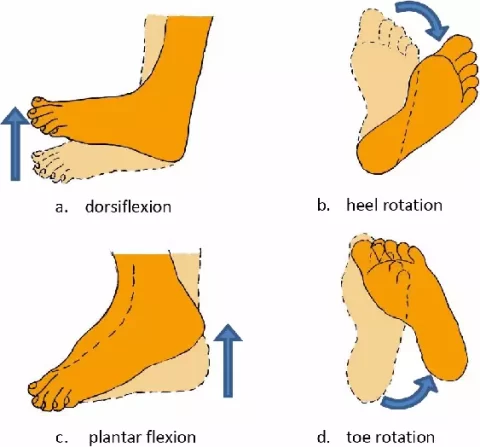
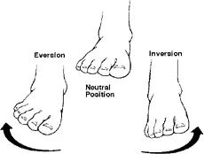
Blog

  • Gastrocnemius Muscle

    Gastrocnemius Muscle

    Anatomy of Gastrocnemius Muscle

    The gastrocnemius is a large, two-headed muscle in the calf that forms the bulk of its shape. It originates from the femur and inserts into the Achilles tendon, working with the soleus muscle to enable plantar flexion of the foot and assist in knee flexion. It plays a crucial role in day-to-day activities such as walking, running, and jumping.

    Origin

    The gastrocnemius muscle, which forms a major portion of the gastrocnemius, has two heads of origin:

    • Medial Head:
      • The posterior aspect of the femur’s medial condyle is where the medial head originates.
      • It also originates from the epicondyle of the femur.
    • Lateral Head:
      • This muscle has its origin on the lateral femoral condyle.

    Insertion

    • The two heads of the gastrocnemius muscle come together and form a tendon. This tendon then joins with the tendon of the soleus muscle (another gastrocnemius muscle) to form the Achilles tendon (also known as the calcaneal tendon). The Achilles tendon then inserts onto the posterior (back) surface of the calcaneus (heel bone).

    Innervation

    The tibial nerve supplies the gastrocnemius’s two heads (S1 and 2).
    L4, 5, and S2 are mostly responsible for cutaneous supply.

    Blood supply

    The lateral and medial sural arteries, which are direct branches of the popliteal artery, supply the medial and lateral heads of the gastrocnemius.

    The popliteal fossa is where the arteries originate, however their level varies; typically, the lateral artery arises more distally and the medial sural artery more proximally. The superior genicular and popliteal arteries may also give rise to minor auxiliary sural arteries. The popliteal vein receives venous drainage via the lateral and medial sural veins.

    Lymphatic drainage

    Lymphatic drainage in the lower limb occurs via two distinct pathways: superficial and deep. The superficial vessels, located just beneath the skin, collect lymph from the skin and are categorized into medial, lateral, and superficial gluteal groups, all of which ultimately drain into the superficial inguinal lymph nodes.

    The deep lymphatic vessels, on the other hand, collect lymph from deeper structures such as bones, muscles, and joints. These vessels follow the path of the deep blood vessels and eventually terminate in the deep inguinal lymph nodes.

    The lumbar lymph nodes, located in the lower back, serve as a major drainage point for lymph from various regions, including the lower limb, pelvic wall, perineal wall, and the sub-umbilical region of the abdominal wall. They also receive lymph from the vascular network supplied by the splanchnic branches of the aorta.

    Functions

    standing-hamstring-curl
    standing-hamstring-curl

    Half of the gastrocnemius muscle is made up of the gastrocnemius and soleus muscles. Its purpose is to flex the leg at the knee joint and plantar flex the foot at the ankle joint.

    Running, leaping, and other “fast” leg motions are the main activities of the gastrocnemius, with walking and standing playing a smaller role. In contrast to the soleus, which has more red muscular fibers (type I slow twitch) and is the main active muscle when standing still, the gastrocnemius has more white muscle fibers (type II rapid twitch), which is linked to this specialization.

    Exercises

    Stretching Exercises of Gastrocnemius Muscle

    1. Standing gastrocnemius Stretch:
    • Place your feet shoulder-width apart and face a wall.
    • Use the wall as a support by placing your hands on it.
    • With one leg, take a step back while maintaining a straight knee and a grounded heel.
    • Until your gastrocnemius starts to stretch, lean forward.
    • Hold the stretch for 30 seconds, then release and repeat the stretch on the opposite side.
    Standing Calf Stretch
    Standing gastrocnemius Stretch
    1. Downward-Facing Dog:
    • To start, go down on your hands and knees.
    • Make an inverted V shape with your body by tucking your toes and raising your hips up and back.
    • Maintain a tiny bend in your knees and an elevated heel.
    • Alternately bend one leg at a time while gently pedaling your feet.
    • For 30 seconds, hold the stretch.
    downward-facing-dog
    downward-facing-dog
    1. Seated gastrocnemius Stretch:
    • Sit down on the floor and extend your legs forward.
    • Wrap a resistance band or cloth around your foot’s ball.
    • While maintaining a straight knee, gently draw your toes towards you.
    • Hold the stretch for 30 seconds, then release and repeat the stretch on the opposite side.
    Seated Calf Stretch
    Seated Calf Stretch

    Strengthening Exercises

    • Gastrocnemius Raises: The most basic and effective calf exercise. Stand with feet flat on the floor. Rise onto the balls of your feet, lifting your heels, and hold briefly at the top. Slowly lower your heels. For a greater range of motion, perform these on a slightly elevated surface (like a step). Increase difficulty by holding dumbbells or a weight plate.
    Calf Raises
    Gastrocnemius Raises
    • Single-Leg gastrocnemius Raises: Perform gastrocnemius raises while standing on one leg. This increases difficulty and engages stabilizing muscles. Use a support for balance if needed.
    Single-leg-calf-raises-with-bent-knee-on-a-step
    Single-leg-gastrocnemius-raises-with-bent-knee-on-a-step
    • Jumping Rope: A dynamic exercise that works the calves and other leg muscles.
    jumping rope
    jumping rope
    • Stair Climbs/Hill Sprints: Excellent for gastrocnemius development. Focus on pushing off with your toes.
    single leg stair jump
    single leg stair jump

    Weighted Gym Exercises:

    • Seated gastrocnemius Raises: This machine exercise effectively isolates the gastrocnemius (calf muscle). The seated position reduces the involvement of the soleus (another calf muscle), emphasizing the gastrocnemius.
    seated-calf-raises
    seated-gastrocnemius-raises
    • Standing gastrocnemius Raises (Machine or Smith Machine): Similar to bodyweight gastrocnemius raises, but with added weight for increased resistance.
    Hack squat with Smith Machine
    Hack squat with Smith Machine
    • Leg Press gastrocnemius Raises: Perform gastrocnemius raises on the leg press machine by extending your ankles.
    Leg press machine
    Leg press machine

    Muscles

    The Myofascial Continuum

    One should consider a myofascial continuum rather than a single area when discussing skeletal muscle. Through its fascial connections—the anatomical interconnections with other tendons (insertion and origin) and the fascia covering muscles—the gastrocnemius muscle stress is sent not only to the foot but also to the knee, hip, and lumbar region.

    Hip anteversion may decrease due to malfunction in the hip’s physiological motions caused by a shortened gastrocnemius muscle.The force generated by the contraction of the muscle’s contractile component is fundamentally transmitted by the fascial system.

    The connective tissue of the muscle stroma is the first tissue to experience the stress created by a muscle contraction. If the muscle has a fusiform architecture, the tension moves longitudinally down the fiber axis via the epimysium and up to the tendon.

    The vector first travels in a transverse direction before subsequently following the epimysium’s longitudinal structures. If the muscle is pennate, such as the gastrocnemius, the force generated follows the interfilament inside the Z line of the sarcomeres, up to the sarcolemma, the extracellular matrix, and the epimysium (and thus the tendon).

    Whereas the pennate muscles in the second scenario have a slower rate of force arrival to the tendons but a wider range of tension values, the fusiform muscles in the first scenario have a faster but less effective rate of force arrival. For instance, the gastrocnemius muscle is stronger than the soleus muscle, which is quicker but weaker, while the biceps brachii is faster but weaker than the deltoid muscle.

    The longest and broadest tendon in the human body is the calcaneal tendon.The tendon’s attachment to the foot’s plantar fascia, which runs from the calcaneus to the metatarsal heads, is thought to be essential to the stability of the various tarsal joints. The plantar fascia’s tension is adversely affected by gastrocnemius muscle dysfunction, which modifies the foot’s physiological support.

    Muscular Phenotypes

    The soleus muscle is rich in aerobic or red fibers, whereas the gastrocnemius muscle is mostly composed of anaerobic or white fibers. This is another significant distinction between the two muscles. The gastrocnemius will be called upon to step in during an action that calls for the rapid expression of high strength. The soleus muscle will be the primary player if walking is the primary physical exertion.

    Anatomical Variations

    The human lower leg, like many parts of the body, exhibits fascinating anatomical variations. The gastrocnemius muscle, a key component of the gastrocnemius, is no exception. Beyond its typical structure, individuals may possess a “gastrocnemius tertius,” an additional muscle slip integrated within the gastrocnemius itself. Furthermore, an extra soleus muscle can also occur.

    In rare instances, both these accessory muscles can be present simultaneously and on both legs (bilaterally), typically without impacting joint function or altering the tibial nerve’s innervation. However, the presence of a gastrocnemius tertius carries potential clinical implications. Specifically, it could potentially compress the popliteal artery within the popliteal fossa, leading to arterial entrapment.

    Anatomical variations of the gastrocnemius muscle are well-documented. One such variant, the “quadriceps gastrocnemius,” has been described, though its functional and pathological significance remains unclear. Importantly, this variation is innervated by the tibial nerve.  

    Furthermore, the gastrocnemius can exhibit a range of anomalies beyond the presence of extra muscle slips. These include the absence of a head, particularly the lateral head, or variations in the origin of the muscle heads. A notable case report details a compression of both the popliteal artery and vein due to an atypical origin of the lateral gastrocnemius head. This anatomical anomaly resulted in the formation of arterial and venous thrombi, ultimately leading to secondary pulmonary hypertension.

    Embryology

    Skeletal muscle formation occurs during the eighth week of pregnancy. The paraxial mesoderm, which originates from the somites in the post-otic area, is the source of the gastrocnemius muscle. The myotomes, sclerotomies, and dermomyotomes of the somites vary.

    The lateral dermatomyotome’s lips are the source of the muscles’ predecessors. Transverse striations, a common indicator of muscle maturity, are present in the last stage of muscle growth. Myotubes, the progenitor cells of muscle fibers, are present at this point.

    Surgical Importance

    Gastrocnemius muscle surgery carries inherent risks, notably the potential for sural nerve injury and suboptimal cosmetic outcomes. To minimize these risks and provide a structured surgical approach, the gastrocnemius region can be divided into five distinct anatomical levels:

    • 1st Level (Distal): This level encompasses the calcaneal tendon’s insertion into the heel, including the portion within the heel itself.
    • 2nd Level : This area involves the combined aponeurosis of the gastrocnemius and soleus muscles, extending proximally from the calcaneal tendon to the point where the muscle fibers transition into the aponeurosis.
    • 3rd Level : This level begins at the musculotendinous junction, where the gastrocnemius muscle fibers merge with its aponeurosis, and extends distally to the point where the gastrocnemius and soleus fascial areas fuse.
    • 4th Level : This level represents the muscular portion of the two gastrocnemius heads, located before the transition into the aponeurosis.
    • 5th Level (Proximal): This level includes the origin of the two gastrocnemius muscle heads within the popliteal fossa.

    This level-based approach provides a clear anatomical framework for surgical procedures, aiding in precise dissection and minimizing complications.

    Gastrocnemius Muscle Flaps in Reconstruction:

    The medial head of the gastrocnemius muscle offers a versatile, innervated flap for reconstructive surgery. This flap can address various clinical conditions, including:

    • Foot drop
    • Muscle defects in the upper limbs
    • Volkmann’s contractures
    • Tongue movement restoration
    • Post-surgical infection management, particularly in knee prosthesis replacements, by promoting improved wound healing.
    • Enhancing cicatrization in transtibial amputation procedures.

    The use of a partial gastrocnemius muscle flap is favored due to its minimal associated complications and efficient surgical application.

    Baker’s Cyst:

    Named after surgeon William Morrant Baker, who first described it in 1877, a Baker’s cyst is a popliteal cyst arising from the knee joint. These cysts typically develop in the context of pre-existing knee pathologies, such as osteoarthritis or meniscal tears. These conditions lead to joint effusions that accumulate in the posterior knee region, specifically within the bursa of the medial head of the gastrocnemius.

    Often, the cyst contains joint material that has migrated from the knee capsule, with fluid flow occurring unidirectionally. In most cases, Baker’s cysts are asymptomatic and are incidentally discovered during magnetic resonance imaging of a knee with known functional issues. Surgical excision, utilizing a posterior approach, is reserved for select cases.

    Gastrocnemius Muscle Tears (Strains):

    Muscle tears, or strains, are common injuries, particularly among athletes. The severity of a gastrocnemius muscle tear can be classified into three grades:

    • First-Degree Strain:
      • This is the mildest form, involving minimal muscle fiber damage (less than 5% of muscle mass).
      • Muscle function is largely unaffected.
      • Individuals may experience a persistent cramping sensation.
      • Conservative treatment is indicated.
    • Second-Degree Strain:
      • This involves a more significant tear, resulting in noticeable pain, especially during movement.
      • Conservative treatment is indicated.
    • Third-Degree Strain:
      • This is the most severe tear, involving at least 75% of the muscle mass.
      • It is characterized by:
        • Intense pain.
        • Hematomas, edema, and significant swelling.
        • Severe functional impairment, rendering the individual unable to move the leg.
        • A palpable depression at the site of the tear, indicating the extent of the muscle disruption.
      • This severe injury may require surgical intervention in some cases.

    Achilles Tendon Disorders:

    Achilles tendon (calcaneal tendon) disorders are prevalent injuries affecting both adolescents and adults, arising from both traumatic and non-traumatic causes. These disorders encompass a range of pathologies, including:

    • Insertional tendinitis (inflammation at the tendon’s insertion point)
    • Intra-substance tears and/or tendinopathy (degeneration within the tendon itself)
    • Partial or complete ruptures

    A significant challenge in Achilles tendon injuries is the potential for misdiagnosis. Acute ruptures are misdiagnosed in up to 25% of cases, with ankle sprains being the most common error. Delayed diagnosis can lead to chronic tendon dysfunction, making surgical repair more complex.

    Key indicators of an Achilles tendon rupture may include:

    • Sudden, acute pain in the calcaneal tendon.
    • Functional impairment (inability to use the leg properly).
    • A palpable gap or discontinuity in the tendon.

    Diagnostic confirmation is typically achieved through ultrasound imaging.

    Achilles tendon ruptures often result from chronic tendinitis that may have gone unnoticed or been underestimated. Contributing factors can include:

    • Systemic diseases such as diabetes or lupus erythematosus.
    • Chronic use of steroid medications.

    These factors can alter the collagen fibers within the tendon, leading to an increase in type III collagen. This results in decreased tendon elasticity and reduced ability to withstand mechanical stress, predisposing it to rupture.

    Achilles Tendon Rupture: Causes and Treatment

    Achilles tendon ruptures are particularly common in athletes involved in jumping and running sports, such as basketball, soccer, and tennis. These injuries typically result from a sudden, forceful muscle contraction.

    Treatment for a complete Achilles tendon rupture is generally surgical. Various tendon suture techniques are employed. Modern surgical approaches favor minimally invasive techniques, using small incisions to minimize scarring and reduce recovery time. In some cases, tendon reinforcement is performed using grafts from the gracilis, plantaris, or peroneus longus tendons.

    Gastrocnemius Muscle Shortening (Equinus Deformity):

    Gastrocnemius muscle shortening can arise from a variety of underlying conditions, including:

    • Central or peripheral nervous system lesions.
    • Hereditary muscle diseases, such as myopathies and dystrophies.
    • Acquired diseases, such as diabetes.

    The most prominent functional consequence of gastrocnemius shortening is equinus deformity, characterized by a plantarflexed foot (the foot pointing downwards) and a toe-walking gait.

    Effective management requires a thorough evaluation to determine the underlying cause of the equinus deformity, assess its impact on gait, and develop an appropriate treatment plan. Treatment options include:

    • Conservative methods:
      • Physical therapy and stretching exercises.
      • Orthotic devices (tutors).
      • Botulinum toxin injections.
    • Surgical methods:
      • Calcaneal tendon lengthening.

    Silfverskiold Test for Equinus Deformity:

    The Silfverskiold test is a valuable clinical tool for differentiating between gastrocnemius and soleus muscle contractures in equinus deformity. By assessing ankle dorsiflexion with the knee in different positions, the test helps pinpoint the primary muscle responsible for the limitation.  

    • Procedure:
      • The test can be performed with the patient either sitting or supine.  
      • First, the examiner attempts to dorsiflex the ankle with the knee fully extended.  
      • The examiner attempts to dorsiflex the ankle with the knee flexed to 90 degrees.
    • Interpretation:
      • If ankle dorsiflexion improves significantly with the knee flexed, but is restricted with the knee extended, it suggests a gastrocnemius muscle contracture.
      • If ankle dorsiflexion is limited in both knee flexed and extended positions, it suggests a soleus muscle contracture, or a combined gastrocnemius and soleus contracture.

    Endoscopic Gastrocnemius Recession:

    Modern surgical techniques for gastrocnemius recession utilize endoscopy to minimize collateral damage. This approach involves a small incision at level 4 of the leg, allowing for targeted release of the gastrocnemius muscle.  

    • Advantages:
      • Reduced scarring compared to traditional open surgery.  
      • Decreased postoperative pain and faster recovery.
    • Caution:
      • Precise surgical technique is crucial to avoid excessive muscle elongation, which could lead to an iatrogenic overcorrection.

    Clinical Importance

    Tennis Leg

    A frequent injury among tennis and squash players is tennis leg. When the myotendinous junction of the medial head of the gastrocnemius ruptures, it often manifests as an abrupt, acute pain toward the back of the gastrocnemius. Less frequently, it involves the plantaris muscle rupturing. It results by dorsiflexing the ankle and completely extending the knee, which overstretches the muscle.

    Achilles tendon tendonitis

    The calcaneal (Achilles) tendon thickening at its insertion causes Achilles tendonitis, a painful condition. The majority of those who experience it are middle-aged or older. It results in bone and cartilaginous metaplasia and is caused by repeated straining of the muscle. A bony growth at the tendon’s insertion onto the calcaneus and soreness in the rear of the heel are typical symptoms.

    Achilles tendon rupture

    Recreational athletes frequently sustain calcaneal (Achilles) tendon ruptures. The tendon’s calcaneal insertion, which is comparatively avascular, is typically 4-6 cm above the rupture, which can be partial or total. The patient may remember the sensation of the muscle rolling up their leg and the snap sound that occurs when a tendon ruptures. Excessive passive dorsiflexion and lack of plantar flexion against resistance are the results of a complete tendon rupture.

    Calcaneal bursitis

    Inflammation of the bursa, which divides the calcaneal tendon from the calcaneal tuberosity, a process of the calcaneus, is known as calcaneal bursitis. The back of the heel may experience discomfort as a result. Inflammation is frequently the result of long-distance running, basketball, and tennis, where the action of the tendon creates significant friction.

    Gastrocnemius Muscle Muscle Pain

    Several factors can cause pain in the gastrocnemius muscle (calf muscle). Muscle strain, the most common cause, happens when the muscle fibers are stretched or torn. Muscle cramps, which are sudden, involuntary muscle contractions, can also cause pain and are often linked to dehydration, electrolyte imbalances, or muscle fatigue.

    Tendinitis, an inflammation of the tendons that connect muscles to bones, is another possibility and is frequently caused by overuse or repetitive movements.

    Finally, although less common, deep vein thrombosis ( DVT ), a blood clot in a deep vein (usually in the leg), can cause gastrocnemius pain, often accompanied by swelling and redness.

    Examination of Gastrocnemius Muscle

    Silfverskiold Test:

    The Silfverskiold test is a valuable clinical tool for differentiating between gastrocnemius and soleus muscle contractures in equinus deformity. By assessing ankle dorsiflexion with the knee in different positions, the test helps pinpoint the primary muscle responsible for the limitation.  

    • Procedure:
      • The test can be performed with the patient either sitting or supine.  
      • First, the examiner attempts to dorsiflex the ankle with the knee fully extended.  
      • The examiner attempts to dorsiflex the ankle with the knee flexed to 90 degrees.
    • Interpretation:
      • If ankle dorsiflexion improves significantly with the knee flexed, but is restricted with the knee extended, it suggests a gastrocnemius muscle contracture.
      • If ankle dorsiflexion is limited in both knee flexed and extended positions, it suggests a soleus muscle contracture, or a combined gastrocnemius and soleus contracture.

    Endoscopic Gastrocnemius Recession:

    Modern surgical techniques for gastrocnemius recession utilize endoscopy to minimize collateral damage. This approach involves a small incision at level 4 of the leg, allowing for targeted release of the gastrocnemius muscle.  

    • Advantages:
      • Reduced scarring compared to traditional open surgery.  
      • Decreased postoperative pain and faster recovery.
    • Caution:
      • Precise surgical technique is crucial to avoid excessive muscle elongation, which could lead to an iatrogenic overcorrection.

    FAQs

    What are the other names of gastrocnemius ?

    The calf muscle is also known as the sural muscle group.
    The gastrocnemius is a major component of the sural muscle group, often referred to as the leg triceps. It’s a large, superficial muscle located on the posterior aspect of the lower leg, forming the bulk of the calf. The name “gastrocnemius” originates from the Greek words “gaster” (stomach or belly) and “kneme” (leg), reflecting its bulging shape.

    Are the gastrocnemius muscles an extensor or a flexor?

    As a biarticular muscle, the gastrocnemius functions as both a knee flexor and a plantar flexor, making it an antagonist during knee extension.

    What signs indicate a weak gastrocnemius?

    Reduced propulsion up and forward when walking and running is a sign of weak gastrocnemius muscles. Prolonged heel contact late in the stance phase of walking or running is the main gait abnormality caused by a weak gastrocnemius muscle.

    Which exercises are beneficial for your gastrocnemius?

    The traditional gastrocnemius-strengthening exercise is the gastrocnemius raise. The gastrocnemius and soleus muscles are toned and strengthened using your body weight. Better still, they don’t need much time and can be done anywhere. To gain equilibrium, stand close to a wall.

    My gastrocnemius muscle hurts; why?

    Numerous factors, such as injury, overuse, or other disorders, might cause pain in your calf’s gastrocnemius muscle.
    Muscle strain injury

    A torn gastrocnemius muscle can happen when you abruptly overstretch it, such when you run or leap.
    Tears in the muscles
    A muscle tear, either partial or whole, may result by abruptly overstretching the muscle.
    Tendinopathy
    Overuse of the muscle can result in microscopic rips in the tendon.
    Overuse
    Fatigue of the muscles: discomfort that may follow vigorous physical exertion or exercise
    Overload of muscles: Pain that may arise from increasing the intensity of your workout, such as jogging farther or more frequently
    Additional circumstances
    Tightness: Muscle tightness caused by cramping or excessive usage
    Vein varicosity: Prolonged standing or walking might cause thick, protruding veins.
    Drugs: Some medicines, including those that decrease cholesterol, might induce discomfort in the calves.
    Other conditions: gastrocnemius discomfort can be caused by conditions including peripheral artery disease (PAD), diabetes, liver illness, renal disease, or hypothyroidism.

    How can discomfort in the gastrocnemius be stopped?

    When a gastrocnemius issue initially appears, you should try to limit your activity but move as much as your symptoms permit over the first 24 to 48 hours.
    When at rep?
    ose, place your gastrocnemius in an elevated posture.
    Every hour while you’re awake, take ten to twenty seconds to gently move your ankle and knee.
    Don’t stand for extended periods of time.

    Which stretch works best for the gastrocnemius?

    Flexibility Stretch for the Gastrocnemius | Saint Luke’s Health System
    Maintaining the left heel on the ground and the left leg straight, lean forward. Wait five to ten seconds. Do this two or three times, or as directed. Another method is to contract the gastrocnemius muscle pressing into the wall for five seconds while maintaining the stretch for five seconds.

    Which deficit results in leg weakness?

    Leg weakness may result from low potassium levels or deficiencies in vitamins B12, D, and magnesium. Intense physical exertion, peripheral vascular disease, and certain drugs are other potential reasons.

    References:

    • Gastrocnemius Muscle , Physiopedia , https://www.physio-pedia.com/Gastrocnemius
    • Gastrocnemius Muscle , NCBI , https://www.ncbi.nlm.nih.gov/books/NBK532946/
    • Physiotherapist, N. P.-. (2023, December 23). Gastrocnemius muscle Origin, Insertion, Function, Exercise. Mobile Physiotherapy Clinic. https://mobilephysiotherapyclinic.in/gastrocnemius-muscles-details/

  • Pyogenic Flexor Tenosynovitis

    Pyogenic Flexor Tenosynovitis

    Introduction

    Pyogenic Flexor Tenosynovitis (PFT) is a bacterial infection of the flexor tendon sheath in the hand, typically caused by Staphylococcus aureus. It results from penetrating trauma, bites, or hematogenous spread.

    The condition is characterized by Kanavel’s four cardinal signs: finger flexion at rest, fusiform swelling, tenderness along the tendon sheath, and pain with passive extension. PFT is a surgical emergency requiring prompt antibiotic therapy and, often, surgical drainage to prevent tendon necrosis and hand dysfunction.

    A closed-space infection of the hand’s flexor tendon sheath, pyrogenic flexor tenosynovitis (PFT), often referred to as septic or suppurative flexor tenosynovitis, continues to be a difficult issue in the field of hand surgery. PFT is still a prevalent issue, accounting for 2.5 to 9.4% of all hand infections, according to research.

    PFT can result in tendon necrosis and finger devitalization, and is responsible for 2.5% to 9.5% of hand infections. This infection causes adhesions within the flexor tendon sheath and changes the gliding mechanism, significantly impairing finger movement.

    Although bloodstream infections can induce PFT, penetrating finger injuries involving the flexor tendon sheath are typically the culprit. The three main symptoms of pyogenic flexor tenosynovitis were identified in 1912: painful passive digital extension, flexor sheath soreness, and a flexed posture of the affected finger. Fusiform swelling of the digit was then included as a fourth sign, making the four cardinal signs.

    Anatomy

    • Although the anatomy of the flexor tendon sheath varies greatly, there are certain commonalities.
    • The distal sheaths in the fingers end at the FDP insertion, which is close to the DIP joint.
    • The sheath in the thumb ends where the FPL is inserted, which is close to the IP joint.
    • The sheaths often reach only proximal to the A1 pulley (close to the MCP joint). Usually, the sheaths of the thumb and small finger connect to the radial and ulnar bursae.
    • A horseshoe abscess may emerge from an infection that travels from the little finger to the thumb or the other way around.

    Pathogenesis

    The pathogenesis of pyogenic flexor tenosynovitis (PFT) involves a bacterial infection within the closed space of the hand’s flexor tendon sheath. This is usually caused by a penetrating injury that allows bacteria to enter the sheath directly, resulting in inflammation, pus accumulation, and possibly tendon damage.

    If treatment is delayed, the infection can spread quickly because the hand’s tendon sheaths are interconnected, and the most common causative bacteria are Staphylococcus aureus and other skin flora.

    Stages of pyogenic flexor tenosynovitis

    Michonne divided PFT into three phases according to the infection’s severity:

    • Stage 1: The flexor sheath begins to fill with serous exudate
    • Stage 2: Purulent fluid fills the sheath.
    • Stage 3: Necrosis is evident in the flexor tendon, flexor sheath, and pulley system.

    Causes

    Although bacteria are typically the source of both infections, the infection pathway may vary. While osteomyelitis can be caused by a direct wound, hematogenous spread (via the circulation), or contiguous transmission from an adjacent infection, pyogenic flexor tenosynovitis frequently results from a penetrating lesion to the hand.

    • Penetrating trauma: PFT can result from bites, puncture wounds, and lacerations to the finger.
    • Bits: PFT can be caused by Pasteurella multocida from an animal bite and Eike Nella corrodes from a human bite.
    • Bloodstream infection: Hematogenous spread may be the cause of PFT.

    Signs and Symptoms

    Kanavel’s signs:

    Clinicians often use the classic “Kanavel signs” to diagnose pyogenic flexor tenosynovitis, including:

    • Fusiform swelling of the finger
    • Tenderness along the flexor tendon sheath
    • Pain with passive extension of the finger
    • Flexed posture of the affected digit
    • Loss of motion
    • Tendon rupture
    • Soft tissue necrosis
    • Amputation

    Risk factor

    Found that the following risk factors, particularly the amputation rate and its impact on total active motion, were associated with deteriorating clinical outcomes in a sample of 75 PFT patients:

    • Diabetes
    • Peripheral vascular disease
    • Renal failure
    • Age over 43 years
    • Subcutaneous purulence
    • Digital ischemia
    • Polymicrobial infection
    • IV drug use
    • Immunocompromised patients

    Diagnosis

    • X-ray
    X-ray for pyogenic flexor tenosynovitis
    X-ray for pyogenic flexor tenosynovitis

    To rule out any retained foreign body, x-rays of the hand taken in the anteroposterior, lateral, and oblique views should be acquired. The acquired x-rays show evidence of osteomyelitis, which points to a persistent infection. Uniform finger swelling did not differentiate PFT from other hand infections, according to a retrospective analysis of adult finger infections. Additionally, they stated that at the level of the proximal phalanx, PFT is distinguished by a difference in radiographic soft tissue thickness between the volar and dorsal aspects.

    • Hand ultrasonography

    Hand ultrasonography is a low-cost, noninvasive technique for confirming the diagnosis of PFT. Ultrasonography can see the flexor tendon and identify any fluid buildup inside the flexor sheath. In addition to assisting the clinical examination in identifying finger infections, ultrasound offers an excellent negative predictive value and specificity for diagnosing PFT. Nevertheless, the ultrasound depends on the operator and cannot distinguish between blood and pus.

    • Magnetic resonance imaging (MRI)

    PFT is infrequently diagnosed with magnetic resonance imaging (MRI). While MRI can determine the degree of the infection, it is unable to distinguish PFT from other inflammatory diseases.

    Because the ratio of tendon sheath to tendon width in the coronal and sagittal planes was shown to increase objectivity, contrast-enhanced computed tomography of the hand has also been claimed to be useful in detecting PFT.

    • Laboratory test

    Patients with PFT typically have higher levels of inflammatory indicators, such as erythrocyte sedimentation rate, C-reactive protein, and white blood cell count. Although these tests cannot specifically diagnose PFT, they can be useful in tracking infection and treatment response.

    To determine the causal organism and guide antibiotic therapy based on sensitivity, pus and necrotic tissue cultures are highly helpful. To isolate the causal organism, get a blood culture if there are indications of sepsis or hematogenous infection spread.

    Differential Diagnosis

    Felon (Distal Pulp Space Infection)

    The thumb or index finger are frequently affected by felon, a closed space infection of the distal finger pulp. Staphylococcus aureus infections resulting from penetrating injury to the fingertip pulp are typically the cause of felon or pulp space infections. However, in immunocompromised people, this illness may be caused by gram-negative bacteria or a variety of species.

    When infected, the fibrous septa, which run from the distal phalanx periosteum to the skin from a confined area, produce excruciating throbbing pain. A surgical incisional drainage is necessary for a felon who presents with a severe, throbbing abscess over the distal finger phalanx.

    Interphalangeal Joint or Metacarpophalangeal Joint Septic Arthritis

    Signs of infection specific to the affected joint are typically evident in these two arthritic diseases. Because of joint effusion and capsular distention, the affected joint’s range of motion is typically limited and painful. Usually lacking are the other cardinal Kanavel symptoms of PFT, such as flexor sheath pain and finger fusiform edema.

    Herpes simplex virus (HSV)

    The herpes simplex virus is the source of this uncommon viral infection of the fingertips. Herpetic whitlow is characterized by painful blisters that may eventually develop bullae and are filled with a clear fluid. Antiviral drugs like acyclovir are necessary for treating herpes whitlow; surgery is not necessary.

    Bacterial infection

    Diffuse hand inflammation without underlying pus accumulation is called hand cellulitis. Hand cellulitis manifests as erythema and widespread swelling without the development of an abscess. Depending on the causative organism, high arm elevation and intravenous antibiotics can be used to treat hand cellulitis.

    Treatment

    Medical treatment

    Cultures and antibiotic treatment beginning initially with a cephalosporin are also required. A cephalosporin should be replaced by trimethoprim/sulfamethoxazole, clindamycin, doxycycline, or linezolid in regions where methicillin-resistant Staphylococcus aureus (MRSA) is common.

    In the end, microbiology and, if available, susceptibility data should inform the antibiotic selection. If a mycobacterial or fungal infection is suspected, the microbiology lab should be notified.

    • Amoxicillin + clavulanic acid: This is often the preferred oral antibiotic for PFT.
    • Cephalosporins: These are often the initial antibiotic used to treat PFT.
    • Trimethoprim: This is sometimes used in areas where methicillin-resistant Staphylococcus aureus (MRSA) is common.
    • Following surgery, oral antibiotics are administered for two to six weeks.
    • To lessen stiffness and adhesion formation, supervised therapy is initiated.

    Physical therapy treatment

    To restore hand function while minimizing additional inflammation and pain, physical therapy treatment for pyogenic flexor tenosynovitis primarily consists of gentle range-of-motion exercises after the acute infection has been controlled with antibiotics and surgical drainage. This often involves splinting to rest the affected tendons and gradually increasing the exercises as tolerated.

    It is important to remember that pyogenic flexor tenosynovitis is a medical emergency that requires immediate surgical intervention and antibiotic treatment; physical therapy should only be started after the acute infection has been managed by a healthcare professional.

    Rest and splinting

    To limit movement and aid in healing, the injured hand will first be immobilized with a splint; the splint may be adjusted as tolerated to permit greater range of motion.

    • Elevation: Swelling can be lessened by keeping the injured hand up above the level of the heart.
    • Ice application: Using ice packs can aid in the management of inflammation and pain.

    Gentle active range-of-motion exercises

    Gentle active range-of-motion exercises
    Active range-of-motion exercises

    With an emphasis on flexion and extension movements, the physical therapist will gradually increase the range of motion in the fingers and hand by introducing mild active exercises once the acute irritation has subsided.

    Tendon gliding exercises

    Tendon gliding exercises
    Tendon gliding exercises

    By gently moving the tendons back and forth within their sheath, these workouts seek to reduce friction within the tendon sheath.

    Phase 1 (Early stage):

    • Gentle finger extensions and flexions within a pain-free range. 
    • Tabletop position with slight finger spread. 
    • Hook position with minimal finger flexion. 

    Phase 2 (Moderate stage):

    • Full range of motion in tabletop, hook, and fist positions. 
    • Adding wrist flexion and extension movements. 

    Phase 3 (Advanced stage):

    • Incorporating light resistance exercises using hand therapy tools. 
    • Functional activities like picking up small objects or gripping a soft ball. 

    Isometric exercises

    In the early phases of rehabilitation, isometric workouts can assist in minimizing joint movement and preserve muscle strength.

    Soft tissue mobilization

    Soft tissue limitations can be addressed and blood flow to the area improved with the use of gentle massage techniques.

    Surgical treatment

    Open Irrigation

    The flexor sheath is exposed and washed out during open surgery for pyogenic flexor tenosynovitis (PFT). Open flexor sheath irrigation and debridement is the term for this process. It is used to treat persistent tenosynovial infections and advanced infection cases.

    Procedure

    • Cut through the injured finger’s palm or mid-axial region.
    • Determine and safeguard the affected finger’s neurovascular bundle.
    • Cut the flexor sheath’s A1 and A5 pulleys.
    • Use saline to debride and rinse the sheath.
    • Prevent the flexor tendon from bowstringing by protecting the A2 to A4 pulleys.

    Closed Tendon Sheath Irrigation

    A cut is performed above the metacarpal neck in the proximal direction. The proximal border of the A1 pulley is where the tendon sheath is transversely sliced. An angiocatheter is placed into the flexor tendon sheath 1–2 cm antegrade.

    Next, on the radial aspect of the thumb or the ulnar aspect of the finger, a distal midfacial incision is made dorsal to the neurovascular bundle at the level of the distal interphalangeal joint.

    Distal to the distalmost pulley, the flexor sheath’s distal edge is exposed and resected. To keep the wound open and for fluid drainage, a Penrose drain can be inserted into the tendon sheath underneath the A4 pulley. It is possible to maintain intermittent bedside irrigation on the floor following surgery.

    Catheter irrigation

    Pyogenic flexor tenosynovitis can be surgically treated using catheter irrigation, which involves flushing the tendon sheath with fluid infused with antibiotics. This serves as a substitute for open surgical drainage.

    Procedure

    • The flexor sheath is accessed through a little incision.
    • The sheath is filled with a catheter.
    • Fluid infused with antibiotics is used to irrigate the sheath.
    • A solution of antibiotics is used to wash the tendons.
    • The wounds are sealed.

    Benefits 

    • Not as painful as open surgical drainage
    • Reduces scarring and produces smaller wounds.
    • It could facilitate a quicker return to normal.

    Prognosis

    If proper treatment is not started as soon as possible, PFT may lead to severe outcomes with finger amputation and spread infection. The full active range of motion may not be regained by 10% to 25% of the affected patients. Following PFT, patients with peripheral vascular disease, diabetes mellitus, and renal failure are more likely to have their fingers amputated.

    Complications

    Fingers Stiffness and Restricted Range of Motion

    Flexor tendon adhesions, larger joint capsules, and damaged pulleys can all be consequences of PFT’s inflammatory process. To reduce the chance of stiffness, suggest early physical workouts for the injured finger. After the inflammatory process has completely subsided, flexor tenolysis of the affected digit could be necessary to restore range of motion.

    Flexor Tendon Scarring and Subsequent Rupture

    Blood flow and nutrients to the flexor tendon are hampered by the high pressure inside the flexor sheath. Scarred, unhealthy flexor tendons become less elastic and can burst with more exercise.

    Soft Tissue Necrosis and Finger Ischemia

    The affected finger may experience critical ischemia as a result of arterial thrombosis caused by the inflammatory process or as a result of the blood flow being interrupted by the high pressure in the finger.

    Hand Horseshoe Abscess

    In patients with communicating ulnar and radial bursae, the infection from PFT of the thumb or little finger may spread, leading to the formation of a horseshoe-shaped abscess.

    Finger Amputation

    A severe infection, extensive soft tissue damage, or a stiff, functionless finger may necessitate amputation at different levels.

    Stiffness

    A bacterial infection known as pyogenic flexor tenosynovitis (PFT) can result in stiffness and decreased finger movement. If treatment is not received, this serious infection may result in amputation.

    Spread of infection

    Because the flexor tendon sheaths are interconnected, pyogenic flexor tenosynovitis (PFT) can spread quickly inside the hand. If treatment is not received, the infection may spread to the palm, forearm, and potentially additional digits.

    Loss of soft tissue

    This illness has the potential to cause severe soft tissue loss as a result of tissue necrosis caused by the infection. If treatment is delayed, this could result in decreased hand function or possibly the amputation of a finger.

    • Tendon necrosis: The flexor tendons may sustain direct harm from the infection, rupturing or losing their ability to function.
    • Adhesions: When scar tissue forms inside the tendon sheath, it can limit the tendon’s range of motion and cause stiffness.
    • Loss of skin: In extreme situations, the infection may spread to the skin, leading to tissue damage and possible skin loss.

    Osteomyelitis

    Both conditions are serious, but pyogenic flexor tenosynovitis mainly affects the tendons and surrounding tissue in the hand, whereas osteomyelitis directly affects the bone structure. Pyogenic flexor tenosynovitis is a bacterial infection within the hand’s flexor tendon sheath, which is regarded as a closed-space infection. Osteomyelitis can infect any bone in the body, whereas pyogenic flexor tenosynovitis affects the tendon sheath in the fingers and hand.

    Conclusion

    A frequent hand infection that can have serious consequences is PFT. The use of intravenous antibiotics or, in more severe cases, surgical irrigation and debridement are necessary for early therapy. But even with quick and careful care, a serious infection may result in permanent function impairment or even amputation of the affected digit.

    It can be difficult to diagnose PFT quickly, but to prevent negative effects and maintain hand function, early detection and treatment are crucial. Broad-spectrum intravenous antibiotics, as well as surgical irrigation and debridement when necessary, are essential components of early treatment. However, a serious infection can still result in decreased function or even amputation of the affected digit even with prompt and comprehensive treatment.

    FAQs

    How is pyogenic flexor tenosynovitis treated?

    It can be difficult to diagnose PFT quickly, but to prevent negative effects and maintain hand function, early detection and treatment are crucial. Broad-spectrum intravenous antibiotics, as well as surgical irrigation and debridement when necessary, are essential components of early treatment.

    Flexor tenosynovitis: what causes it?

    Although bloodstream infections can induce PFT, penetrating finger injuries involving the flexor tendon sheath are typically the culprit. For fingers to remain viable and functional, urgent care techniques are essential, with a focus on surgical washout.

    How does pyogenic flexor tenosynovitis manifest itself?

    The Ka navel symptoms of FTS include symmetrical swelling of the affected finger, holding the affected finger flexed, pain when passive digit extension is attempted, and tenderness along the flexor tendon sheath.

    How can flexor tenosynovitis be managed at home?

    Rest: for two to three days, try not to move the tendon.
    Ice: apply a tea towel-wrapped ice pack (or a bag of frozen peas) on the tendon for up to 20 minutes every two to three hours.
    Support options include using a soft brace, a tube bandage, or an elastic bandage across the affected area.

    Is it possible to heal a flexor tendon without surgery?

    It is unlikely that a cut tendon will mend without surgery because the cut ends typically split after an injury. When a flexor tendon is cut, your doctor will advise you on when surgery is necessary. A cut tendon can be repaired in a variety of methods, and some cuts require a particular kind of healing.

    How long does tenosynovitis treatment last?

    Reducing inflammation, maintaining thumb mobility, and avoiding recurrence are the goals of treatment for de Quervain’s tenosynovitis. Your symptoms should go away in four to six weeks if you begin treatment early.

    Is surgery necessary to treat flexor tenosynovitis?

    To cleanse the tendon sheath, incisions will be made on the front of the hand and finger during surgery if the ailment has progressed past its very early stages. More than one surgery is typically needed in moderate and severe instances.

    Which therapy is most effective for tenosynovitis?

    Treatment options for tenosynovitis include rest and cessation of the activity that triggered the condition.
    Putting on a splint or brace to lessen tendon stress.
    To lessen pain or inflammation, apply heat or ice to the affected area.
    Steroid injections or oral steroids to reduce inflammation.

    Can tendinitis be completely recovered from?

    Following rest and therapy, the majority of patients with tendinitis have quite good prognoses. Depending on the extent of your injury, recovering from tendinitis could take a few weeks to several months. Hold off on starting your usual physical activity until your doctor gives you the “all clear.”

    Reference

    • Sarpatwari, R. (2018, July 2). Getting a hand on pyogenic flexor tenosynovitis — BROWN EMERGENCY MEDICINE BLOG. BROWN EMERGENCY MEDICINE BLOG. https://brownemblog.com/blogposts/2018/6/20/pyogenic-flexor-tenosynovitis
    • Yoon, R., MD. (n.d.). Pyogenic flexor tenosynovitis – hand – orthobullets. https://www.orthobullets.com/hand/6105/pyogenic-flexor-tenosynovitis
    • Chan, E., Robertson, B. F., & Johnson, S. M. (2019). Kanavel signs of flexor sheath infection: a cautionary tale. British Journal of General Practice69(683), 315–316. https://doi.org/10.3399/bjgp19x704081
    • Crowe, C. S., MD. (n.d.). Tenosynovitis: Practice essentials, pathophysiology, etiology. https://emedicine.medscape.com/article/2189339-overview
  • What Is The Difference Between Bone Pain and Muscle Pain?

    What Is The Difference Between Bone Pain and Muscle Pain?

    Introduction:

    Bone pain is usually deep, dull, or aching and often persists at rest or night, potentially indicating fractures, infections, or conditions like osteoporosis. In contrast, muscle pain tends to be sore, cramping, or throbbing, often worsening with movement and improving with rest or stretching. Muscle pain is commonly due to strain, overuse, or inflammation, while bone pain may signal more serious underlying conditions.

    If the pain is severe or last for more than 48 hours, they should see a professional. Orthopedists and orthopaedic surgeons are medical professionals that specialize in the musculoskeletal system.

    A dull, aching feeling is a common description of bone pain, which is felt in the bones or joints. Numerous condition, including cancer, osteoporosis, arthritis, and injuries, can result in this kind of pain. It can be challenging to move the injured limb due to severe pain and swelling from injuries like fractures or fractured bones. Because osteoporosis weakens bones, it increases the risk of fractures and other injuries. Because tumors can develop in the bones and exert pressure on the surrounding tissues, cancer can also result in bone pain.

    Contrarily, muscle pain is experienced in the soft tissues or muscles and is sometimes characterized as an aching or sore feeling. Injury, misuse, condition, or underlying medical issues can all contribute to this kind of pain, which can vary in intensity from mild to severe. Strains and sprains, for example, might result in localized pain and make it difficult to move the injured muscle.

    Pain and weakness can result from small tears in the muscle fibers caused by overuse, such as repetitive actions. Conditions like polymyalgia rheumatica and fibromyalgia can result in widespread muscular weakness and pain. Muscle inflammation caused by infections like the flu can also result in muscle pain.

    What is Bone Pain?

    Soreness or pain in one or more of the body’s bones is known as bone pain. The severity of this pain might vary, and it can be either acute (transient) or chronic (persistent). Numerous things, such as disease, degenerative disorders, or injuries, can result in bone pain.

    A fracture can happen when a bone is subjected to excessive force, like in a fall or car accident, while a sprain or strain occurs when the muscles and ligaments supporting a joint are overstretched or torn. In these situations, the pain is typically localized to the area of the injury and may be accompanied by bruising, swelling, and trouble moving the affected limb.

    A disease is another prevalent cause of bone pain. A persistent autoimmune disease called rheumatoid arthritis results in joint inflammation, which can cause pain, stiffness, and swelling.

    Bone pain can also be caused by infections like osteomyelitis. In addition to fever and exhaustion, this infection may cause redness, swelling, and pain in the affected area.

    Benign bone tumors can hurt as they grow and put pressure on nearby tissues, but they don’t spread to other areas of the body. However, as they infiltrate and destroy surrounding tissues, malignant bone tumors can migrate to other parts of the body and cause pain.

    Other conditions like hormonal imbalances, nutritional deficiencies, and certain drugs can also result in bone pain. In women going through menopause, for instance, a drop in estrogen levels can cause bone loss and consequent bone pain. Deficits in certain nutrients, including calcium or vitamin D, can also cause pain and weak bones. Glucocorticoids are one example of a medicine that can weaken bones and cause pain.

    Injuries, condition, and other underlying medical issues are some of the causes of bone pain. For an accurate diagnosis and treatment plan, it’s critical to consult a physician if you’re having bone pain. Among the available treatment options include prescription drugs, physical therapy, lifestyle modifications, and, in certain situations, surgery. Finding the source of your bone pain and getting the right care will help you manage it and enhance your quality of life.

    What is Muscle Pain?

    Myalgia, another name for muscular pain, is a frequent condition caused by soreness, aching, or pain in the muscles. The duration of this kind of pain can vary from a few days to many weeks, and it can be mild to severe. Numerous things, such as condition, injury, overuse, or underlying medical issues, can result in muscle pain.

    Injury is a common cause of muscle pain. A direct injury, like a strain or sprain, or overexertion, like during physical exercise, can cause this. In these circumstances, the pain is usually restricted to the area of the injury and may be followed by edema, bruising, and difficulty moving the affected muscle.

    Overuse is another prevalent cause of muscle soreness. When a muscle is worked repeatedly, tiny rips in the muscle fibers may result. These tears may eventually result in the affected muscle becoming weak, swollen, and painful. People who perform repetitive tasks, like typing or playing an instrument, frequently experience this kind of muscle soreness.

    Diseases can also cause muscle pain. Muscle pain can result from conditions including dermatomyositis, polymyalgia rheumatica, and fibromyalgia. A chronic condition called fibromyalgia results in tenderness, exhaustion, and extensive muscle pain in particular body parts. An inflammatory disease that affects the muscles and joints, polymyalgia rheumatica can result in muscle pain, stiffness, and weakening.

    Muscle pain can also be caused by infections like the flu or a viral disease. Muscle inflammation caused by these infections may result in soreness and weakness. Additionally, muscle pain is an adverse effect of various drugs, including statins.

    Muscle soreness can also result from nutrient deficiencies, such as a lack of calcium or vitamin D. Pain may result from these inadequacies when the muscles weaken and become more prone to damage. Additionally, muscle weakness and soreness can be caused by hormonal imbalances such low testosterone or thyroid disorders.

    Numerous things, such as injury, overuse, condition, and underlying medical disorders, can result in muscle pain. It’s critical to consult a physician for a correct diagnosis and treatment plan if you’re having muscle pain. Medication, physical therapy, lifestyle modifications, and, in certain situations, surgery are all possible forms of treatment. You can assist control your pain and enhance your quality of life by determining the source of your muscle soreness and pursuing the right treatment.

    What differentiates muscle pain and bone pain?

    Two different types of pain that can arise in various bodily areas are bone pain and muscle pain. Although their symptoms may be identical, bone pain and muscle pain can be distinguished from one another thanks to a few important distinctions. The correct diagnosis and treatment of the underlying condition depend on an understanding of these distinctions.

    This type of pain is often described as a dull, aching sensation and can be felt in a specific area of the body or spread throughout the bones. Injuries, such as fractures or broken bones, can cause intense, sharp pain that may be accompanied by swelling and difficulty moving the affected limb. Osteoporosis is a condition that results in the loss of bone density, making bones more prone to fractures and other injuries. Arthritis is a condition that causes inflammation in the joints, leading to pain, stiffness, and difficulty moving. Because tumors can develop in the bones and exert pressure on the surrounding tissues, cancer can also result in bone pain.

    This type of pain can range from mild to severe and is often described as aching or soreness in the affected area. Many factors, such as injury, overuse, illness, and underlying medical disorders, can result in muscle pain. Overuse, such as from repetitive motions, can cause microscopic tears in the muscle fibers, leading to pain and weakness. Infections, such as the flu or a viral condition, can also cause muscle pain, as they can cause inflammation in the muscles. Conditions like polymyalgia rheumatica and fibromyalgia can result in widespread muscular weakness and pain. Muscle inflammation caused by infections, such the flu or a viral disease, can also result in muscle pain.

    Whereas muscular pain is felt in the muscles or soft tissues, bone pain is usually felt in the bones or joints. Swelling and trouble moving the affected limb can accompany bone pain. On the other hand, weakness or pain in the affected area may accompany muscle pain. Additionally, a cracking or popping sound may accompany bone pain. However, there are typically no sounds associated with muscle pain.

    The underlying reason will determine how to manage muscle and bone pain. Immobilization with a cast or brace may be required for injuries such fractures or fractured bones in order to promote appropriate bone healing. Pain can also be managed with medications, such as anti-inflammatory or pain medicines. For pain management and movement restoration, physical therapy and exercises can also be beneficial. In order to control pain and enhance joint function, treatments for diseases like osteoporosis or arthritis may involve prescription drugs, lifestyle modifications, and physical therapy. Physical therapy, rest, and drugs like muscle relaxants or painkillers can all be used to alleviate muscular pain.

    In conclusion, there are two different types of pain that can arise in various bodily parts: muscle pain and bone pain. The correct diagnosis and treatment of the underlying condition depend on an understanding of the distinctions between the two. Usually located in the bones or joints, bone pain is frequently accompanied by swelling and trouble moving the affected limb. Usually, soft tissues or muscles are where muscular pain is felt. Tenderness or weakness in the affected area may accompany it. Depending on the underlying reason, treatment for both kinds of pain may involve medication, physical therapy, and lifestyle modifications.

    Causes of bone pain:

    Some possible reasons of bone pain include the following.

    • As the term implies, osteoporosis affects “porous bones.” It is more prevalent in older persons and is caused by a lack of calcium and vitamin D. Menopause, hyperthyroidism, and a family history of osteoporosis are further reasons.
    • It’s crucial to remember that osteoporosis doesn’t hurt unless it results in a fracture or compressed vertebrae.
    • Bones that have osteoporosis are brittle, weak, and thin. Bone injuries are more likely as a result.

    Look out for early indicators like:

    • severe back pain
    • loss of height
    • a slumped or stooping position,
    • a bone damage from a small fall, or bruises.

    If osteoporosis is identified early by a physician, it can be treated. Medications that promote bone growth and slow down bone loss are part of the treatment. A balanced diet, exercise, and nutritional supplements may be used in conjunction with this.

    The bone may break completely or partially as a result of an injury. We refer to this as a fracture. Bones can fracture longitudinally, crosswise, or into two or more pieces, depending on the cause and severity of the injury.

    The three most frequent reasons for fractures are as follows, per the American Academy of Orthopedic Surgeons:

    trauma caused by a car crash, a severe tumble, or a sports injury
    osteoporosis bone tension caused by excessive muscle contraction (also known as a stress fracture).

    The following are signs of a fracture:

    • severe, sharp pain at the injury site, restriction of movement, and bruises
    • deformity (a limb that sticks out from the skin or appears out of position).

    To determine whether a bone is shattered, the doctor will perform an X-ray. Cast immobilization, which involves wrapping the wounded area with a fiberglass or plaster cast, is one method of treating a fracture. This aids in the bone’s healing process, which may take a few weeks. Invasive surgery can be necessary for some persons in order to internally realign the bone pieces.

    Many forms of cancer start in the bone’s surrounding tissues or cells. Among the forms of bone cancer are:

    • Osteosarcoma
    • Non-Hodgkin lymphoma
    • Multiple myeloma
    • Chondrosarcoma
    • Giant cell tumor of the bone.

    Adults rarely develop bone cancer. The American Cancer Society states that younger individuals between the ages of 10 and 30 are more likely to develop osteosarcoma. People in their 60s and 70s account for little over 10% of instances.

    Hereditary disorders can cause bone cancer in certain persons. Additional causes include Paget’s disease of the bone, radiation therapy, and prior anticancer medication treatment for cancer.

    Bone cancer symptoms include:

    • Pain in a bone or bone region (arms, legs, back, ribs, or pelvis) that feels deep and dull;
    • swelling or inflammation,
    • as though there is a lump or mass there; abrupt and inexplicable weight loss
    • exhaustion, particularly when the cancer spreads.

    The most common therapy for bone cancer is surgery, in which the entire tumor is removed by the physician. A combination of chemotherapy, radiation, and targeted therapy may be necessary for different forms of bone cancer.

    Common Causes of bone pain:

    One or more of the body’s bones may feel sore or uncomfortable, which is a common symptom of bone pain. It can be a crippling, severe pain that is hard to control. Sharp, deep-seated pain could be a sign of an underlying condition or condition. If you have bone pain, you should contact a doctor very away since the underlying reason must be found and addressed to avoid more problems. Bone pain can be managed and additional bone deterioration can be avoided with early diagnosis and treatment.

    Osteoporosis:

    A reduction in bone mass and mineral density, along with alterations to the structure and strength of the bone, are the hallmarks of osteoporosis, a chronic and progressive bone disease. In addition to increasing the risk of fractures, particularly in the wrist, hip, and spine, this can cause bone pain. Certain drugs, hormonal changes, genetics, and lifestyle choices like smoking and binge drinking can all play a role in the development of osteoporosis.Although it can affect people of any age or gender, it is more prevalent in older persons, especially women.

    You will feel pain when your bones become less able to support the rest of your body due to their increased porosity and loss of mass and density. According to research, osteoporosis-related fractures will occur in half of all white women over 50. Since there are no symptoms, it is challenging to identify this condition. Before suffering a fracture, many people are unaware that they have osteoporosis. Early warning indicators, however, include stooped posture, height loss, severe back pain, and a propensity to tumble. For those who have osteoporosis, falls can be more deadly, and many require hip, knee, and other replacement surgery.

    Osteoarthritis:

    The most common type of arthritis that typically affects the hands, hips, and knees is osteoarthritis, also known as wear and tear or degenerative joint disease. It appears as a result of the cartilage, which acts as a cushion between the joints, gradually breaking down. The affected area may experience soreness and bone ache as a result of this disintegration. The joint degenerates as a result of alterations in the bone that supports the cartilage. Despite the fact that osteoarthritis is frequently described as joint wear and tear, it is not caused by simple mechanical stress on the joints. Rather, the breakdown process might be triggered by changes in the tissue, like inflammation or cartilage injury.

    Bone fracture:

    Bone fractures happen when a bone breaks or cracks as a result of trauma, mishaps, or sports injuries. Extreme and ongoing bone pain, edema, and trouble moving the affected area are all possible outcomes of fractures. However, repetitive activities like running or leaping, as well as certain medical disorders like osteoporosis or bone cancer, can also raise the chance of bone fractures.

    The majority of folks observe swelling or bruises where their injury occurred. The area may be extremely sensitive or painful, and they might have heard a snap-like sound during the event that resulted in the break. But occasionally, people break bones without realizing it for a few days. It’s a good idea to get in touch with a doctor if you’ve recently had a fall or other physical trauma so they can perform an examination and most likely an x-ray to rule out the potential of a fracture.

    Pregnancy:

    Hormonal shifts during pregnancy cause major physical changes in women. During pregnancy, bone pain, especially in the pelvic region, is a frequent problem that can be uncomfortable. The pelvic girdle may become uncomfortable as a result of the ligaments holding the pelvic bones together becoming more flexible, which is necessary during labor.

    Bone infection:

    The hallmark of osteomyelitis, sometimes referred to as a bone infection, is an inflammatory process that can be either acute or chronic and is mostly caused by bacteria. The affected area may experience severe bone pain as a result of this condition. Usually, the infection happens when bacteria or other pathogens from affected skin, muscles, or tendons close to the bone travel to the bone. Infections can also occasionally result from trauma, surgery, or the implantation of medical devices like screws or artificial joints. Bone infections can also be caused by other microbes, such viruses or fungi.

    Bone Bruising:

    An injury to the bone that does not result in a complete fracture is called a bone contusion or bruise. The most common cause of bone bruising is a direct trauma or impact that ruptures the blood vessels inside the bone, resulting in internal bleeding and swelling. A hematoma may result from this internal bleeding, which exerts pressure on the outer layer of the bone and causes pain.

    Bone bruises are frequently sustained in falls, high-impact sports, and accidents. Depending on the amount of the damage and the bone involved, a bone bruise can range in severity and take weeks to months to heal. Because of collisions and falls, athletes who play contact sports like football, basketball, or hockey are more likely to get bone bruises. Bone bruises can result from unintentional falls, particularly when they occur from a height. In addition to compressing the bone, impacting a hard surface can result in internal bleeding and bone bruising.

    Bone Cancer:

    Numerous types of bone cancer, or cancer that starts in the cells of the bone, can be extremely painful. Leukemia, osteosarcoma, multiple myeloma, chondrosarcoma, and non-Hodgkin lymphoma are among the most prevalent. tumors like osteosarcoma are most common in those between the ages of 10 and 30, and these tumors hardly ever occur in older folks. Among the symptoms to watch out for are:

    Anywhere there are bones, there is a dull, deep, aching pain.
    Inflammation or swelling that cannot be described
    Unexpected and inexplicable weight reduction
    Fatigue that progressively worsens, as it usually does when cancer starts to spread to other body regions

    The type of cancer you have and how far along it has spread throughout your body will determine how you are treated for bone cancer. Surgery is frequently an option for its removal, but you might also need to combine a number of additional treatments, including chemotherapy, radiation, and other focused measures.

    Leukaemia:

    One kind of cancer that affects the bone marrow, which produces blood cells, and the blood itself is called leukemia. The inner portion of the bones contains the bone marrow, which is essential for the production of healthy blood cells. It arises from the production of abnormal blood cells that malfunction, resulting in a number of issues. Because the cancer cells may impact the bone marrow, causing inflammation and pain, people with leukemia may have bone pain, especially in the legs.

    Sickle cell disease:

    A genetic disorder known as sickle cell disease causes red blood cells to change from their typical round shape to a crescent one. Because of its irregular shape, red blood cells have a harder time passing through blood vessels, which lowers the amount of oxygen that reaches the body’s tissues and organs. Severe bone pain, especially in the long bones of the arms and legs, can result from a shortage of oxygen reaching the bone tissue. Physical exertion, stress, or temperature changes can all cause this pain, which can be either acute or persistent.

    Causes of muscle pain:

    Though its cause isn’t always obvious, muscle pain is more prevalent than bone pain. Muscle aches and pains, often known as myalgia, can affect the joints, tendons, ligaments, and soft tissues that connect them to the bones and organs.

    Athletes and participants in high-demand sports frequently sustain muscle injuries. Ten to fifty-five percent of all acute sports injuries are muscle injuries.

    A muscle can sustain damage from a fall, an external force like a sports tackle, or an automobile accident. Additionally, muscles can stretch, which is what most people refer to as a muscular strain. Depending on the impact power and the kind of muscle affected, the injury could be mild, moderate, or severe.

    The following are signs of a muscle injury:

    • soreness
    • inflammation or swelling
    • redness
    • stinging sensation at the wounded location that can go away over time
    • decreased mobility in the impacted area.

    A physical examination is used to identify a muscle injury, and imaging tests like an ultrasound or magnetic resonance imaging (MRI) scan may be necessary. Muscle injuries are treated with rest, over-the-counter painkillers, mild stretches if the pain is manageable, and heat or cold therapy to relax the injured muscle.

    An increasingly prevalent form of muscle pain that produces severe pain all over the body is fibromyalgia. It is known to create mental and emotional anguish and to have an impact on a person’s sleeping patterns.

    Over 4 million persons in the US, or roughly 2% of the adult population, suffer from fibromyalgia, according to the Centers for Disease Control and Prevention (CDC). The majority of diagnoses occur in middle age.

    Fibromyalgia is more common in those with lupus and rheumatoid arthritis. Obesity, a family history of fibromyalgia, and certain viral infections are additional risk factors.

    Typical signs and symptoms include:

    • body pain that lasts more than 3 months
    • frequent fatigue and tiredness
    • anxiety
    • depression
    • sleep problems
    • headaches
    • migraines
    • tingling feeling in the hands or feet

    Prescription medications, painkillers, muscle-strengthening exercises, and stress-reduction techniques including yoga, massages, and meditation are all used to treat fibromyalgia.

    These are a collection of condition that result in chronic pain, weakness, and inflammation of the muscles. Polymyositis, dermatomyositis, inclusion body myositis, and necrotizing autoimmune myopathy are the four myopathies that have been discovered thus far.

    All of the inflammatory myopathies are autoimmune diseases, despite their nomenclature appearing to be misleading. When the body’s own tissues, muscle fibers, and blood vessels are attacked by immune cells that are meant to fight pathogens, an autoimmune condition results.

    This results in symptoms like:

    • muscle weakness
    • muscle pain
    • drained or exhausted muscles that are sensitive to the touch after standing or walking.

    Inflammatory myopathies do not yet have a known cure, however certain medications may lessen symptoms. To reduce symptoms and enhance quality of life, the National Institute of Neurological Disorders and Stroke suggests physical therapy, exercise, medicine, heat therapy, and rest.

    Common Causes of muscle pain:

    Autoimmune conditions:

    People may experience muscle pain as a result of autoimmune diseases such as lupus, multiple sclerosis, myositis, and inflammatory myopathies. These disorders arise when the body’s healthy cells and tissues, including the muscles, are mistakenly attacked by the immune system. The immune system therefore damages muscles and induces inflammation, which results in pain, weakness, and other symptoms.

    Muscle strain:

    Muscle strain or damage can result from using the same muscles repeatedly without getting enough rest. Overuse or participation in physical activities that call for repetitive actions, including jogging, jumping, or lifting weights, might cause this. When muscle fibers rip, it results in a strain, which causes pain, edema, and trouble moving the injured muscle. The strain can be low, moderate, or severe, and in certain situations, medical treatment may be necessary.

    Strains are among the most frequent causes of muscle soreness. In contrast to sprains, which typically involve the ligament, strains relate to injury to the soft tissue surrounding the bone, including the muscle and tendon. Athletes and those whose works require a lot of repetitive motions, such as those who lift large boxes all day, are prone to strains. They may show up gradually or quite suddenly, like in the case of an injury. You might also notice muscle weakness, pain, decreased range of motion, and edema when muscle strains do manifest. The RICE method rest, ice, compression, and elevation is the most effective way to manage muscular strain.

    Muscle Tears:

    A muscle tear, in which the muscular fibers are partially or totally torn, is a more serious type of muscle injury than a strained muscle. Based on their severity, muscle rips are divided into three grades.

    • Grade I (Mild): Only a few muscle fibers are ripped.
    • Grade II (Moderate): The muscle is not entirely ruptured, but a considerable number of muscle fibers are ripped.
    • Grade III (Severe): There is a full tear or rupture in the muscle.

    High-impact activities, such as those involving extreme force or impact, can result in muscle tears. Severe muscular tears can also result from accidents like crashes, falls, or direct strikes. Another frequent reason is overexertion, which occurs when too much force is applied beyond the muscle’s limit.

    Muscle tears frequently cause strong, instant symptoms. The affected area may expand quickly and significantly, and the pain is severe and acute. If there is internal bleeding, bruises are common. Your ability to use the muscle is impaired, and your muscle strength is drastically diminished. A perceptible gap in the muscle may be felt in the event of a complete tear.

    Fibromyalgia:

    Myalgia, or muscle pain, is a misdiagnosed condition that affects people all throughout the body. According to certain study, 90% of fibromyalgia sufferers are female. This indicates that women are the ones who are diagnosed with it the most frequently. Usually, there is no explanation for the pain. Headaches and persistent weariness are two other common symptoms of this condition. This condition has no known cure, but pain can be controlled with over-the-counter drugs and various therapy, such as headache chiropractic care.

    Neuromuscular disorders:

    The peripheral nervous system, which includes the motor and sensory nerves that connect the brain and spinal cord to the body, is impacted by a class of diseases known as neuromuscular disorders. The most prevalent neuromuscular conditions are muscular dystrophy, amyotrophic lateral sclerosis, and myasthenia gravis. These conditions mostly result in muscle weakening. These conditions can have a major influence on a person’s day-to-day activities and necessitate a multidisciplinary approach to treatment, which frequently includes assistive technology, physical therapy, and medicine.

    Infections:

    Bacterial infections:

    • Tuberculosis
    • Lyme disease
    • Streptococcal infections
    • Staphylococcal infections

    Viral infections:

    • Influenza (flu)
    • Common cold
    • Dengue fever
    • Chikungunya fever
    • Epstein-Barr virus (EBV)
    • Cytomegalovirus (CMV)
    • Zika virus.

    muscular inflammation caused by these condition or the immune system’s reaction to the infection can both result in muscular pain.

    Other causes:

    Other medical diseases that can induce muscle pain include leukemia, chronic fatigue syndrome, fibromyalgia, hypothyroidism, stress, tension, and cancer, especially sarcomas, which are cancers that start in the soft tissues of the body or in the bone. One or more body muscles may experience pain and discomfort as a result of these disorders. Apart from these condition, certain drugs may also result in inflammation of the muscle cells, which can cause pain in the muscles.

    Symptoms of Bone Pain:

    Depending on the source, bone pain symptoms might vary, but generally speaking, they include:

    • Deep, aching pain: Bone pain is sometimes more severe and persistent than muscle pain.
    • Localized pain: Following an accident or fracture, you may experience pain in a particular bone or region.
    • Getting worse with movement: When you move or put pressure on the affected bone, the pain could get worse.
    • Swelling or tenderness: The bone region may swell and feel painful to the touch in situations including infection or trauma.
    • Chronic pain: Pain that doesn’t go away for extended periods of time might be caused by diseases like cancer or osteoporosis.

    To identify the underlying problem, it’s critical to get evaluated by a doctor if you’re exhibiting these symptoms, especially if there isn’t a known explanation.

    Symptoms of Muscle Pain:

    You can detect muscle pain early if you know the signs, which can manifest in a variety of ways. Here are a few typical indicators:

    • Aching or throbbing: After vigorous exercise or hard lifting, you may have a dull aching or tenderness in a single muscle or group of muscles.
    • Stiffness: Have you ever woken up with tense muscles that make movement difficult? This is a typical condition that is frequently caused by poor posture or overuse.
    • Sharp pain during movement: A muscle strain or injury may be the cause of sharp pain during specific actions, such as reaching or bending.
    • Localized soreness: Muscle pain, such as a sore shoulder after a long day at a desk, tends to remain in one place rather than radiating like nerve pain does.
    • Fatigue: Conditions like fibromyalgia can cause muscles to feel weak or worn out after performing ordinary tasks.

    Now that you are aware of the signs, let’s look at some quick recovery solutions for muscle pain.

    Treatment of Bone Pain:

    In order to effectively address severe and chronic bone pain, a multimodal strategy is frequently necessary. The underlying cause and intensity of bone pain determine the best course of treatment. Pain and inflammation can be lessened using over-the-counter (OTC) pain relievers such acetaminophen or nonsteroidal anti-inflammatory medicines (NSAIDs) like ibuprofen. Stronger drugs may be prescribed by doctors for more severe bone pain.

    After fractures or operations, for example, physical therapy can help you regain your strength and mobility. Exercises customized for your condition will speed up your recuperation and lessen pain. In certain instances, bone pain may require surgical intervention. Internal fixation using plates, screws, or rods is one surgical technique used to stabilize and mend shattered bones. Joint replacement surgery could be required to relieve pain and restore function in cases of severe joint injury.

    Your doctor could suggest lifestyle changes in addition to treatment to help you feel less pain and discomfort during and after your recovery. A healthy, well-balanced diet high in calcium and vitamin D helps promote bone health and lower the risk of pain associated with osteoporosis. Particularly in weight-bearing regions like the hips and knees, maintaining a healthy weight lessens the strain on bones and joints.

    Treatment of Muscle Pain:

    Generally speaking, myalgia, or muscle pain, is less severe than bone pain and frequently improves with conservative measures. Rest and recuperation are two important tactics in the management of muscle pain. It’s critical to give the impacted muscles time to rest and heal. keep away of activities that can make the pain worse. Resuming physical activity gradually aids in healing from tears and strains and helps avoid re-injury. Particularly in the first 48 hours following an injury, using ice packs to the injured area can also lessen inflammation and numb pain. Applying heat can assist relax muscles and promote blood flow, which will relieve muscle pain once the acute inflammation has gone down.

    As you undergo treatment for muscle pain, your doctor might also suggest topical analgesics or specific painkillers. Over-the-counter painkillers, including acetaminophen or ibuprofen, can help control inflammation and muscle soreness. Additionally, there are gels and lotions with capsaicin or menthol that help relieve pain locally.

    Physical therapy, chiropractic adjustments, and lifestyle changes are examples of other therapeutic options and methods. Certain stretching exercises that can help reduce muscle tension and increase flexibility may be suggested by your Atlanta physical therapist or chiropractor. Additionally, strengthening activities can enhance general muscle health and help avoid further muscle injuries.

    Maintaining proper hydration helps preserve muscle function and avoid cramps, while therapeutic massage can assist ease stiff muscles, increase circulation, and reduce pain. Additionally, a diet high in vital nutrients such as proteins, vitamins, and minerals supports the health and recuperation of muscles.

    Summary:

    • Muscle and bone pain can result from a number of acute and chronic condition.
      Simple over-the-counter medications, heat therapy, and rest can be used to treat many of these disorders.
    • Some, though, might need immediate medical care. It’s critical to keep a watchful eye on symptoms for 24 to 48 hours. See a physician if the pain continues.

    FAQs

    How can bone pain be different from muscle pain?

    Important Distinctions:
    Locate-ability: While muscle pain is more diffuse and difficult to locate, bone pain is easily located. Sensation: Muscle pain is dull and aching, but bone ache is deep and intense. Duration: While muscle pain is typically transient, bone pain typically lasts longer, especially while at rest.

    What is the most effective way to relieve muscular soreness?

    The source and intensity of muscular pain determine the best course of action. Painkillers, massage, exercise, cold, heat, and rest are all used as treatments.

    How is bone muscle checked?

    The three main parts of your body fat, muscle, and bone can be precisely analyzed using a DXA (Dual-Energy X-ray Absorptiometry) scan. An extensive, multi-page report with percentages, mass, and annotated photos demonstrating the gathered data will be sent to you after your scan.

    What is the best medication for bone pain?

    How to treat bone pain
    Advil, ibuprofen, or acetaminophen are examples of over-the-counter medications that may be used. For moderate to severe pain, prescription medications like paracetamol or morphine may also be utilized.

    How can one differentiate between a broken bone and muscle pain?

    A fracture occurs when a bone cracks or breaks, as opposed to sprains and strains. When a fracture happens, the ligaments and joints may also sustain damage. The inability to move or bear weight on the affected area of your body, together with abrupt and intense pain, swelling, and bruises, are signs of a fractured bone.

    How can I determine whether shoulder pain is bone or muscle?

    Any damage to the bones that comprise your shoulder joint will be visible on an X-ray. ultrasonography and magnetic resonance imaging (MRI) scans. Compared to soft tissue X-rays, these imaging procedures produce better images. Your doctor may be able to detect damage to the ligaments and tendons surrounding your shoulder joint with the use of an MRI.

    How can I tell whether my hip pain is coming from a bone or muscle?

    Issues with the hip joint itself typically cause pain in the groin or inside the hip, while issues with the muscles, ligaments, tendons, and other soft tissues that surround the hip joint are typically the cause of hip pain on the outside of the hip, upper thigh, or outer buttock.

    How can I determine whether my back pain is muscular or bone?

    Since your spinal disc is located at the base of your back, you could think that a slipped disc in your lumbar spine is the cause of your lower back pain. Additionally, the two will experience pain in different ways. While disc pain can feel crippling and tingling, muscle strains will feel like soreness after working out.

    How can I tell the difference between muscle and bone pain?

    Compared to muscular pain, bone pain is typically more concentrated, deeper, and sharper. Additionally, bone pain usually lasts longer than muscle pain.

    References

    • Patel, D. (2023, February 10). What Is The Difference Between Bone Pain and Muscle Pain?  Samarpan Physiotherapy Clinic. https://samarpanphysioclinic.com/difference-between-bone-pain-and-muscle-pain/
    • D’Souza, G. (2023, November 14). What are the differences between bone pain and muscle pain? https://www.medicalnewstoday.com/articles/difference-between-bone-pain-and-muscle-pain
    • Patil, S. (2023, October 4). Bone Pain vs. Muscle Pain: Understanding The Differences. Polarishealthcare. https://www.polarishealthcare.in/post/difference-between-bone-pain-and-muscle-pain
    • Aicastaging. (2024, July 22). Difference between bone pain and muscle pain. AICA Atlanta. https://chiropractoratlanta.com/difference-between-bone-pain-and-muscle-pain/
    • MaryT. (2024, October 24). Bone Pain Vs Muscle Pain Vs Nerve Pain – Spot & Heal Faster. Innovative Therapy PC. https://innovativetherapypc.com/bone-pain-vs-muscle-pain-vs-nerve-pain/
  • Pubic Symphysis

    Pubic Symphysis

    The pubic symphysis is a cartilaginous joint located between the left and right pubic bones of the pelvis. It provides stability while allowing slight movement to accommodate activities such as walking and childbirth. The joint is reinforced by strong ligaments and contains a fibrocartilaginous disc that helps absorb shock.

    Introduction

    The hip bones’ left and right superior rami of the pubis form a secondary cartilaginous joint called the pubic symphysis (plural: symphyses). It is located beneath and in front of the bladder. The pubic symphysis is where the penis’ suspensory ligament connects in males.

    The suspensory ligament of the clitoris is where the pubic symphysis is connected in females. Most people can rotate it by one degree and move it about two millimeters. For women, this rises when they give birth.

    The Greek word symphysis, which means “growing together,” is where the name originates.

    Biomechanics

    The pubic symphysis is exposed to several forces during daily activities. These include shearing and compression during single-leg stance, compression during sitting, traction on the inferior portion of the joint, and compression of the superior region during standing (Meissner et al. 1996). Despite its high resistance to separation, the healthy joint may occasionally burst during childbirth (Boland, 1933).

    It is not surprising that not many biomechanical studies have been conducted given the location of this joint. Furthermore, it is challenging to compare various studies due to the inconsistent methodology. Steel pins were placed into the superior pubic ramus on either side of the symphysis in a study involving 15 healthy young adults (six men, six nulliparous women, and three multiparous women). Movement in certain postures and vertical and sagittal movements were measured (Walheim et al. 1984).

    The anteroposterior sagittal movements were similar in both sexes at about 0.6 mm but larger (up to 1.3 mm) in multiparous women as a result of the joint’s morphology. The mean lateral movement of each pin was 0.5 mm for men and 0.9 mm for women when they were in the supine position with their hips flexed 90° and maximally abducted. When standing on alternate legs, the contralateral side pin’s mean vertical descent was 2.1 mm for multiparous women, 1.3 mm for nulliparous women, and 1 mm for men.

    The direction of maximum symphyseal movement was noted. In a follow-up experimental investigation using ten new cadavers, Meissner et al. (1996) calculated that 120 N in the vertical direction and 68 N in the sagittal direction would be needed to generate such joint movements. When multiparous women’s mobility was at its highest, radiographic studies revealed similar values for vertical shift at the pubic symphysis (Garras et al. 2008).

    In healthy young adults, rotation at the pubic symphysis was also examined by Walheim et al. (1984). The joint rotated less than 1° in both a sagittal plane about a horizontal axis and a coronal plane about a sagittal axis. Another study of two healthy young adults, a male, and a multiparous female, found that the young woman rotated up to 2° in the sagittal plane and up to 3° in the coronal plane at various points after steel pins were inserted and tantalum balls were implanted into her pubic bone (Walheim & Selvik, 1984).

    Four groups of adult cadavers were used in one poorly documented study to test the strength of the pubic ligaments: males, nulliparous females, non-pregnant multiparous females, and, somewhat unsettlingly, primigravidae in the final trimester of pregnancy (Ibrahim & El-Sherbini, 1961). It was established how much force was needed to rupture the remaining pubic ligament in each group after one was left intact and the others were divided.

    Before the inferior and superior ligaments, the anterior ligament was the strongest. For the posterior pubic ligament, no information was given. Each ligament exhibited the same pattern in the final trimester of pregnancy: it was weakest in primigravidae, slightly stronger in nulliparous compared to multiparous women, and strongest in men (Ibrahim & El-Sherbini, 1961).

    These studies’ data suggest that the pubic symphysis can experience small-magnitude, multidirectional movements, with slightly larger ranges possible in postpartum women.

    Structure

    The pubic symphysis is an amphiarthrodial joint that is not synovial. It is 3 to 5 mm wider at the front of the pubic symphysis than it is at the rear. This joint is made of fibrocartilage and may have a cavity filled with fluid. The core of the joint is avascular, which could be caused by the compressive forces that travel through it and result in dangerous vascular disease. Attached to the fibrocartilage is a thin layer of hyaline cartilage that covers the ends of both pubic bones. Numerous ligaments support the fibrocartilaginous disk. These ligaments adhere to the fibrocartilaginous disk until fibers start to mingle with it.

    These ligaments are weaker than the anterior and posterior ligaments, but the superior and inferior pubic ligaments offer the greatest stability. The tendons of the abdominal external oblique muscle, the rectus abdominis muscle, the gracilis muscle, and the hip muscles all support the sturdy and thicker superior ligament. The superior pubic ligament joins together the two pubic bones superiorly, reaching laterally as far as the pubic tubercles.

    A thick, triangular arch of ligamentous fibers, the inferior ligament in the pubic arch is also referred to as the arcuate pubic ligament or subpubic ligament. It forms the upper border of the pubic arch and connects the two pubic bones below. Below, it is free and separated from the fascia of the urogenital diaphragm by an opening through which the deep dorsal vein of the penis enters the pelvis. Above, it is merged with the interpubic fibrocartilaginous lamina; laterally, it is joined to the inferior rami of the pubic bones.

    Fibrocartilage

    Small, linked bundles of thick, well-defined type I collagen fibers make up fibrocartilage. These bundles of fibrous connective tissue include cartilage cells, which are somewhat similar to tendon cells. Normally, the collagenous fibers are arranged in a systematic pattern parallel to the tissue’s tension. It has a modest quantity of glycosaminoglycans (2% of dry weight).

    Consisting of repeating disaccharide units, glycosaminoglycans are long, unbranched polysaccharides that are relatively complex carbohydrates. There is no perichondrium around fibrocartilage. The perichondrium, which has a layer of thick, asymmetrical connective tissue and aids in cartilage formation and repair, envelops the cartilage of growing bone.

    Hyaline cartilage

    The white, glossy gristle at the end of long bones is called hyaline cartilage. Attempts to encourage this cartilage to mend itself often result in a similar but inferior fibrocartilage because of its low capacity for healing.

    Development

    The newborn’s symphysis pubis has thick cartilaginous end plates and is 9 to 10 mm in breadth. The adult size is attained by the middle of adolescence. As the organism reaches adulthood, its end plates get thinner. The symphysis pubis degenerates with age and after giving birth. The pubic disc is thicker in women, which permits the pelvic bones to move more freely, increasing the pelvic cavity’s diameter during birthing.

    Articular surfaces

    The pubic bones have oval, slightly convex articular surfaces that run posteroinferiorly in a craniocaudal direction and are oriented obliquely in the sagittal plane (Knox, 1831; Luschka, 1864; Fick, 1904; Testut & Latarjet, 1928). According to Testut and Latarjet (1928), the articular surfaces’ mean length is between 30 and 35 mm, and their mean width is between 10 and 12 mm.

    Although the surfaces are parallel on the back, they typically diverge on the front, top, and bottom (Aeby, 1858; Fick, 1904). Most adult men have the pubic symphysis’s upper and lower borders at the same horizontal level, but 16% of the upper margins and 5% of the lower margins were uneven in a random sample of adult women of unknown parity (Vix & Ryu, 1971).

    Hyaline cartilage covers the articular surfaces and ranges in thickness from 1 to 3 mm (Aeby, 1858; Luschka, 1864; Fick, 1904; Frazer, 1920; Sutro, 1936; Frick et al. 1991). However, Gamble et al. (1986) reported that the hyaline cartilage was only 200–400 μm thick in adults. As people aged, their hyaline cartilage became thinner, according to Loeschcke (1912).

    In young adults, the subchondral bony surfaces are irregular (Luschka, 1864; Fick, 1904), but they become smoother and straighter on radiography around the age of 30 (Todd, 1930). Degenerative changes, such as joint narrowing, subchondral sclerosis, and irregularity, begin to appear around the age of six (Todd, 1930). According to Todd (1920), Gilbert and McKern (1973), Brooks and Suchey (1990), and White and Folkens (2005), biological anthropologists use these characteristics to help determine age and sex.

    According to Putschar (1976), 8–12 subchondral transverse bony ridges were present in young people, but by the time they were 25 years old, they had progressively vanished. According to Sutro’s (1936) radiographic analysis of cadavers, the subchondral bone became more porous after the age of fifty.

    There is no evidence of bony fusion of the pubic symphysis in healthy adult humans, but it has been reported in some adult primates, including the red-leaf monkey (Presbytis rubicunda) (Tague, 1993).

    Ligaments

    Although the pubic symphysis is reinforced by four ligaments, Terminologia Anatomica only lists the superior and inferior pubic ligaments (Federative Committee on Anatomical Terminology, 1998).

    Superior pubic ligament

    The pubic crest is connected to the superior pubic ligament as far laterally as the pubic tubercles, which spans the superior margins of the joint (Fick, 1904; Gamble et al., 1986). According to various descriptions, this ligament is connected to the pectineal ligament (Fick, 1904), the linea alba (Luschka, 1864; Testut & Latarjet, 1928), the interpubic disc (Testut & Latarjet, 1928; Frick et al. 1991), and the periosteum of the superior pubic ramus (Frick et al. 1991).

    Although early reports noted a yellowish color (Fick, 1904; Testut & Latarjet, 1928), indicating the possibility of elastic fibers, Luschka (1864) claimed that the ligament was made up of irregular fibrous tissue. There is disagreement regarding the ligament’s strength and importance; some contend that it is crucial for strengthening the joint (Luschka, 1864), while others maintain that it has no functional significance (Rosse & Gaddum-Rosse, 1997).

    Inferior pubic ligament

    The inferior pubic ligament spans the inferior pubic rami in an arch (Fick, 1904; Frazer, 1920; Rosse & Gaddum-Rosse, 1997) and is also known as the subpubic (Gray, 1858; Frazer, 1920) or arcuate (Frick et al. 1991; Standring, 2008) pubic ligament. Its upper fibers are short and transverse, blending with the posterior pubic ligament (Luschka, 1864; Fick, 1904) and interpubic disc (Gray, 1858; Testut & Latarjet, 1928; Frick et al. 1991). Only its inferior fibers are connected to the inferior pubic rami, according to Luschka (1864) and Testut & Latarjet (1928).

    According to reports, the inferior pubic ligament is stronger than the superior one (Knox, 1831; Testut & Latarjet, 1928), and it has once more been noted to have a yellowish appearance (Knox, 1831; Gray, 1858). There aren’t many quantitative studies on the ligament. With a height of 10–12 mm for both sexes, its maximum width has been measured to be 25 mm for men and 35 mm for women (Fick, 1904; Testut & Latarjet, 1928). The deep dorsal vein of the clitoris, or penis, is transmitted through a tiny opening between the anterior margin of the perineal membrane and its sharp inferior edge (Fick, 1904; Standring, 2008).

    Anterior pubic ligament

    The pubic bones are joined anteriorly by the anterior pubic ligament, which laterally combines with their periosteum (Knox, 1831; Luschka, 1864; Fick, 1904). According to reports, this ligament is a thick, resilient structure that is second only to the interpubic disc in preserving the pubic symphysis’ stability (Fick, 1904; Testut & Latarjet, 1928). It is made up of multiple layers of collagen fibers with different orientations. The deeper layers are more transversely aligned (Knox, 1831; Gray, 1858; Testut & Latarjet, 1928) and may blend with the interpubic disc (Aeby, 1858; Sutro, 1936).

    On the other hand, the more superficial layers cross obliquely, connecting with the tendinous insertions of the pyramidal (Fick, 1904; Testut & Latarjet, 1928) and the rectus abdominis and oblique abdominal muscles (Gray, 1858; Luschka, 1864; Gamble et al., 1986). The tendinous insertions of the adductor muscles, namely the adductor longus, adductor brevis, and gracilis, have also been reported by several authors to contribute to the anterior pubic ligament (Luschka, 1864; Fick, 1904; Testut & Latarjet, 1928). The ligament’s vertical fibers that connect to the corpora cavernosa and ischiocavernosus muscles were only observed by Testut & Latarjet (1928).

    The rectus abdominis and adductor longus muscles were consistently connected to the anterior pubic ligament and interpubic disc, according to Robinson et al.’s 2007 microdissection research of 17 old cadavers. There were both tendinous and muscular attachments to the adductor longus in nine of the specimens, while only muscle fibers were present in the other eight. Seven specimens had the adductor brevis muscle fibers blending with the anterior aspect of the pubic symphysis, but only one had the gracilis attached.

    Anterior pubic ligament thickness has been reported to range from 5 to 12 mm on average (Aeby, 1858; Luschka, 1864; Fick, 1904; Testut & Latarjet, 1928). In 1904 and 1912, two authors referred to tiny perforating vessels in the ligament.

    Posterior pubic ligament

    The posterior pubic ligament, which spans the posterior aspect of the pubic symphysis and is thought to be composed of only a few thin fibers, is a relatively poorly understood structure (Aeby, 1858; Gray, 1858). Luschka (1864) said that this ligament blended with the pubic rami’s periosteum, but Testut & Latarjet (1928) were more explicit about where it was attached, mentioning the posterior edges of the pubic articular surfaces. There are oblique fibers that cross over and merge with the inferior pubic ligament and transverse fibers that blend with the superior pubic ligament (Luschka, 1864; Testut & Latarjet, 1928). In multiparous women, the ligament is thicker (Sutro, 1936; Putschar, 1976).

    Blood supply and innervation

    The blood supply and innervation of the pubic symphysis have not been extensively studied by authors. According to reports, a pubic branch of the obturator artery and a branch of the inferior epigastric artery supply blood to the joint. The medial circumflex femoral artery and branches of the external and internal pudendal arteries contribute less and vary more (Fick, 1904; Gamble et al., 1986). There are small blood vessels in the interpubic disc (Loeschcke, 1912), and as people age, they may become more noticeable (Putschar, 1976). The inferior epigastric arteries and pubic branches of the obturator supply an anastomotic arterial circle that vascularizes the interpubic ligaments and fibrous rim of the disc in rats (da Rocha & Chopard, 2004).

    There are several different descriptions of the innervation of the joint, including branches of the iliohypogastric, ilioinguinal, and pudendal nerves (Standring, 2008) and the pudendal and genitofemoral nerves (Gamble et al., 1986). The innervation pattern and the branches that supply particular joint parts are not further explained, though.

    Function

    Your pelvis is strong enough to support your body and flexible enough to stretch during childbirth, thanks to the pubic symphysis, which connects your left and right abdominal bones. Each pelvic bone is connected to the others by a joint, making them nearly identical. The pelvic bones function in tandem to transfer weight from the upper body to the legs and feet.

    You can rotate your pubic symphysis joint one degree and move it up to two millimeters. When you walk or run, this movement helps your pelvis absorb shock. This joint is particularly crucial during pregnancy. It becomes more flexible to allow for the widening of your pelvic bones and the passage of a baby through the birth canal.

    Clinical significance

    Injury

    When the legs are stretched widely apart, the pubic symphysis slightly widens. These movements are frequently performed in sports, which increases the risk of a blockage of the pubic symphysis. In this scenario, the bones at the symphysis may not realign properly after the movement is finished, resulting in a dislocated position. The ensuing pain may be excruciating, particularly if the afflicted joint is subjected to additional strain. Most of the time, only a qualified medical practitioner can successfully reduce the joint to its normal position.

    Disease

    Widening is caused by metabolic disorders like renal osteodystrophy, whereas calcific deposits in the symphysis are the result of ochronosis. Inflammatory conditions like ankylosing spondylitis cause the symphysis to fuse bony. The most prevalent inflammatory condition in this region, osteitis pubis, is managed with rest and anti-inflammatory drugs. Symphysis degenerative joint disease, which can cause groin pain, is caused by instability or aberrant pelvic mechanics.

    The symphysis slipping or separating is known as symphysiolysis. It is thought to happen in 0.2% of pregnancies.

    Pregnancy

    Human hormones like relaxin remodel this ligamentous capsule during pregnancy, making the pelvic bones more flexible for delivery. The symphysis pubis gap is normally 4–5 mm, but during pregnancy, it will increase by at least 2–3 mm. As a result, it is thought that a pregnant woman should have a total width of up to 9 mm between the two bones.

    During childbirth, the symphysis pubis partially separates. This separation may develop into a diastasis of the symphysis pubis in certain females. The diastasis might be a prenatal condition, the result of a forceps delivery, or a result of a rapid birth. The cause of pelvic girdle pain (PGP) is a diastasis of the symphysis pubis. In total, PGP affects roughly 45% of pregnant women and 25% of postpartum women.

    Symphysiotomy

    A symphysiotomy is a surgical procedure used to widen the pelvis and enable childbirth in cases of mechanical issues. It involves dividing the cartilage of the pubic symphysis. When a cesarean section is not an option, it enables the safe delivery of the fetus. For women in remote locations who are having obstructed labor and no other medical intervention is available, symphysiotomy is advised.

    Before the Caesarean section was invented, this procedure was used in Europe. In the past, the fetus’s skull was also, at least sometimes, crushed during obstructed labor to aid in the delivery process.

    FAQs

    Do fibrous joints make up the pubic symphysis?

    One of the secondary cartilaginous joints situated in the midline between the pubic bone bodies is the pubic symphysis. The interpubic disc’s fibrocartilage and the surrounding ligaments form a robust fibrous sheet that makes up the symphysis.

    Does pubic symphysis exist?

    The connection at the front of the pelvis that joins the left and right pelvic bones is called the pubic symphysis. Between the pubic bones lies a fibrocartilaginous disc that makes up this structure.

    Why does the pubic symphysis separate?

    The pubic joint dislocates with a diastasis or separation. This can also occur as a result of a fall, a motor vehicle accident, a sports injury, or the stress of childbirth, as well as hormonal changes and pressure during pregnancy.

    What feature distinguishes pubic symphysis?

    A fibrocartilaginous joint, or symphysis, is a joint where the physis of one bone joins the body of another. Fibrocartilage is a constituent tissue of all but one of the symphyses, which are located in the vertebral (spinal) column.

    How big is the pubic symphysis normally?

    The pubic symphysis’s breadth varies with age. A newborn’s is 9–10 mm, and as they become older, they gradually lose it. In adults, the pubic symphysis typically measures 3–6 mm in width, with the anterior portion being larger than the posterior.

    What is discomfort from the pubic symphysis?

    Pregnant women may experience pelvic pain. This is sometimes referred to as symphysis pubis dysfunction (SPD) or pregnancy-related pelvic girdle discomfort (PGP). PGP is a group of painful symptoms caused by either your pelvic joints moving unevenly in the front or back of your pelvis or by your pelvic joints becoming tight.

    References

    • Wikipedia contributors. (2024, April 2). Pubic symphysis. Wikipedia. https://en.wikipedia.org/wiki/Pubic_symphysis
    • Professional, C. C. M. (2025, February 25). Pubic symphysis. Cleveland Clinic. https://my.clevelandclinic.org/health/body/23025-pubic-symphysis
    • Pubic symphysis. (2023, October 30). Kenhub. https://www.kenhub.com/en/library/anatomy/pubic-symphysis

  • Systemic Juvenile Idiopathic Arthritis

    Systemic Juvenile Idiopathic Arthritis

    Systemic Juvenile Idiopathic Arthritis: What is It?

    Systemic Juvenile Idiopathic Arthritis (sJIA) is a rare but severe form of juvenile arthritis that affects children. Unlike other types, it involves joint inflammation and systemic symptoms such as high fevers, rash, and inflammation of internal organs.

    Flare-ups of arthritis, fever, and rash in children characterize systemic juvenile idiopathic arthritis (systemic JIA), which could also include issues with the heart, lungs, and blood, as well as enlarged glands.

    Treatments can alleviate symptoms, allowing kids to lead active, fulfilling lives. The symptoms can go away for a while (called remission). Some children have a lasting cure for the disease.

    Systemic Juvenile Idiopathic Arthritis: What Causes It?

    It’s unclear exactly What causes SJIA. It is often believed that a kid has a genetic predisposition and that an environmental factor causes the condition to manifest. “Idiopathic” describes an unidentified cause or trigger. Researchers are learning more about SJIA and how it is different from other forms of juvenile arthritis, but there are still unanswered questions.

    Innate and adaptive immunity are the two forms of immunity the body possesses. The initial line of defense against infection is the innate immune system, which is active from birth. The adaptive immune system changes during an individual’s lifetime. Its function is to detect and destroy bacteria and viruses that evade the innate immune system.

    The majority of JIA types arise when healthy cells and tissues are attacked by an overactive adaptive immune system. We refer to these conditions as autoimmune illnesses. According to research, SJIA is unique. The innate immune system may be triggered by this autoinflammatory state even in the absence of an infection. Researchers believe that SJIA is an autoinflammatory illness because of several variables.

    For starters, unlike many children with other types of JIA, children with SJIA typically do not have autoantibodies in their blood. When the adaptive immune system is hyperactive, autoantibodies are produced. Additionally, two inflammatory proteins (cytokines) known as interleukin-1 (IL-1) and interleukin-6 (IL-6) are elevated in the blood of children with SJIA. These proteins are thought to promote inflammation in SJIA and are known to cause inflammation in other autoinflammatory disorders.

    What are the signs and symptoms of systemic juvenile idiopathic arthritis?

    Children who suffer from systemic JIA have:

    • Arthritis that lasts longer than six weeks in one or more joints, commonly the ankles, knees, and wrists.
    • Elevated temperature that often lasts longer than two weeks.
    • A rash.

    They could also have:

    • Decreased red blood cell count, or anemia.
    • An enlarged lymph node, spleen, or liver.
    • Joint injury.
    • Inflammation of either the lung or heart lining.

    Fever

    One of the initial symptoms of SJIA is a high, recurrent fever, frequently accompanied by a rash. A youngster with a fever often exhibits a pattern whereby their temperature rises to 103 degrees or higher, usually in the evening, and then falls off in a few hours. Studies have revealed that the pattern can vary, even though one of the criteria for identifying SJIA is a daily, climbing fever, usually in the evening. Sometimes the fever happens twice a day or in the morning, and sometimes it lasts all day. However, nearly all children with untreated SJIA eventually experience the usual pattern of a daily fever that eventually subsides.

    Joint Pain

    The second most prevalent early symptom of SJIA is arthritis. In the morning and following a nap or extended periods of immobility, the symptoms of joint swelling, discomfort, stiffness, and warmth are more severe. Joint issues, in contrast to other types of pediatric arthritis, might appear weeks or even months after systemic symptoms.

    Children, especially very young ones, frequently don’t complain of joint discomfort with SJIA, but parents usually detect the development of arthritis when a kid starts to limp, feels stiff in the morning, or suddenly becomes less active. The knees, wrists, and ankles are the most often affected joints, however occasionally just one is. Children with SJIA may also develop arthritis in the hip, jaw, and spine (around the neck).

    Symptoms of SJIA might sometimes appear and go away. Flares are times when symptoms intensify and there is a lot of inflammation. Days or months may pass during a flare.

    A rash.

    Depending on the child’s skin tone, a flat, pale, or pink rash typically develops on the trunk, arms, or legs, however, it can spread to other areas of the body. The rash is often not irritating, though it can be. It is linked to temperature rises and usually lasts a few minutes to a few hours.

    Diagnosis of Systemic Juvenile Idiopathic Arthritis:

    High fever for at least two weeks and arthritis (pain and inflammation in one or more joints) in one or more joints for at least six weeks are prerequisites for a diagnosis of SJIA, according to the diagnostic criteria for the condition created by the International League of Associations for Rheumatology (ILAR). Thankfully, pediatric rheumatologists are adept at recognizing SJIA and managing the occasionally contradictory difficulties associated with the diagnosis.

    Since there are no particular tests for SJIA, clinicians must use their knowledge and experience in addition to the child’s medical history and a thorough physical examination to diagnose the condition. In addition to ruling out illnesses that cause similar symptoms, laboratory and other investigations can assist confirm a diagnosis of SJIA.

    SJIA testing

    Medical Background:

    To learn about prior diseases, current medications, and specifics of present symptoms, such as how long a kid has had them, doctors take a patient’s medical history. It is easier to rule out infections and other issues that might momentarily impact the joints when you know how long the symptoms of SJIA have been present.

    Physical Examination:

    Even if they don’t exhibit any overt joint complaints, children with chronic fever and rash should have a comprehensive musculoskeletal examination. During the physical examination, physicians search for signs of pain, warmth, edema, and decreased range of motion, particularly in the jaw and neck as well as the knees, wrists, ankles, and hips, which are the joints most frequently impacted by SJIA.

    Doctors measure limb length and overall growth because joint inflammation can cause the growth centers in bones to become shorter than usual and perhaps unequal from side to side in some youngsters. Additionally, doctors could check for enlarged lymph nodes in the neck, behind the ears, or in the groin.

    Blood examinations:

    The following are a few of the most popular blood tests for suspected cases:

    ESR, or erythrocyte sedimentation rate. The pace at which your red blood cells sink to the bottom of a blood tube is known as the sedimentation rate. Inflammation may be indicated by an increased rate. The main purpose of measuring the ESR is to assess the level of inflammation.

    C-reactive protein. Although it uses a different scale than the ESR, this blood test also gauges the body’s overall degree of inflammation.

    Antinuclear antibody. The immune systems of patients with several autoimmune disorders, such as arthritis, frequently create proteins called antinuclear antibodies. They indicate a higher risk of inflammation of the eyes.

    Rheumatoid factor. This antibody may indicate a greater risk of arthritis-related damage and is infrequently detected in the blood of children with juvenile idiopathic arthritis.

    CCP, or cyclic citrullinated peptide. Similar to the rheumatoid factor, children with juvenile idiopathic arthritis may have the CCP antibody in their blood, which may be a sign of increased damage risk.

    These blood tests will not reveal any notable abnormalities in a large number of children with juvenile idiopathic arthritis.

    Imaging scans:

    To rule out further problems such as fractures, tumors, infections, or congenital anomalies, X-rays or magnetic resonance imaging may be performed.

    Following the diagnosis, imaging may also be done sometimes to track bone growth and identify joint deterioration.

    What is the treatment for juvenile systemic idiopathic arthritis?

    SJIA has no known cure. Treatment aims to address the underlying inflammation and lessen the symptoms.

    Children with juvenile idiopathic arthritis are treated with drugs that reduce pain, enhance function, and reduce the risk of joint deterioration.

    Typical drugs consist of:

    NSAIDs, or nonsteroidal anti-inflammatory medications. These drugs, which include naproxen sodium (Aleve) and ibuprofen (Advil, Motrin, and others), lessen pain and swelling. An unsettled stomach and, less frequently, liver and kidney issues are side effects.

    DMARDs, or disease-modifying antirheumatic medications. When NSAIDs alone are unable to reduce joint pain and swelling symptoms or when there is a significant chance of further harm, doctors turn to these drugs.

    DMARDs are used to halt the progression of juvenile idiopathic arthritis and can be given in conjunction with NSAIDs. Methotrexate is the most often prescribed DMARD for kids (Trexall, Xatmep, etc.). Methotrexate side effects can include liver issues, low blood counts, nausea, and a slightly elevated risk of infection.

    Biological substances. TNF blockers, such as etanercept (Enbrel, Erelzi, Eticovo), adalimumab (Humira), golimumab (Simponi), and infliximab (Remicade, Inflectra, and others), are part of this more recent family of medications, also referred to as biologic response modifiers. These drugs can help avoid joint injury and lower systemic inflammation. They can be taken alongside other drugs, including DMARDs.

    Other biologics, such as abatacept (Orencia), rituximab (Rituxan, Truxima, Ruxience), anakinra (Kineret), and tocilizumab (Actemra), suppress the immune system in slightly different ways. Every biologic has the potential to raise infection risk.

    Corticosteroids. Until another drug takes effect, symptoms may be managed with medications like prednisone. When inflammation occurs outside of the joints, like in the sac surrounding the heart, they are also utilized to treat it. Generally speaking, these medications should be taken for as little time as possible because they can disrupt normal growth and make a person more vulnerable to infection.

    Therapies

    To maintain range of motion, muscular tone, and joint flexibility, your doctor might advise your child to see a physical therapist.

    Additional suggestions about the ideal exercises and safety gear for your child may come from an occupational therapist or physical therapist.

    To help protect joints and maintain a good functional position, a physical or occupational therapist could also advise your kid to utilize splints or joint supports.

    Surgery

    Surgery may be required to enhance joint function in extremely severe cases.

    What Other Issues Might Occur?

    Rarely, children with JIA may develop macrophage activation syndrome, a potentially fatal condition. It occurs when the body’s organs are harmed by an overactive immune system. Seizures, hearing loss, severe diarrhea, and confusion might result from it.

    Treatment for macrophage activation syndrome must begin immediately:

    If your child appears extremely tired or has severe diarrhea bleeding, confusion, or seizures, you should take them to the emergency hospital.

    How Can Parents Assist?

    Although systemic JIA is typically a lifelong condition, some treatments can reduce discomfort, encourage physical activity in children, and stop long-term joint degeneration.

    To assist your child:

    • Make sure your child takes all of their medications as prescribed.
    • Work with the physical therapist to develop a regular exercise routine. This will keep your child’s muscles strong and flexible.
    • With your child, learn as much as you can about systemic JIA.

    FAQs

    What causes juvenile systemic idiopathic arthritis?

    The innate immune system, which is the portion of the immune system that is present from birth, is hyperactive in children with sJIA. This causes inflammation throughout the body, including in the joints. Although the exact etiology of this overactivity is unknown, a mix of environmental and hereditary factors is most likely to blame.

    What is the severity of idiopathic arthritis in children?

    Inflammation that restricts movement can impact a child’s physical and mental development, and because JIA mostly affects the joints, it can impede bone formation. The entire body may be impacted by autoimmune disease, and in rare instances, JIA may result in issues with other organs.

    What criteria are used to diagnose juvenile systemic idiopathic arthritis?

    A child must be under 16 years old, have “arthritis in one or more joints with or preceded by the fever of at least 2 weeks’ duration that is documented to be daily (“quotidian”) for at least 3 days, and be accompanied by one or more of the following symptoms to meet the criteria for systemic juvenile idiopathic arthritis (sJIA).”

    What symptoms appear initially in children with arthritis?

    The initial symptoms of juvenile arthritis (JIA) are warmth, stiffness, edema, and joint discomfort. Usually, these symptoms are worse in the morning, after a lengthy period of sitting, or after a nap.

    Is it possible to cure juvenile arthritis?

    While certain forms of juvenile arthritis may go away in youth, others may continue into maturity. Sometimes a child’s joint symptoms go away and they enter remission, but years later, the disease flares up again.

    In what ways might JIA be avoided?

    Exercise, including physical and occupational therapy, can help maintain muscle tone, lessen pain, increase mobility (the capacity to move), and avoid long-term issues. Splints or braces may occasionally be used by a provider to help safeguard your child’s developing joints.

    References

    • Systemic juvenile idiopathic arthritis (for parents). (n.d.). https://kidshealth.org/en/parents/systemic-jia.html
    • Systemic Juvenile Idiopathic arthritis | Arthritis Foundation. (n.d.). https://www.arthritis.org/diseases/systemic-juvenile-idiopathic-arthritis
    • Systemic Juvenile idiopathic arthritis | HSS Pediatrics. (n.d.). Hospital for Special Surgery. https://www.hss.edu/conditions_systemic-juvenile-idiopathic-arthritis.asp
    • Juvenile idiopathic arthritis – Diagnosis and treatment – Mayo Clinic. (n.d.). https://www.mayoclinic.org/diseases-conditions/juvenile-idiopathic-arthritis/diagnosis-treatment/drc-20374088

  • 19 Best Exercises For Osteoarthritis Of Knee

    19 Best Exercises For Osteoarthritis Of Knee

    Introduction:

    Osteoarthritis (OA), a degenerative joint disease, is characterized by knee pain, stiffness, and swelling. It develops as the cartilage cushioning the knee’s bones decreases, resulting in pain and reduced range of motion. Although there isn’t a cure for osteoarthritis in the knee, regular exercise can help control symptoms, better joint function, as well as improve overall quality of life.

    Knee osteoarthritis (OA) is a common condition that makes the joint painful and stiff. Exercises For Osteoarthritis Of Knee can help with stability by strengthening the muscles surrounding the knee, reducing pain, and increasing mobility.

    When exercising, especially in the beginning, you should be mindful of how much pain you are experiencing. In the beginning, these exercises might make your symptoms a little higher. They should, however, become easier with time and may help in improving knee movement with regular exercises.

    Causes:

    • Age: As you get older, your chance of getting osteoarthritis increases. Over time, cartilage naturally decreases, and joint degeneration is more common in older persons.
    • Gender: Osteoarthritis is more common in women than in men, particularly after menopause. This elevated risk may be related to hormonal changes.
    • Genetics: An important factor may be family history. You may be at a higher risk of developing osteoarthritis if either your parents or your siblings already have it.
    • Previous Injuries: Fractures, ligament tears, and meniscus tears are examples of previous knee injuries that can raise the chance of getting osteoarthritis (OA) in the future. Arthritis may result from cartilage damage, even if the injury heals.
    • Inflammation: Prolonged knee joint inflammation might cause cartilage degradation. This may be associated with rheumatoid arthritis or other autoimmune illnesses.
    • Weak or Unbalanced Muscles: Weak or unbalanced knee muscles can contribute to abnormal cartilage wear and change how the joint operates.
    • Joint Alignment Problems: Uneven wear on the cartilage and an increased risk of osteoarthritis can result from misalignment or abnormalities in the knee joint, such as bow legs or knock knees.
    • Obesity: Carrying too much weight strains the knees and may cause cartilage degeneration. Inflammatory chemicals that might worsen joint injury are also produced by fat cells.
    • Metabolic Disorders: Inflammatory conditions that raise the risk of knee OA include metabolic syndrome and diabetes.
    • Repetitive Stress: Jobs or activities that require heavy lifting or repetitive motions can raise your chance of developing osteoarthritis in your knees. Over time, athletes or those who participate in high-impact activities (such as running or jumping) may become more at risk.

    Signs and symptoms:

    There are many different signs and symptoms of osteoarthritis (OA) in the knee, and these symptoms usually get worse over time. Typical symptoms and signs include:

    • Knee Pain

    The main symptom is pain, which is frequently experienced during or after an activity. It can also happen after prolonged periods of inactivity, including prolonged sitting. From mild to severe, the pain may get worse when you move, as when you walk, climb stairs, or squat.

    • Swelling

    Inflammation within the joint may cause the knee to swell. This may be an effect of fluid buildup (effusion) in the knee brought on by inflammation and degradation of cartilage.

    • Reduced Range of Motion

    As the issue worsens, you could find it harder to fully bend or straighten your knee. The joint’s degeneration and the formation of bone spurs or other abnormalities are the causes of this movement restriction.

    • The sensation of grating or crunching

    When you move your knee, you might feel like it’s grinding, cracking, or popping. This is commonly known as “crepitus” and happens because the bones’ rough surfaces rub against one another as a result of cartilage loss.

    • The knee’s weakness

    Disuse or changed movement patterns can weaken the muscles surrounding the knee, which can lead to joint instability.

    Particularly after extended periods of inactivity or right after waking up in the morning, the knee joint may feel stiff. It may be more difficult to bend or fully extend the knee due to this stiffness.

    • Deformity

    In more severe cases, the knee joint can show obvious deformity. As a result of bone changes in the joint, this may include bowing inwards or outwards (varus or valgus deformity).

    • Pain Following Extended Activity

    After engaging in lengthy physical activity, such as long walks, exercise, or prolonged standing, you might have pain. With relaxation, the pain might go away, but with more activity, it might come back.

    •  Instability.

    Particularly when walking or standing, the knee may feel unstable or as though it would give out. This happens because the knee joint cannot be properly supported by the injured muscles and cartilage.

    Depending on the osteoarthritis stage, these symptoms might vary in intensity, and they frequently worsen as the disease progresses. For a proper diagnosis and treatment, it’s important to speak with a healthcare professional if you have knee pain or other symptoms that point to osteoarthritis.

    Benefits of exercise:

    One of the best ways to manage osteoarthritis (OA) in the knee is to exercise. Even though it would seem strange to exercise a stiff and sore joint, doing so can significantly reduce pain and enhance knee joint function. The following are some main advantages of exercise for osteoarthritis in the knee:

    • Pain Relief

    By increasing the strength and flexibility of the muscles surrounding the knee, regular, moderate exercise can help lessen pain and improve joint support. By releasing endorphins, the body’s natural painkillers, it may also help in pain reduction.

    • Controlling Weight

    As being overweight puts more strain on the knees, maintaining or reaching a healthy weight becomes essential for controlling knee Osteoarthritis. Exercise helps in weight loss or maintenance, which relieves knee joint pressure and lowers inflammation and pain.

    • Increased Mobility

    The range of motion in the knee can be preserved or even improved with regular exercise. Better flexibility and ease of movement may result from this, making it simpler to carry out everyday duties and participate in physical activities.

    • Better Joint Performance

    Exercise helps increase the knee joint’s flexibility and range of motion. The stability and functionality of the knee joint can be improved by strengthening the muscles surrounding it, which will make daily tasks like standing, walking, and climbing stairs simpler.

    • Improved Mental Wellness

    Exercise helps elevate mood and lessen anxiety and depression symptoms, which are prevalent in people with osteoarthritis-related chronic pain. Through the production of endorphins and the promotion of better sleep, exercise itself can improve mental health.

    • Building Muscle

    Managing osteoarthritis requires strengthening the quadriceps, hamstrings, and other knee-related muscles. Strong muscles can lessen the strain on the cartilage and assist stop more cartilage degradation by absorbing the pressures applied to the knee joint.

    • Improved Circulation

    Exercise increases blood flow, which stimulates the tissues surrounding the knee, helps in healing, and lessens stiffness. The elimination of waste materials from the joint may also benefit from this improved circulation.

    • Stopping Additional Joint Deterioration

    Exercise can help delay the progression of osteoarthritis, but it cannot undo the damage already done. Exercise can stop additional knee joint degradation by preserving joint function, strength, and mobility.

    • Increased Stability

    Improving your balance and strengthening the muscles surrounding your knee will help keep it from feeling unstable or “giving way.” This is particularly beneficial for avoiding injuries or falls.

    • Decreased Inflammation

    It has been shown that regular exercise lowers inflammation throughout the body, particularly in the knee joint. It can help in reducing edema and improving the joint’s general functionality.

    Exercises For Osteoarthritis Of Knee:

    The exercises that follow focus on the knee’s surrounding muscles to increase the range of motion and lessen pain. To make sure the workouts are safe for your particular condition, it’s a good idea to speak with your doctor before beginning any new exercise program.

    Walking

    One of the best low-impact exercises for knee osteoarthritis (OA) is walking. It can lessen stiffness, strengthen the muscles surrounding the knee, and increase joint mobility.

    • On a level surface, begin by walking slowly for five to ten minutes, then progressively increase the speed.
    • Make an effort to keep your shoulders back and your abdominal muscles contracted while keeping proper posture.
    • Make an effort to move through to your toes after landing on your heel.
    • This lessens the strain on the knee joint and helps distribute the load more evenly.
    • Try for a constant moderate pace.
    • Talking should be possible, but it ought to feel like you’re working hard.
    • If you initially find regular walking too challenging, consider dividing it up into shorter breaks.
    • Take three to five minutes to walk at a moderate speed.
    • Take 1-2 minutes to rest if necessary.
    Brisk Walking
    Brisk Walking

    Ankle pump exercise

    • You can either lie down with your legs outstretched or sit comfortably on a chair.
    • Keep your toes pointed directly in front and your feet relaxed.
    • Stretch your calf muscles by pointing your toes as far down (towards the floor) as you can.
    • Pull your toes toward your shin by flexing your foot upward.
    • Your lower leg’s front muscles should be stretched as a result.
    • Take your time and be gentle as you move.
    • Then return to your neutral position.
    • Then relax.
    • Repeat these exercises 5 to 10 times.
    Ankle Pumps
    Ankle Pumps

    Straight leg raise

    This is a low-impact workout that strengthens the quadriceps without bending the knee.

    • On a cozy surface, like a carpet or mat, lie flat on your back.
    • One leg should be bent while the other remains straight.
    • The straight leg ought to be level on the ground and fully stretched.
    • With your hands facing down, place your arms at your sides.
    • Pressing the back of your knee gently into the floor can help you tighten the thigh muscles, which are located in your straight leg.
    • Keeping your knee straight, slowly lift your straight leg 6–8 inches (15–20 cm) off the floor.
    • When the movement reaches the highest point, hold the position for a few seconds.
    • Return the leg to the floor slowly and securely.
    • Then return to your neutral position.
    • Then relax.
    • Repeat these exercises 5 to 10 times.
    Straight-leg-raise
    Straight-leg-raise-

    Quad Sets

    Strengthen the front thigh muscles, or quadriceps, which provide knee stability.

    • Either lie on your back with your legs straight or sit with one leg out in front of you.
    • To keep your knee slightly raised, place a tiny pillow or rolled towel underneath it.
    • This offers a bit more support.
    • The rear of your knee should be pressed down toward the floor or mat to tighten your quadriceps, which are the muscles at the front of your leg.
    • Squeeze the muscles on the front of your thigh while maintaining a straight leg to do this.
    • The muscles at the top of your thigh and surrounding your knee should feel tighter.
    • Take a few seconds to hold the contraction, being careful not to hold your breath.
    • Each time you contract your muscles, carefully relax them.
    • Then return to your neutral position.
    • Then relax.
    • Repeat these exercises 5 to 10 times.
    static-quadriceps-exercise
    static-quadriceps-exercise

    Heel slide

    The Heel Slide is a quick and efficient workout that works the quadriceps, hamstrings, and knee muscles. It is frequently used in rehabilitation to strengthen the muscles that surround the knee joint, increase knee mobility, and lessen stiffness. People who are managing knee diseases like osteoarthritis (OA) can particularly benefit from it.

    • On a comfortable surface, like a bed or mat, lie flat on your back.
    • Keep your legs out in front of you and your knees straight.
    • Ensure that your feet are level with the floor.
    • With your hands facing down, keep your arms by your sides.
    • Slide the heel of one knee toward your buttocks while bending it slowly.
    • To move your leg, concentrate on activating the muscles surrounding your hip and knee.
    • Keep your foot flat on the floor as you slide your heel toward your body.
    • When your foot is as close to your buttocks as it feels comfortable, hold this for a few seconds.
    • Slide your heel back to the beginning position as you slowly extend your leg again.
    • Then return to your neutral position.
    • Then relax.
    • Repeat these exercises 5 to 10 times.
    Heel slide exercise
    Heel slide exercise

    Leg slides (abduction/adduction)

    • On a level, comfortable surfaces, such as a bed or mat, lie on your back.
    • Stretch your legs out in front of you while maintaining a straight posture.
    • You should keep your arms by your sides, palms down.
    • Make sure your feet are flat on the floor and your hips and knees are in line.
    • To stay stable during the activity, contract your core.
    • Maintaining a straight knee, slowly slide one leg outward, away from the center of your body.
    • The movement ought to be regulated and fluid.
    • You want to slide your leg as far as you can without straining or hurting yourself.
    • Instead of moving up, the leg should move sideways.
    • When your leg is out to the side, hold for a few seconds.
    • Return your leg slowly to the beginning position by sliding it back in
    • As you slide the active leg back inward, keep your other leg level and straight on the floor.
    • Move your foot back toward the other leg as you carefully slide your leg toward the midline of your body.
    • When one leg is near or touches the other, hold for a few seconds.
    • Return the leg to its initial position gradually.
    • Then return to your neutral position.
    • Then relax.
    • Repeat these exercises 5 to 10 times.
    leg-slide
    leg-slide

    Standing Quadriceps Stretch

    • With your arms at your sides and your feet hip-width apart, take an upright posture.
    • To stay stable, keep your shoulders loose, your back upright, and your core active.
    • As though you were attempting to touch your heel to your buttocks, bend one knee and move your heel up toward your glutes.
    • Using the hand on the same side, grasp your foot or ankle (left hand for left leg, right hand for right leg).
    • Ensure that your knee is pointed directly down toward the floor rather than sideways.
    • To increase the stretch on the front of your thigh, gently pull your ankle toward your glutes.
    • Avoid bending forward or arching your back; instead, keep your hips level. Maintain an erect posture.
    • For a few seconds, hold the stretch.
    • The front of your thigh should feel slightly stretched.
    • Return your foot to the initial position gradually.
    • Stretch the other leg as well.
    • Then relax.
    • Repeat these exercises 5 to 10 times.
    Quadriceps stretching exercises
    Quadriceps stretching exercises

    Sit-to-stands

    • With your feet shoulder-width apart and flat on the floor, take a seat in a comfortable chair.
    • Your hips should be level with or slightly higher than your knees, and your knees should remain bent at a 90-degree angle.
    • For an extra challenge, place your hands on your thighs or cross your arms over your chest.
    • Maintain your chest up and your back straight.
    • Shift your weight toward your feet and lean forward a little.
    • To stand up, use your legs and push through your heels rather than your toes.
    • As you raise your body, keep your back straight and your chest open using your core.
    • When you are completely upright, take time to rest.
    • Push your hips back, not your knees, and gently drop yourself onto the chair to sit back down.
    • Using your core to keep from dropping down, control your movement.
    • Then return to your neutral position.
    • Then relax.
    • Repeat these exercises 5 to 10 times.
    Sit-to-stand
    Sit-to-stand

    Side-Lying Quadricep Stretch

    • On a comfortable surface, such as a carpet or mat, lie on your side.
    • From head to toe, maintain a straight bodily position.
    • You can either put your arm by your side for stability or bend your lower arm to support your head.
    • For balance, place your upper arm on the floor in front of you.
    • Move your heel closer to your glutes while slowly bending your top knee, or the leg on top.
    • A slight stretch should be felt at the front of your thigh.
    • As much as you can, try to keep your hips arranged in a or one on top of the other.
    • Keep your top hip from rolling forward.
    • Grab your top ankle or foot (whatever is available) with your top hand behind your body.
    • For a deeper stretch, hold your ankle with your hand and slowly draw it toward your glutes.
    • Don’t allow your bent knee to lean to the side; instead, keep it in line.
    • Breathe regularly and deeply while holding the stretch for a few seconds.
    • Return your leg to its initial position by gradually releasing your ankle.
    • Then return to your neutral position.
    • Then relax.
    • Repeat these exercises 5 to 10 times.
    • Repeat the stretch on the opposing leg after switching to the other side.
    side-lying-quadriceps-stretch
    side-lying-quadriceps-stretch

    Standing calf stretch

    • Stand upright with your feet hip-width wide.
    • For balance, place your hands on a wall, a chair’s back, or another stable surface.
    • With both feet flat on the ground, take a single-leg step back.
    • As your front leg bends at the knee, make sure your back leg stays straight.
    • You should press your rear heel against the floor.
    • Both of your toes should point forward rather than outward.
    • Gently push your hips forward while keeping your back heel on the floor and your back leg straight.
    • In the straight back leg, you should feel a stretch around the back of your calf.
    • You can bend your front knee slightly while maintaining a straight rear leg to increase the stretch’s depth.
    • It helps in targeting the soleus muscle, which is located in the lowest portion of your calf.
    • Keep your hips square and refrain from bending forward or putting too much strain on your back.
    • For a few seconds, hold the stretch.
    • Take a deep breath and let the stretch relax you.
    • Then return to your neutral position.
    • Then relax.
    • Repeat these exercises 5 to 10 times.
    • Repeat the stretch on the other leg after holding it on the first one.
    Standing Calf Stretch
    Standing Calf Stretch

    knee-extension

    Build up your quadriceps and strengthen your knees.

    • With your knees bent at a 90-degree angle and your feet flat on the floor, take a seat on a stable chair.
    • Straighten your knee as much as you can as you slowly stretch one leg in front of you.
    • As you straighten your leg, contract your quadriceps and maintain your foot flexed (toes pointed upward).
    • At the highest point, hold the extended position for a few seconds.
    • Return to the starting position by lowering your leg slowly while maintaining control of the movement.
    • To maximize the benefits, keep the leg drop slow and controlled rather than letting it happen too quickly.
    • Then return to your neutral position.
    • Then relax.
    • Repeat these exercises 5 to 10 times.
    knee-extension
    knee-extension

    Step-Ups

    Improve knee joint function by strengthening the calves, hamstrings, and quadriceps.

    • Place your feet hip-width apart and stand in front of a stable step or platform (a bench, box, or stair can be used).
    • Maintain a straight posture, with your core active and your chest raised.
    • Make sure your entire foot, not just the toes, is on the step as you place one foot fully on the platform or step.
    • As you raise yourself onto the step, straighten your leg and encourage your body upward by pressing through your heel.
    • You should not lock your back leg, but it should stay straight.
    • Avoid bending forward and maintain an upright upper body posture.
    • Bend your knee on the leg on the step and let your rear leg follow, then slowly drop your body back down.
    • Use the same leg that started the movement to step down.
    • For both the step-up and step-down, concentrate on making calm, careful movements.
    • Do not jump or hurry.
    • Then return to your neutral position.
    • Then relax.
    • Repeat these exercises 5 to 10 times.
    step-ups
    step-ups

    Seated Calf Stretch

    • Take a seat on the floor with your legs bent out in front of you.
    • For extra comfort, you can sit on a towel or mat.
    • Maintain a straight back and relaxed shoulders.
    • Wrap a towel, belt, or yoga strap around the ball of one foot (the foot you wish to stretch).
    • You can wrap the strap around your hands for a more secure hold, or hold it with both hands while keeping your arms out in front of you.
    • Pull the strap gently so that the ball of your foot is closer to the center of your body.
    • Keep your knee straight and your toes pointed toward the ceiling while you perform this.
    • The calf muscle of the extended leg should feel stretched. Maintain a straight leg and a flexed foot (toes pointed upward) while you hold the stretch.
    • For a few seconds, hold the stretch.
    • Relax into the stretch and concentrate on taking deep, calm breaths.
    • Refrain from jerking or twisting.
    • Hold the stretch on one leg, then slowly release it and repeat the process with the other leg.
    • Then return to your neutral position.
    • Then relax.
    • Repeat these exercises 5 to 10 times.
    Long sitting Calf Stretching
    Long sitting Calf Stretching.

    Hamstring Stretch

    • Position yourself on your back on a comfortable surface, such as a carpet or mat.
    • Keep your arms loose at your sides and your legs outstretched.
    • Wrap the heel of one foot (the leg you wish to stretch) with a yoga strap, belt, or towel. In your hands, hold the strap at both ends.
    • Ensure that the strap is comfortable but not too tight.
    • Keeping your knee straight, slowly elevate your leg toward the ceiling.
    • As you stretch your leg, the strap will help you in keeping control.
    • Your hamstrings will stretch more deeply if you keep your foot flexed, with the toes facing toward your body.
    • To move your leg closer to your torso, gently tug on the strap.
    • Avoid locking the joint and maintain a straight knee.
    • From the hamstring to the calf, the back of your leg should feel somewhat stretched.
    • For a few seconds, hold the stretch while taking slow breaths and concentrating on letting go of tension.
    • Avoid arching your back by keeping your hips and lower back on the floor.
    • Hold the stretch for a moment, then slowly return your leg to the initial position.
    • Then return to your neutral position.
    • Then relax.
    • Repeat these exercises 5 to 10 times.
    • Repeat the procedure with the opposite leg.
    lying-hamstring-stretch-with-band
    lying-hamstring-stretch-with-band

    Hamstring Curls

    To improve knee stability, strengthen the hamstrings, which are the muscles in the rear of the thigh.

    • Place yourself face down on a carpet or mat that feels comfortable.
    • Keep your arms relaxed by your sides and fully extend your legs.
    • Move your heels up to your glutes and slowly bend both knees.
    • As you bend your knees, concentrate on using your hamstrings.
    • Be sure to regulate the movement and move your legs slowly.
    • Squeeze your hamstrings and hold the position for a moment when your feet are near your glutes.
    • To avoid arching your back, make sure your hips remain stable and your back stays neutral.
    • Maintaining control of the action as your feet return to the floor, slowly lower your legs back to the beginning position.
    • For maximum muscle engagement, concentrate on a gradual, careful drop.
    • Then return to your neutral position.
    • Then relax.
    • Repeat these exercises 5 to 10 times.
    standing-hamstring-curl
    standing-hamstring-curl

    Calf Raises

    To improve the knee joint’s support, strengthen your calf muscles.

    • Place your feet hip-width apart and stand upright.
    • In front of you, place a strong chair or other supporting item.
    • For balance, rest one or both hands lightly on the chair’s back.
    • Make sure your shoulders are relaxed, your chest is open, and your posture is straight.
    • By pushing through the heels of your feet and tensing your calf muscles, slowly raise both heels off the ground.
    • When lifting, maintain an upright posture and refrain from bending forward or backward.
    • Try for a complete extension at the top by lifting your heels as high as you can.
    • To get the greatest benefit out of your calves, hold the elevated position for a few seconds at the top.
    • For added activation, squeeze your calf muscles at the highest point of the exercise.
    • Lower your heels gradually to return to the starting position in a controlled manner.
    • For a complete range of motion, make sure you lower them all the way.
    • To maximize the stretch and calf muscle engagement, concentrate on a controlled drop.
    • Maintain an active core.
    • Then return to your neutral position.
    • Then relax.
    • Repeat these exercises 5 to 10 times.
    calf-raise
    calf-raise

    Wall Sits

    Improve knee stability and quadriceps strength.

    • Place your feet about two feet away from a wall and stand with your back flat against it.
    • Ensure that your feet are squarely beneath your knees and shoulder-width apart.
    • Bend your knees and slide your back down the wall to slowly lower your body.
    • Keep lowering your body until your thighs are parallel to the floor, or as near to that position as you can manage.
    • Your knees should be exactly over your ankles and at a 90-degree angle, or slightly less.
    • Refrain from extending your knees above your toes.
    • To stay stable, keep your back flat against the wall and contract your core.
    • For added support, rest your arms on your thighs or keep them loose at your sides.
    • Maintain this posture while preventing your knees from sliding inward and keeping your lower back firm against the wall.
    • Depending on your degree of fitness, try to keep the posture for a few seconds.
    • To help you keep the position, concentrate on taking deep breaths.
    • Don’t let your lower back arch; instead, keep your core active.
    • Finish by carefully sliding back up the wall to a standing posture while applying pressure to your heels.
    • To keep control and avoid getting hurt, don’t push too fast or get up too suddenly.
    • Then return to your neutral position.
    • Then relax.
    • Repeat these exercises 5 to 10 times.
    Wall-sit-exercise
    Wall-sit-exercise

    Seated Hip March

    • With your feet hip-width apart and flat on the floor, take a seat in a comfortable chair.
    • Maintain a straight back, loose shoulders, and an active core.
    • For support, you can place your hands on the chair’s sides or your thighs.
    • Look straight ahead and maintain your head in line with your spine.
    • Start by raising your right leg off the ground and lowering it up to your chest.
    • Pay attention to moving your thigh with your hip flexor muscles as you raise your leg.
    • Try for a 90-degree angle at the knee if at all possible, and keep your foot flexed.
    • At the highest point of the movement, hold the position for a short while.
    • Resuming control of the movement, slowly lower your right leg back to the starting position
    • Either let your foot tap the ground lightly or keep it hanging just above it.
    • Now raise your left leg in the same way, lifting your knee to your chest.
    • Once more, concentrate on raising your leg with your hip flexors while maintaining an upright position and a tight core.
    • Then return to your neutral position.
    • Then relax.
    • Repeat these exercises 5 to 10 times.
    seated hip march
    seated hip march

    Stationary Cycling

    Strengthen the muscles surrounding the knee while providing a low-impact cardiovascular workout.

    • When the pedal is at its lowest position in the rotation, your knee should be slightly bent (approximately 10 to 15 degrees) while you are sitting on the bike.
    • Make sure the handlebars are at a height that suits your posture so you can sit up straight without putting excessive strain on your shoulders or back.
    • People with knee problems will find that riding a recumbent bike offers a more supported, reclining posture, which will make the position feel more comfortable and relaxed.
    • Start with a very low-resistance warm-up that lasts 5 to 10 minutes.
    • To increase blood flow to your muscles and joints, pedal slowly and gently.
    • Increase your resistance gradually until it reaches an average level.
    • Maintaining a constant speed that is both comfortable and offers enough resistance to work your muscles is the goal.
    • Avoid jerky or sudden motions that can strain your knee and instead concentrate on using fluid, smooth pedal movements.
    • When cycling, make sure your knees and feet are in line.
    • To avoid putting excessive strain on your knee, try not to rotate it too far inward or outward.
    • Then relax.
    • Do these exercises ten to twenty times.
    indoor-cycling
    indoor-cycling

    Which safety measures should be followed when working out?

    It’s important to follow specific instructions when exercising if you have osteoarthritis (OA) in your knees in order to prevent injury, control symptoms, and guarantee that the exercise is successful.

    Here are some important safety recommendations to remember:

    • Speak with a Medical Professional

    It’s crucial to speak with your physician or physical therapist before beginning any fitness plan, particularly if you have severe osteoarthritis. They may help you in choosing safe activities that are personalized for your particular situation.

    • Properly Warm Up

    To get your joints and muscles ready for exercise, always begin with a little warm-up. To improve blood flow and lower the chance of injury, this could involve dynamic stretching (such as hip circles or leg swings) or mild walking.

    • Make muscle strengthening a top priority.

    Improving the strength of the quadriceps, hamstrings, and calf muscles that surround the knee can improve joint support and lessen cartilage stress. Include strength training, but make sure to perform it correctly to avoid strain.

    • Keep Your Form Correct

    To prevent undue strain on your knees, proper technique is essential. Make sure you’re using the proper alignment and posture whether you’re doing cardio or strengthening workouts. You can get form guidance from a physical therapist.

    • Don’t Put Too Much Stress on Your Knee Joint

    Don’t put too much effort on yourself. As your body adjusts to the activities, gradually increase the length and intensity of your workouts. Knee injury and increased pain can result from overworking the knee.

    • Consider Pain

    There shouldn’t be any severe pain while exercising. Sharp or intense pain may be a sign that you are overtaxing the joint, but some pain (such as slight muscle soreness) is typical. Stop the workout right away and change the activity if you feel any pain.

    • Give Attention to Flexibility

    Exercises for flexibility and stretching can increase the range of motion and avoid knee stiffness. Stretching gently after exercise can help reduce knee joint strain and increase flexibility.

    • Make Time for Regular Rests

    The muscles and joints can recuperate and overuse can be avoided by taking breaks in between sets or exercises. Take breaks to rest your knee if you’re exercising for prolonged periods of time.

    • Stay away from Extended High-Impact Activities.

    Running and sports that require quick direction changes or jumping can put an undue amount of strain on the knee joint. Avoid engaging in such activities, especially while episodes are prevalent.

    • Put on supportive footwear.

    The key to controlling arthritis in the knees is wearing the right shoes. To help reduce shock and lessen knee strain, shoes should have enough arch support and cushioning. Stay away from shoes with insufficient cushioning or high heels.

    • Keep an eye out for inflammation or swelling.

    After working out, lower the intensity or take a break if you experience increasing knee swelling, warmth, or stiffness. After working out, applying ice can help lower inflammation.

    • If required, make use of assistive devices.

    To help stabilize the knee and lessen strain during exercise, you might want to think about using a knee brace or other supportive device if your knee Osteoarthritis is more severe. Never be afraid to ask your healthcare physician if this is right for you.

    • Keep Yourself Hydrated

    Maintaining joint mobility through proper hydration can be especially beneficial for people with Osteoarthritis.

    • Be patient but consistent.

    Exercise for osteoarthritis of the knee requires regularity but doesn’t rely on immediate relief. You should have patience with your body because progress could be slow.

    You can lessen pain, improve the health of your joints, and successfully manage your knee osteoarthritis by taking these safety measures.

    When should you stop exercising?

    When exercising with osteoarthritis (OA) in your knees, it’s important to pay attention to your body. Even while exercise helps manage OA, there are some symptoms and indicators that tell you to pause and reassess your activity.

    The following are the main situations in which you should stop exercising:

    • Severe or Sharp Pain

    Stop the workout right once if you feel sudden, severe pain that doesn’t go away after a little rest. This can mean that the activity is not suitable for your condition or that you are overworking the joint.

    • Inflammation or swelling

    You should stop exercising and give your knee some time to heal if it becomes swollen or irritated. Swelling may be a sign of irritation or damage to the knee’s cartilage or other components.

    • Increased or Chronic Pain Following Exercise

    It’s common to experience minor pain, particularly after working out, but if your pain continues or does not go away with rest, you may have overexerted yourself. It’s beneficial to pause and review your fitness regimen in these situations.

    • “Giving Way” or instability.

    Stop exercising right once if your knee “gives way” or feels unstable. This may indicate that your knee is more prone to injury because the muscles surrounding it are too weak or because you are overworking the joint.

    • Tingling or numbness

    A more significant problem or nerve involvement may be indicated by numbness or tingling in the lower leg or knee. Stop exercising and get medical help if this happens.

    • Unable to Complete Exercises with the Correct Form

    You should stop if knee pain or weakness is making it difficult for you to maintain proper form. Inadequate form can make the issue worse and cause more harm.

    • Catching or Locking the Joint

    If your knee feels like it’s locking or catching, it could be a sign of cartilage damage or joint dysfunction. This is a warning sign that you should stop working out and see a doctor.

    • Weakness or Fatigue

    It’s recommended to quit exercising if you begin to feel extremely weak or exhausted. Pushing beyond exhaustion can place more strain on the knee joint and raise the chance of injury or falling.

    • Breathing problems or feeling lightheaded

    If you feel lightheaded, have trouble breathing, or have any other cardiovascular symptoms, stop right away and get help. These can indicate a more significant underlying problem.

    • Following an Acute Injury or Recurrence

    You should cease exercising until the pain and swelling of your knee Osteoarthritis go away if you’ve experienced an acute injury or flare-up. Only use mild motions, and before starting your usual workout program again, speak with your doctor.

    Summary:

    The most common type of arthritis, osteoarthritis, also referred to as degenerative joint disease, mainly affects older persons.  Cartilage is the tissue that cushions and protects the ends of bones in a joint.  That tissue has been affected by this illness. Osteoarthritis causes the cartilage to break down over time. Pain, stiffness, swelling, and a limited range of motion (the joint’s ability to bend and move freely) are all symptoms of knee osteoarthritis.

    One of the best strategies to treat osteoarthritis in the knee is to exercise regularly. You can lessen pain, increase mobility, and improve the general function of your knee joint by including strength-building and flexibility workouts in your program. Think about working with a physical therapist to create an individualized plan that meets your needs if you’re new to exercising or have knee concerns.

    An important component of treating arthritis is exercise. However, as arthritis can change over time, it can be required to modify the program of exercise as well as every other therapy strategy. It is important to maintain communication with a doctor because of this. Keep in mind that the effects of exercise will become more noticeable with time, and regularity is important.

    FAQ:

    Which exercises are most effective for osteoarthritis in the knee?

    As they lessen joint stress, low-impact workouts are perfect for those with osteoarthritis in their knees. Among the suggested workouts are:
    Walking: Maintains mobility and is low-intensity.
    Water supports the body and eases knee strain while encouraging movement, as in swimming or water aerobics.
    Cycling: Increases joint mobility and is easy on the knees.
    Strength training: Quadriceps and hamstring strengthening exercises can help stabilize the knee joint.
    Exercises for the range of motion: Increased flexibility can be achieved by moderate motions and stretching.

    How frequently should someone with osteoarthritis in their knees exercise?

    On most days of the week, it is generally advised to try for 30 minutes of exercise. This can be divided into small phases, like three 10-minute sessions a day. For specific recommendations, always speak with your physician or physical therapist.

    Can my knee osteoarthritis worsen with exercise?

    In order to effectively manage osteoarthritis, exercise is essential, but excessive exertion should be avoided. The knee joint may be strained by intense or high-impact exercises. To prevent future pain or damage, stick to low-impact workouts and progressively raise the intensity. Stop and speak with your healthcare professional if you have sudden pain or swelling.

    Should you stretch before or after working out?

    Stretching is best done while your muscles are warmed up from exercise. It may be helpful to stretch before working out, but it should be mild and gentle, particularly if your knees are stiff. Focus on static stretches after working out and mild, active stretches before.

    If I have knee pain, is it safe for me to exercise?

    Exercise shouldn’t cause sudden or severe pain, but mild to moderate pain during or after is usually normal. Take some time to rest and speak with a healthcare professional if you are experiencing severe pain, swelling, or irritation. To avoid overtaxing the joint, it’s important to pay attention to your body and modify activities accordingly.

    Can osteoarthritis in the knee be treated with physical therapy?

    Indeed, physical treatment has several advantages. To help strengthen the knee muscles, increase joint flexibility, and lessen pain, a physical therapist can create a customized workout regimen. They can also teach you how to lessen the strain that everyday activities put on your knee.

    If I have osteoarthritis in my knees, are there certain workouts I should avoid?

    Running, jumping, and other high-impact exercises that set excessive stress on the knee joint should be avoided. Avoid sports that require sudden turns, stops, or strong impacts, such as basketball and tennis. These activities may speed up the degeneration of joints and worsen symptoms.

    How does managing weight affect osteoarthritis in the knee?

    When it comes to knee osteoarthritis, maintaining a healthy weight is important. Increased body weight strains the knee joints, speeding up the start of osteoarthritis and worsening pain. A healthy weight can be maintained or attained with exercise and a balanced diet, which will lessen knee pain.

    If I have osteoarthritis in my knees, can I still walk?

    For knee osteoarthritis, walking is typically an excellent form of exercise. It is low-impact and can support the maintenance of muscular strength and joint flexibility. Walking on level ground and at a comfortable speed is important for lowering the chance of joint strain or injury. Try using a walker or cane for additional support if walking hurts.

    Which symptoms indicate osteoarthritis in the knee?

    Pain: Particularly when moving or right after.
    Stiffness: The knee’s inability to bend or straighten, especially after sitting or resting.
    Swelling: Particularly after exercise, around the knee joint.
    Crunching or grinding sensation: When you move your knee, you might hear or feel a grating sound.
    Reduced range of motion: Inability to bend or completely extend the knee.

    What is the reason behind knee osteoarthritis?

    Age: Because cartilage naturally decreases with age, the risk rises with age.
    Previous injury: A history of knee injuries, such as ligament damage or fractures, may raise the risk.
    Obesity: Carrying too much weight strains the knee joint.
    Genetics: The onset of OA may be influenced by family history.
    Overuse: Constant strain on the knee joint, particularly in physically demanding jobs or athletic activities.

    Is there a cure for osteoarthritis in the knee?

    As of right now, osteoarthritis in the knee cannot be cured. Medication, physical therapy, lifestyle modifications (such as exercise and weight loss), and in certain situations, surgical procedures like knee replacement, can all be used to control symptoms.

    How can osteoarthritis in my knees be managed?

    Over-the-counter medications: such as NSAIDs or acetaminophen.
    Physical therapy: Pain-relieving stretches and exercises.
    Using heating pads or ice packs to relieve pain and inflammation is known as cold or heat treatment.
    Supports or braces for the knee: To stabilize the joint and lessen discomfort when exercising.
    Injections: Hyaluronic acid or corticosteroids may offer short-term relief.

    Can someone with osteoarthritis in their knees still be active?

    Of course! There are many methods to be active, even though high-impact activities should be avoided. You may maintain your fitness level without overworking your knees by engaging in low-impact exercises like yoga, cycling, swimming, and walking. Always pay attention to your body and modify it as necessary to prevent severe pain.

    References:

    • WebMD. Library of Osteoarthritis Slideshows, n.d. WebMD. Osteoarthritis/ss/ https://www.webmd.com/
    • Harmer, A. R., Spiers, L., Kimp, A., Dell’Isola, A., Hinman, R. S., Lawford, B. J., Hall, M., Van Der Esch, M., & Bennell, K. L. (2024). Exercise for knee osteoarthritis. Library Cochrane, 2024(12). cd004376.pub4 https://doi.org/10.1002/14651858
    • NHS inform. February 7, 2025. Activities to aid with knee osteoarthritis | NHS provides information. Exercises for osteoarthritis of the knee, NHS Inform. https://www.nhsinform.scot/illnesses-and-conditions/muscle-bone-and-joints/leg-and-foot-problems-and-conditions/
    • Exercises for the Knee | Arthritis Foundation, n.d. https://www.arthritis.org/well-being/healthy-living/physical-activity/beginning/your-exercise-solution/knee-movements/lower-body-movements
    • Y. Brown (n.d.). Knee osteoarthritis exercises from Royal Orthopaedic Hospital. Exercises for osteoarthritis of the knee: https://roh.nhs.uk/services-information/therapy or exercises
    • Higuera, V. January 22, 2024. Ten different sorts of workouts for knee arthritis. Articles/311138: https://www.medicalnewstoday.com
    • Vandever, L. (April 29, 2023). Simple knee arthritis exercises. Healthline. Easy Knee Exercises https://www.healthline.com/health/osteoarthritis
    • Osteoarthritis knee strengthening exercises | Micah Adamson, MD, Denver, Wheat Ridge, Westminster, CO, orthopedic surgeon, hip and knee replacement expert (n.d.). Knee-strengthening exercises for osteoarthritis: orthopedic hip replacement Denver Westminster, Colorado https://www.micahadamsonmd.com/
    • Five exercises for knee discomfort caused by osteoarthritis Medibank, n.d. Best exercises for osteoarthritis knee pain. Medibank. https://www.medibank.com.au/health-support/joint-health/article/
    • Image 18, Easy exercises for knee arthritis | Physical treatment for arthritis | Easy exercises for knee arthritis. (December 1, 2020). Knee-arthritis-pain exercises https://www.myarthritisrx.com/blog/2020/12/01/
  • Inferior Gluteal Nerve

    Inferior Gluteal Nerve

    Introduction

    The inferior gluteal nerve is also a motor branch of the sacral plexus, coming from the posterior divisions of the L5, S1, and S2 anterior roots. It flows inferiorly and exits the pelvic cavity via the larger sciatic foramen while going inferiorly to the piriformis muscle.

    The gluteus maximus muscle receives motor innervation from the branches of the inferior gluteal nerve.

    Structure

    The dorsal branches of the ventral rami of the fifth (L5), first (S1), and second (S2) sacral nerves give birth to the inferior gluteal nerve, which supplies the muscle.

    The lumbar and sacral plexuses are efficiently connected by the lumbosacral trunk, which is composed of L5 and a minor branch of L4. The sacral plexus is where the lower branches of the L4 and L5 nerves enter.

    The lumbosacral trunk, the first through third sacral ventral rami, and a portion of the fourth, with the remaining portion connecting the coccygeal plexus, make up the sacral plexus. In front of the piriformis muscle, the sacral plexus forms in the pelvis.

    or femoral cutaneous nerves originate from the sacral plexus, which is created anterior to the piriformis muscle.

    However, because they exit the pelvis through the larger sciatic foramen, most sacral plexus nerves are hardly identifiable. A mass of nerves on the piriformis and the anterior main branches of the nerves entering the plexus—the first sacral nerve being a particularly big one—can be identified from the pelvis.

    Function

    Extending the flexed thigh and aligning it with the trunk is the gluteus maximus’ primary job. In a bipedal walk, it could stop the trunk’s forward motion from causing bending at the supporting hip. When the knee extensors are relaxed, they stabilize the femur on the tibia and are continually active in strong lateral rotation and abduction of the thigh. It is also sporadically active during the walking cycle and stair climbing. Furthermore, the gluteus maximus plays a significant part in some activities, such as standing or running.

    A strong extensor of the thigh or trunk when the lower limbs are in a fixed position is the gluteus maximus, a big muscle with many attachments. Another powerful hip stabilizer is the gluteus maximus. It helps to laterally rotate the thigh and expands it at the hip. When the heel strikes, it contracts, stopping hip flexion and starting extension, which slows the trunk’s forward motion. The trunk doesn’t collapse forward because of this action.

    Surprisingly, though, the gluteus maximus is not used much when walking, is relaxed when standing, and has little postural significance. It is used for running, climbing, and getting up from a seated or hunched posture. Additionally, it regulates hip flexion when seated.

    Course

    The larger sciatic foramen is where the inferior gluteal nerve exits the pelvis, going beneath the piriformis muscle. After there, it splits into muscular branches that nourish the gluteus maximus and enter the deep surface of the muscle posteriorly.

    The sciatic nerve is superficial to the inferior gluteal nerve. According to some descriptions, it has several branches, and the gluteus maximus above them is innervated.

    Very inferiorly, the inferior gluteal nerve penetrated the deep surface of the gluteus maximus. The nerve splits into upward and downward diverging branches at the lower border of the piriformis muscle, and these branches then enter the gluteus maximus. The posterior femoral cutaneous nerve may get a branch from the nerve as well.

    The piriformis muscle consistently appeared superior to the inferior gluteal nerve. The nerve’s branching properties may be divided into two major groups. One group consists of small stalks that originated underneath the piriformis and gave rise to all of the nerve’s terminal branches that extended across the gluteus maximus muscle. Four to six branches were growing from the stem. The second kind is characterized by a partial split of the stalk that happens close to the piriformis’s covering. The inferior gluteal nerve had two or three divisions beneath the piriformis, which would split again near where the nerve entered the muscle belly.

    When the nerve exited the sacral plexus inferior to the piriformis, it was usually observed medially and near the sciatic nerve. The nerve in each specimen passed through the inferior third of the muscle belly and into the deep surface of the gluteus maximus, around 5 cm from the tip of the greater trochanter of the femur.

    The inferior gluteal artery, a branch of the internal iliac artery’s anterior trunk, runs across the inferior gluteal nerve.

    It was discovered that the inferior gluteal nerve and artery had an unexpected interaction. However, recent research has shown that there is no reliable correlation between them.

    External anatomic markers and the common stalk of the inferior gluteal nerve are related. The targeted area should be positioned at the depth of the posterior border of the proximal femur, medial to the landmark of the ischial tuberosity, and inferior to the most conspicuous part of the greater trochanter. The inferior gluteal nerve’s origin may be consistently reached by triangulating these three positions. When employing electrical stimulation to avoid pressure ulcers, will produce the greatest amount of stimulation of the gluteus maximus muscles.

    The larger sciatic foramen, located under the piriformis, is where the sciatic nerve (L4 to S3), the biggest nerve in the body, exits the pelvis right away. Above the piriformis, the superior gluteal nerve travels backward via the larger sciatic foramen, whereas the inferior gluteal nerve travels backward through the same foramen but below it.

    Muscle Supply

    Returning from a squat and extending the hips and torso are the main functions of this muscle. The gluteal tuberosity of the femur and parts of the iliotibial fascia are where the gluteus maximus muscle inserts itself. Its broad base of origin extends from the posterior surface of the iliac crest to the lateral border of the coccyx, where it fascially attaches to the sacral multifidus and gluteus medius.

    When comparing the gluteus maximus muscle during various physical actions, Bartlett and colleagues discovered that sprinting significantly outperformed walking, running, and climbing. When they contrasted this muscle pattern with that of apes, they discovered that humans exhibited more gluteus maximus activation during running with varying insertional patterns, indicating an evolutionary adaptation.

    At the hip, the gluteus maximus is a strong thigh extensor. Gluteus maximus pathology will appear in two distinct circumstances. Patients will show signs of trouble climbing stairs. Leaning the patient up to the next stair level is the function of the gluteus maximus. The femoral nerve innervates the quadriceps femoris, which flexes the thigh at the hip and the leg at the knee to raise the foot to the next stair level. When getting out of a chair, the gluteus maximus is used in a second scenario. These movements make effective use of the thigh’s capacity to expand at the hip.

    The gluteus maximus’ function in power extension during pelvic thrusting during sexual activity is another frequently overlooked feature.

    Embryology

    Neural crest cells create the inferior gluteal nerve, which is part of the peripheral nervous system. During the early stages of vertebrate development, neural crest cells form in the space between the neural plate and the non-neural ectoderm. The ectoderm will ultimately develop into the epidermis, the neural plate into the central nervous system, and the neural crest into the peripheral nervous system. The inferior gluteal nerve is part of the sacral plexus and transports the fifth lumbar division through the second sacral.

    Anatomical Variation

    Variations in the inferior gluteal nerve’s path have been the subject of some research. Similar to the variances observed with the sciatic nerve, Tillman discovered that 17 out of 112 people had the inferior gluteal nerve travel through the piriformis. They found that men were more likely to have this variant.

    The gluteus maximus was innervated by a division of the common peroneal nerve in a rare example from Sumalatha et al., where the inferior gluteal nerve was missing. In around 0.2% to 4.4% of adults, the inferior gluteal nerve will pass superiorly to the piriformis, even though most anatomy textbooks depict it going below the piriformis.

    Variations in the Inferior Gluteal Nerve

    A variation of the inferior gluteal nerve and sciatic nerve has been reported. The inferior gluteal nerve on the right was quite thick. The common fibular nerve gave rise to the inferior gluteal nerve. These differences make injections in the sciatic and inferior gluteal nerves potentially highly crucial and even harmful when used intramuscularly to treat a spastic piriformis muscle for piriformis syndrome.

    There has been an instance of the inferior gluteal nerve being absent from birth. The tiny medial trunk and the much thicker lateral trunk were formed by the innervation of the piriformis muscle, which was punctured by the common fibular nerve. Once more, these differences are crucial for deep injections of the gluteal region, including piriformis muscle injections. Nerve block damage during posterior hip surgeries is another factor to take into account.

    There have been reports of bilateral variants in the sciatic nerve, where the tibial nerve division exited below the piriformis on the right and subsequently joined the tibial division, whereas The piriformis muscle is where the common fibular nerve leaves the body.

    Clinical Importance

    Sciatica

    Piriformis syndrome usually affects the sciatic nerve, although it can affect any structure that passes through the infra-piriform foramen, including the inferior gluteal nerve. Gluteus maximus atrophy is the predominant symptom of inferior gluteal nerve entrapment.

    One useful method for evaluating a neurological injury is the Seddon and Sunderland categorization of peripheral nerve impairment. Neurapraxia, axonotmesis I–III, and neurotmesis are the five primary categories of nerve impairment established by Seddon and Sunderland. Neurapraxia is characterized by localized demyelination. The likelihood of a full recovery is often highest in this condition, which is the least severe. A more advanced stage of nerve damage that affects the axon is called axonotmesis.

    The surrounding endoneurium and perineurium are involved in the progression of axonotmesis grade I to III. Although it is less common, this stage can recover. The most severe stage is neurotmesis. Post-surgical compromise is commonly present at this point, which usually entails total transection. Without surgical repair, the majority of these disorders do not completely heal. An end-to-end repair of the damaged nerve is the recommended therapy for these problems. 70% of patients with iatrogenic nerve injury recovered well after surgery, according to Kretschmer et al.

    Sciatica is a type of pain that radiates down the back of the lower limb from the sacral plexus. A herniated lumbar disc can cause compression of the sciatic nerve’s extra-spinal segments, which can mimic sciatica. Lumbar discomfort may result from pathology affecting the hip and sacroiliac joints. Sciatica was long believed to be caused by a herniated lumbar disc. This is undoubtedly the case, although sciatic neuropathy might be mistaken for a piriformis condition.

    Damage to the sciatic nerve causes paresthesias in the leg and foot, radicular pain in the hip or lower back, and pain when sitting. There may be motor weakness or abnormal reflexes.

    Piriformis Syndrome

    The anterior sacrospinous ligament, the sacroiliac joint, the sacrotuberous ligament, and segments S2–S4 of the sacrum are the origin of the piriformis muscle. On the greater trochanter, it inserts onto the femur after leaving the greater sciatic foramen.

    Buttock discomfort, sciatic notch soreness, pain that worsens when sitting and goes away when standing, and higher pain when doing certain tests, including the FAIR test and Pace test, are the most typical signs of piriformis syndrome. The inferior gluteal nerve may be the origin of certain myogenic buttock discomfort.

    There are two distinguishable forms of piriformis syndrome: primary piriformis syndrome, which is caused by the piriformis itself, and secondary piriformis syndrome, which is caused by a difference in leg length, fibromyalgia, congenital variation in the piriformis and sciatic nerve length, or spinal stenosis. Wallet neuritis, extra-spinal sciatica, and deep gluteal space syndrome are synonyms for piriformis syndrome.

    Injections of corticosteroids reduce piriformis syndrome discomfort.

    Wallet Neuritis Syndrome

    Despite the absence of neurological tests for piriformis syndrome, such as flexion, adduction, internal rotation (FAIR test), and pace sign, wallet neuritis (sitting with a fat wallet in the back pocket) might resemble the condition. An extra-spinal neuropathy called wallet neuritis affects the ipsilateral side of the body and results in lower limb and gluteal discomfort.

    Deep Gluteal Space Syndrome

    In deep gluteal syndrome, non-discogenic sciatic nerve entrapment causes gluteal discomfort from extra pelvic sciatic nerve entrapment. The diagnosis of sciatic nerve lesions may be complicated by extra-spinal lesions of the sciatic nerve. Cluneal condition, osteitis ilii, quadratus lumborum syndrome, and piriformis syndrome are examples of these extra-spinal lesions. Therefore, whereas some authors describe the piriformis as a distinct structure, others claim that the phrase “deep gluteal space” now includes the piriformis.

    The following are the limits of the deep gluteal space.

    • The proximal femur, posterior acetabular column, and hip joint capsule form the anterior border.
    • The Gluteus maximus and inferior gluteal nerve form the posterior border.
    • Lateral boundary: gluteal tuberosity and the lateral lip of the linea aspera
    • Medial border: sacrotuberous ligament and falciform fascia
    • The Sciatic notch and the surrounding osseous edge form the superior boundary.
    • On the inferior side, the hamstring muscles originate from the ischial tuberosity.
    • The vascular system, the Gemelli-obturator complex, the piriformis muscle, and the hamstring muscles are all found in the deep gluteal area. Ischiofemoral impingement, fibrous bands connected to the sciatic nerve, and piriformis syndrome are issues related to the deep gluteal region.

    Some of the tissues seen in the larger sciatic foramen may be connected to the deep gluteal space syndrome. These include the sciatic nerve, the quadratus femoris, the obturator internus, the posterior femoral cutaneous nerve, the superior and inferior gluteal nerves, and the pudendal nerve.

    Buttock and sciatic discomfort, aversion to sitting, loss of lower extremity feeling, limping, lumbar pain, and nocturnal pain are the most typical signs of deep gluteal space syndrome.

    The anterior/seated piriformis test, Lasegue test, Freiberg sign, Beatty sign, Pace sign, and the FAIR test are some of the tests that might help identify deep gluteal space syndrome.

    Both endoscopic and open surgical techniques can be used to reach the deep gluteal region.

    Surgical Importance

    Total Hip Arthroplasty (THA)

    Posterolateral Approach

    The posterolateral approach is one of the most popular surgical techniques used by total joint replacement surgeons. A real inter-nervous plane is not used in this dissection. The gluteus maximus fibers are bluntly dissected proximally and the fascia lata is sharply incised distally during the intermuscular gap. The hip joint capsule and the short external rotators are carefully dissected during the thorough dissection. These structures, which are subsequently restored to the proximal femur via trans-osseous tunnels, require caution to preserve.

    Peripheral Nerve Injury in THA

    The incidence rates and total risk of peripheral nerve damage with the most popular THA techniques have been documented in several of studies. The literature reports a wide range of incidence rates. Up to 8% of THA patients had the condition, according to reports from the 1980s and 1990s. According to more recent research, THA patients had a 0.6% to 3.7% chance of suffering nerve damage, with the incidence rate at least doubling in the revision situation. Since the early 1970s, the latter has been extensively documented in the literature.

    Inferior Gluteal Nerve Considerations

    The inferior gluteal nerve receives far less attention in the literature than injuries to the sciatic, femoral, obturator, superior gluteal, and lateral femoral cutaneous nerves. The precise “definition” of a clinical result varies greatly from one research to the next. Thankfully, most nerve injuries (i.e., moderate neuropraxia) are not severe. The actual frequency of neurologic damage after THA, however, is probably underestimated by the postoperative clinical assessment; this is especially true for the inferior gluteal nerve.

    The inferior gluteal nerve’s architecture and path to its anatomic vulnerability during the posterolateral approach to the hip have been described by several cadaveric investigations. Apaydin et al. created a triangle landmark for the inferior gluteal nerve using 36 cadavers, which included the greater trochanter, ischial tuberosity, and posterior inferior iliac spine.

    Other Technical Considerations

    The potential advantages of minimally invasive THA procedures can only be realized clinically with a thorough understanding of nerve anatomy, even though many authors advocate for their use. This is because the advantages of improved visualization during these procedures may be outweighed by the possibility of iatrogenic neurovascular injuries.

    There are ways to lessen the possibility of nerve damage. One method is a less invasive total hip replacement, in which the surgeon removes less tissue and does not cut the gluteus maximus tendon, greatly minimizing nerve damage. Nevertheless, mastery is necessary for greater comprehension of the nerve’s path.

    Marcy et al. outline an alternative method for treating the hip. Instead of splitting the gluteus maximus, they only pull the muscle back with minimal nerve injury. However, there are drawbacks to this method. It is labor-intensive as it necessitates a large amount of muscular mobilization. The gluteus maximus muscles are divided in the more popular method. The incision must run about 10 cm down the iliotibial tract and be 5 mm or less from the greater trochanter’s apex. The inferior gluteal nerve will be safeguarded by this path.

    FAQs

    What is the difference between superior and inferior gluteal nerves?

    The sciatic notch above the piriformis muscle is where the superior gluteal nerve (L4, L5, S1) travels. It feeds the muscles of the tensor fasciae latae and gluteus medius minimus. The inferior gluteal nerve supplies the gluteus maximus (L5, S1, S2).

    What is entrapment of the inferior gluteal nerve?

    One uncommon (or underappreciated) cause of low back and buttock discomfort is inferior gluteal nerve entrapment. The discomfort that results from the piriformis muscle trapping is sometimes mistaken as myofascial pain.

    How do you treat gluteal nerve pain?

    Treatment for Deep Gluteal Pain Syndrome
    Anti-inflammatory drugs are prescribed to alleviate pain and swelling.
    Muscle relaxants to help tight muscles relax.
    Stretching and strengthening the gluteal muscles is accomplished through physical therapy.
    Muscle spasms can be prevented and extreme pain can be lessened by electrotherapy (TENS).

    What is the inferior gluteal nerve used for?

    The primary motor neuron innervating the gluteus maximus muscle is the inferior gluteal nerve. It controls how the gluteus maximus moves during tasks like ascending stairs that call for the hip to stretch the thigh.

    What occurs if there is injury to the inferior gluteal nerve?

    Alteration in the gait cycle (gluteus maximus ‘lurch’) and wasting of the ipsilateral gluteus maximus muscle, which results in the loss of the defined form of the affected buttock, are the main sequelae of inferior gluteal nerve injuries.

    References

    • Gluteal nerves. (2023, October 30). Kenhub. https://www.kenhub.com/en/library/anatomy/gluteal-nerves
    • Gluteal nerve. (2023, July 30.) StatPearls.https://www.ncbi.nlm.nih.gov/books/NBK532884/
    • Wikipedia contributors. (2024b, July 27). Inferior gluteal nerve. Wikipedia. https://en.wikipedia.org/wiki/Inferior_gluteal_nerve

  • Distal Radioulnar Joint

    Distal Radioulnar Joint

    The distal radioulnar joint (DRUJ) is a synovial pivot joint located between the distal ends of the radius and ulna. It allows for forearm rotation (pronation and supination) by enabling the radius to move around the fixed ulna.

    Stability is provided by the triangular fibrocartilage complex (TFCC), along with ligaments and muscle support. The DRUJ plays a crucial role in wrist and hand function, facilitating smooth and coordinated movement.

    The palm faces up when it is supinated and down when it is pronated. These movements are specific to the upper limb’s forearm inside the human body.

    Introduction

    A synovial pivot joint connecting the radius and ulna, the distal radioulnar articulation is also referred to as the inferior or distal radioulnar joint. Along with the proximal radioulnar articulation, it is one of two joints that connect the radius and ulna. The dorsal radioulnar and palmar ligaments strengthen the joint, which has an articular disc.

    The distal radioulnar joint (DRUJ) maintains the integrity of the articulation between the distal ulna and distal radius, which is crucial for anteroposterior translational stability, forearm pronation, and supination. A vital supporting component of the ring, the distal radioulnar joint stabilizes the ulna and radius. Disorders affecting this joint can affect the elbow, wrist, and hand.

    The distal radioulnar joint is of great clinical significance because it can sustain an acute injury or develop chronic degeneration as a result of long-term aftereffects of prior traumas. Significant wrist pain, dysfunction, degeneration, and mild to severe instability are caused by this spectrum of pathology.

    Structure

    During pronation and supination, the distal radioulnar joint’s main function is to support the radius’s articulation around the immobile ulna.

    Osteology

    There is an ulnar head and a styloid process on the distal ulna. Between them lies a fovea, or depression. The ulnar head is situated inside the radius’s sigmoid notch. They attach to the radioulnar ligaments near the styloid process. The styloid process, the sulcus of the extensor carpi ulnaris tendon, the fovea, and the form of the joint cartilage all influence the distal end of the ulna’s shape.

    According to an anatomical analysis of this osseous structure, the sigmoid notch’s length increases from volar to dorsal. Significant variance also exists in the ulnar length, which was found to be negatively correlated with the sigmoid notch’s mid-coronal length.

    The ulnar variance was also observed to correspond with the obliquity of the distal radioulnar joint. The sigmoid notch was seen to be angled in a proximal-ulnar direction in patients with a typical negative ulnar variation. However, as the ulnar variance increased, this became less angled, ultimately leading to a reverse obliquity.

    Articular disc

    The joint has a triangular articular disc whose base is connected to the inferior margin of the ulnar notch and whose apex is connected to a fossa at the base of the ulna’s styloid process. The distal extremities of the two bones are securely joined by the articular disc.

    Articular surfaces

    The concave ulnar notch of the radius and the crescent-shaped convex distal head of the ulna articulate to form the distal radioulnar joint. The hyaline cartilage lines both surfaces.

    A triangular fibrocartilaginous articular disc is present in the joint. The base of the disc is fixed to the inferior edge of the ulnar notch of the radius, and the tip is connected to the lateral surface of the styloid process of the ulna.

    In addition, the disc’s inferior surface contributes to the radiocarpal joint and the distal radioulnar joint. Since the disc’s center is narrower than its periphery, a possible perforation would allow the distal radioulnar and radiocarpal joints to communicate.

    Joint Capsule

    The “L” form represents the radioulnar joint cavity. The fibrous ring that surrounds the distal radioulnar joint is known as the radioulnar joint capsule. To allow for the twisting of the capsule with pronation, it features a sack-shaped depression.

    Though less research has been done on their roles in preserving joint stability, the articular disc, joint capsule, and interosseous membrane are all included in the sectioning sequences employed in biomechanical studies and have not always been linked to the joint’s overall instability.

    Ligaments

    The primary stabilizers of the distal radioulnar joint (DRUJ) are the radioulnar ligaments. These are the triangular fibrocartilage complex’s (TFCC) proximal condensations.

    The ulna is prominent on the styloid process’s dorsolateral aspect. This is the location where the radioulnar ligaments attach. The articular surface of the fovea and the styloid process of the distal ulna are joined by the radioulnar ligaments. The radius and ulna are joined to form the radioulnar joint by the volar and dorsal radioulnar ligaments. Stabilizing the DRUJ during dynamic loading requires the radioulnar ligaments, which join the distal ulna and articular disc. The distal radioulnar joint and the wrist joint cavity are divided by the articular disc.

    The triangular fibrocartilagenous complex (TFCC) and the volar and dorsal radioulnar ligaments (RULs) combine to form the structures that support and stabilize the distal radioulnar joint (DRUJ).

    The primary stabilizers of the distal radioulnar joint are the radioulnar ligaments. The triangle fibrocartilage complex has these proximal condensations. According to the current theory, the palmar radioulnar ligament becomes relaxed during supination while the dorsal radioulnar ligament gets tight during pronation.

    There have also been several studies on the role of the radioulnar ligaments in preserving the translational stability of the distal radioulnar hip. The dorsal radioulnar ligament is particularly important for dorsal translational stability during forearm pronation, according to these findings. The stability of volar translational stability with the forearm in supination is correlated with the palmar radioulnar ligament. Furthermore, in the biomechanical investigation conducted by Ward et al., it was discovered that radioulnar ligament disruption increased the rotational range of motion. According to this new report, the ligamentous complexes are crucial for the distal radioulnar joint’s rotational stability.

    Triangular Fibrocartilage Complex (TFCC)

    Lesions of the triangular fibrocartilage complex (TFCC), which is essential for maintaining the distal radioulnar joint, are the most frequent cause of DRUJ instability. In addition to the articular disc, proximal and distal laminae, volar (anterior) radioulnar ligament, dorsal radioulnar ligament, ulnar collateral ligament, ulnolunate ligament, ulnotriquetral ligament, ulnocollateral ligament, and the insertion of the extensor carpi ulnaris tendon on the fovea, the triangular fibrocartilage complex is made up of these elements.

    A component of the triangle ligament is the fovea. The distal ulna is where a radioulnar ligament is located. The distal radioulnar joint’s ligamentous stability is mostly attributed to the foveal connection. Under pressure on the wrist, the ulnar variation may result from the disruption of the triangular fibrocartilage complex.

    Interosseous Membrane

    The radioulnar joint is stabilized by the interosseous membrane, particularly when it is supinated. The interosseous membrane and the triangular fibrocartilage complex join the ulna and radius. The central, proximal, and distal bundles of the forearm are the constituents of the interosseous membrane of the forearm. Stability over time is influenced by the interosseus membrane.

    Both the triangular fibrocartilage complex and the central band of the interosseous membrane play a crucial role in providing the radius with axial stability in situations of proximal radial head fracture. A distal radioulnar joint fracture can cause instability in a variety of ways, including anterior, posterior, and intermediate.

    Innervation

    The anterior and posterior interosseous nerve branches provide innervation for the distal radioulnar joint. The radial nerve is the origin of the latter, whereas the former is a branch of the median nerve.

    Blood Supply and Lymphatics

    The anterior interosseous artery’s palmar and dorsal branches supply blood to the distal radioulnar joint. A lesser degree of joint vascularization is contributed by the ulnar and posterior interosseous arteries.

    The palmar and dorsal branches of the anterior interosseous artery provide the majority of the arterial supply to the distal radioulnar joint. At the pronator quadratus’ proximal boundary, these arteries split off to form an anastomotic complex. The dorsal interosseous artery to the dorsal branch of the anterior interosseous artery, the ulnar artery to the palmar branch of the anterior interosseous artery, and collateral circulation from the dorsal and palmar carpal arches are further contributions produced by anastomoses.

    Although the upper extremity’s lymphatic channels are not well understood, some routes have been identified, such as perforators that enable access to the wrist compartment. These flow inconsistently from deep to superficial or superficial to deep, draining from distal to proximal. In the upper extremities, the cubital and axillary regions are where primary lymph node collections are found.

    Related Muscles

    The wrist, hand, and finger flexors and extensors give the distal radioulnar joint more soft tissue support and stability. In particular, the pronator quadratus is noteworthy. The pronator quadratus plays a part in tensioning the distal radioulnar joint, according to a biomechanical study.

    The pronator quadratus’s superficial and deep heads both attach to the palmar aspect of the radius after emerging from the dorsoulnar aspect of the ulna. By allowing the distal radioulnar joint to be tensioned during pronation, this posture helps to avoid capsule interposition and joint diastasis.

    Function

    As a load-bearing joint, the radioulnar joint’s function is to raise and move weight from the distal radioulnar joint and distribute it across the ulna and radius of the forearm. The radioulnar joint can range from 0 degrees neutral to about 80-90 degrees in supination and 0 degrees neutral to about 70-90 degrees in pronation.

    The palms are facing up (supination) as opposed to down (pronation). All pronator muscles (brachioradialis, pronator quadratus, and pronator peres) and supinator muscles (biceps brachii, brachioradialis, and supinator) have a role in this function.

    Movements

    The distal radioulnar joint, in conjunction with the proximal radioulnar joint, allows the forearm to rotate around a sagittal axis. The distal radioulnar joint has one degree of flexibility and is uniaxial;

    • Pronation (61-66°) – supination (70-77°)

    The distal end of the radius revolves around the ulna’s head during these motions. The rotational axis is dynamic and varies according to the location of the forearm. The axis in supination travels through the ulnar attachment of the articular disc in the distal radioulnar joint as well as through the center of the head of the radius proximally. The distal point of the axis travels medially during pronation, passing through the ulna’s head.

    The distal radioulnar joint assumes a closed, packed form at 5° of supination. The forearm is in the open-packed (resting) position when it is 10° supinated. The distal radioulnar joint has a broad range of motion in its capsular arrangement, with discomfort at excessive rotation. The ulna’s head can glide anteroposteriorly against the radius thanks to the distal radioulnar joint.

    Muscles acting on the distal radioulnar joint

    The pronator quadratus and pronator teres are the muscles that cause the forearm to pronate at the distal radioulnar joint. When the action is not resisted, the pronator quadratus can perform it; however, rapid motions and movements against resistance require the pronator teres.

    The supinator muscle produces supination when the forearm is stretched and not under strain. The biceps brachii muscle serves as an accessory supinator during resistance exercises and/or forearm flexion.

    Embryology

    Although the triangular fibrocartilagenous complex has been the subject of extensive embryologic research, few studies focus on other wrist ligamentous structures. According to reports, the ligamentous structures start to form in O’Rahilly stage 23 embryos and are finished by week 14.

    The onset of wrist joint creation is indicated by the presence of the Interzone, a section of the condensed mesenchymal structure. The growth of the wrist’s ligamentous structures is believed to be mostly dependent on the intermediate layer, one of the three layers that make up the Interzone.

    Clinical significance

    The distal radioulnar articulation is frequently injured when someone falls onto an outstretched hand. Injury may be solitary or may coexist with a distal radius or ulna fracture. The distal radioulnar distance’s upper limit has an origin range of 2 to 5 mm. A classification framework has been provided by Estaminet and companions.

    Estaminet Classification

    Two subtypes of distal radioulnar articulation injuries were identified by Estaminet: those that were solely ligamentous (subclass A) and those that had accompanying bony injury (subclass B).

    • Attenuation on MRI alone (Estaminet I)
    • The Volar Distal Radioulnar Ligament is implicated (Estaminet II). Supination instability. In pronation, the fixation should be.
    • The dorsal distal radioulnar ligament is implicated (Estaminet III). unstable when prone. Supination is the proper fixation position.
    • The two ligaments are affected by estaminet IV. Pronation and supination are both unstable. Fixation is in a neutral state.

    FAQs

    The distal radioulnar is what kind of joint?

    The ulnar notch of the distal radius and the head of the ulna are separated by the distal radioulnar joint, a pivot-type synovial joint.

    How does the distal radioulnar joint look, and how does it function?

    Significant rotational and translational motion is possible at the distal radioulnar joint (DRUJ), a complicated articulation. Bony contact, the triangular fibrocartilage complex’s intrinsic stabilizers, and the distal forearm’s extrinsic stabilizers are all necessary for the DRUJ to remain stable.

    Which pair of bones make up the distal radioulnar joint?

    A synovial pivot joint connecting the radius and ulna, the distal radioulnar articulation is also referred to as the inferior or distal radioulnar joint. Along with the proximal radioulnar articulation, it is one of two joints that connect the radius and ulna.

    In what way can a distal radioulnar joint be fixed?

    When instability occurs, the patient must either undergo open reduction through an incision centered over the DRUJ to rejoin the TFCC and repair the capsule and ECU or extra-articular pinning in the decreased position for six weeks while immobilization captures the elbow. If there is no instability in the ECU, arthroscopy can be used to repair it.

    What kind of motion takes place at the distal radioulnar joint?

    During pronation and supination, the distal radioulnar joint’s main function is to support the radius’s articulation around the immobile ulna. The ulnar head and styloid process are features of the distal ulna. Between them lies a depression, or fovea.

    What is the typical distance between the distal radioulnar joints?

    In volar ulna dislocation, the distal radius and ulna overlap, although normally, there is a difference of 2-3 mm between them.

    What is the main way that the distal radioulnar joint is stabilized?

    The palmar radioulnar ligament was found to be more crucial than the dorsal radioulnar ligament in stabilizing the distal radioulnar joint in pronation, and the dorsal radioulnar ligament was more crucial than the palmar radioulnar ligament in pronation when the interosseous membrane was first disrupted.

    What are the distal radioulnar joint stabilizers?

    Primary and secondary stabilizers of the DRUJ have been identified in some anatomical soft tissue structures. The pronator quadratus, flexor carpi ulnaris (FCU), and extensor carpi ulnaris (ECU) muscles provide dynamic stability (Johnson and Shrewsbury, 1976; Linscheid, 1992).

    DRUJ injuries: What are they?

    An irregular orientation or movement of the radius and ulna bones at the wrist relative to each other is known as distal radioulnar joint instability. Damage to the muscles, ligaments, and/or tendons that support the joint can result in partial or total dislocation.

    References

    • Distal radioulnar joint. (2023, November 3). Kenhub. https://www.kenhub.com/en/library/anatomy/distal-radioulnar-joint
    • Wikipedia contributors. (2025, January 4). Distal radioulnar articulation. Wikipedia. https://en.wikipedia.org/wiki/Distal_radioulnar_articulation

  • Psoriatic Arthritis

    Psoriatic Arthritis

    Psoriatic Arthritis: What Is It?

    Psoriatic Arthritis (PsA) is a chronic inflammatory condition that combines joint inflammation (arthritis) with skin lesions of psoriasis. It is an autoimmune disorder where the immune system mistakenly attacks healthy tissues, leading to joint pain, stiffness, and swelling, often in the fingers, toes, and spine.

    One frequent condition that affects your joints is arthritis. It makes the area surrounding your joints painful and inflamed. Psoriasis causes inflammation of the skin. Scale-covered areas of discolored skin are known as psoriasis rashes. Plaques are these thick, scaly patches. Additionally, it may cause your toenails and fingernails to thicken and seem pitted or flaky (covered with small indentations).

    Psoriasis and arthritis are both autoimmune diseases. You might develop an autoimmune illness when your immune system unintentionally targets your body rather than defending it. When you experience joint inflammation and psoriasis symptoms on your skin, you have psoriatic arthritis.

    The skin and joint symptoms of psoriatic arthritis often flare up and subside throughout these intervals. A medical professional will provide therapies to alleviate your symptoms during a flare.

    If you have any new skin problems or joint pain, edema, or stiffness, see a doctor.

    Causes of Psoriatic Arthritis:

    When your body’s immune system targets healthy cells and tissue, psoriatic arthritis develops. The immunological reaction results in an excess of skin cells and joint inflammation.

    This immune system response appears to be influenced by both environmental and genetic variables. Many psoriatic arthritis sufferers have a family history of the condition or psoriasis. Certain genetic markers that seem to be connected to psoriatic arthritis have been found by researchers.

    People with a genetic predisposition to psoriatic arthritis may develop it as a result of environmental factors, such as bacterial or viral infections, or physical trauma.

    Which five types of Psoriatic Arthritis exist?

    A medical professional may categorize psoriatic arthritis according to the joints it affects or the side of the body where symptoms are felt. The following are the five types of psoriatic arthritis:

    Distal interphalangeal predominant psoriatic arthritis: The joints close to the tips of your fingers and toes (phalanges) are affected by distal interphalangeal predominance psoriatic arthritis. Your fingernails and toenails are affected by the most prevalent kind of psoriatic arthritis. Your nails may appear pitted, flaking, or discolored.

    Symmetric polyarthritis: Polyarthritis that affects five or more joints simultaneously is known as symmetric polyarthritis. The same joints on both sides of your body are impacted by symmetric polyarthritis. For instance, both of your elbows and both of your knees. This kind of psoriatic arthritis is among the most prevalent.

    Asymmetric oligoarticular psoriatic arthritis: One knee and one elbow, for instance, are affected by asymmetric oligoarticular psoriatic arthritis, which affects two to four joints on both sides of the body. Along with symmetric polyarthritis, it is the other most prevalent kind of psoriatic arthritis.

    Spondylitis: The vertebrae (plural of vertebra) are the 33 bones that make up your spine. Inflammation and other symptoms in the joints between your vertebrae are caused by spondylitis. Additionally, it may result in shoulder and hip discomfort.

    Arthritis mutilans: Your hands and feet may experience significant symptoms from arthritis mutilans. Usually, the inflammation is so severe that it results in osteolysis or bone loss. This kind of psoriatic arthritis is the rarest. Arthritis mutilans affect less than 5% of psoriatic arthritis patients.

    What are the symptoms of Psoriatic Arthritis?

    Any joint in your body may be impacted by psoriatic arthritis. There may be minimal indications of psoriasis on your skin, along with moderate discomfort and stiffness. In addition to causing bigger psoriasis patches on your skin, more severe flare-ups of psoriatic arthritis can make it difficult or impossible to move and utilize joints.

    The following are the most typical signs of psoriatic arthritis:

    • Joint pain.
    • Stiffness.
    • Redness or discoloration of the area around your affected joints.
    • Where tendons and ligaments connect to your bones, such as close to your Achilles tendon, you may experience pain or soreness.
    • Dactylitis, sometimes known as “sausage fingers,” is swelling in your fingers and toes.
    • Silver or grey scaly areas on your skin, particularly on your scalp, elbows, knees, and lower back, are known as psoriasis rash.
    • Discoloration or pitting on your fingernails or toenails are signs of nail psoriasis.
    • Exhaustion.

    How often does it occur?

    Psoriatic arthritis affects less than 1 percent of the population. However, those who have psoriasis are far more likely to have it. Psoriatic arthritis affects around 30% of individuals with psoriasis at some time in their lives.

    Diagnosis:

    Throughout the examination, your physician may:

    • Look for indications of soreness or swelling in your joints.
    • Examine your fingernails for any irregularities, such as pitting or flaking.
    • To feel for any sore spots, apply pressure to the soles of your feet and the region surrounding your heels.

    A diagnosis of psoriatic arthritis cannot be verified by a single test. However, certain tests can rule out other conditions like gout or rheumatoid arthritis that can cause joint discomfort.

    Imaging examinations:

    X-rays. These can assist in identifying joint abnormalities that are specific to psoriatic arthritis but absent from other arthritic diseases.

    MRI. This creates fine-grained pictures of your body’s soft and hard tissues using radio waves and a powerful magnetic field. An MRI can be used to look for issues with your lower back and foot tendons and ligaments.

    Laboratory tests:

    Rheumatoid factor (RF). People with rheumatoid arthritis frequently have rheumatoid factor (RF), an antibody, in their blood, whereas those with psoriatic arthritis typically do not. Your doctor can use this test to help you differentiate between the two disorders.

    Joint fluid test. The doctor may take a little sample of fluid with a needle from one of your affected joints, usually the knee. If you have crystals of uric acid in your joint fluid, you may have gout instead of psoriatic arthritis. Additionally, psoriatic arthritis and gout can coexist.

    Treatment of Psoriatic Arthritis:

    There is no recognized cure for psoriatic arthritis. Treatment focuses on controlling skin involvement and lowering inflammation in the affected joints to prevent joint pain and disability. Among the most popular treatments are prescription medications referred to as disease-modifying antirheumatic medicines (DMARDs).

    The way you are treated depends on the extent of your disease and the affected joints. You might need to try a few different therapies before you discover one that suits you.

    Medication:

    The following medications are used to treat psoriatic arthritis:

    NSAIDs. For those with moderate psoriatic arthritis, nonsteroidal anti-inflammatory medications (NSAIDs) help decrease inflammation and alleviate discomfort. Ibuprofen (Advil, Motrin IB, and others) and naproxen sodium (Aleve) are examples of nonsteroidal anti-inflammatory medications (NSAIDs) that are accessible without a prescription. Prescriptions are available for stronger NSAIDs. Liver and kidney damage, cardiac issues, and stomach discomfort are possible side effects.

    Conventional disease-modifying antirheumatic medications (DMARDs). These medications can prevent irreversible damage to joints and other tissues by slowing the course of psoriatic arthritis.
    Methotrexate is the most widely used disease-modifying antirheumatic medication (DMARD) (Trexall, Otrexup, etc.). Sulfasalazine (Azulfidine) and leflunomide (Arava) are two more. Liver damage, bone marrow suppression, lung inflammation, and scarring (fibrosis) are possible side effects.

    Biologic agents. This type of DMARD, sometimes referred to as a biological response modifier, targets several immune system pathways. Ustekinumab (Stelara), secukinumab (Cosentyx), ixekizumab (Taltz), guselkumab (Tremfya), etanercept (Enbrel), golimumab (Simponi), infliximab (Remicade), adalimumab (Humira), certolizumab (Cimzia), and abatacept (Orencia) are examples of biologic drugs. These medications may make infections more likely.

    Targeted synthetic DMARDs. If biologic medicines and traditional DMARDs have not worked, tofacitinib (Xeljanz) may be taken. Increased tofacitinib dosages may raise the risk of cancer, major heart-related events, and pulmonary blood clots.

    Newer oral medication. Apremilast (Otezla) reduces the body’s activity of an enzyme that regulates cell-level inflammation. People with mild to severe psoriatic arthritis who cannot or do not wish to use DMARDs or biologic medicines are treated with apremilast. Headaches, nausea, and diarrhea are possible adverse effects.

    Therapies

    Occupational and physical therapy may reduce discomfort and facilitate daily activities. Consult your physician for recommendations. Additionally, massage treatment may assist.

    Surgery and other procedures:

    Steroid injections. Inflammation can be decreased using injections into the affected joint.

    Joint replacement surgery. Artificial joints composed of plastic and metal can be used to replace some joints that have been badly destroyed by psoriatic arthritis.

    Risk factors of Psoriatic Arthritis:

    Your chance of developing psoriatic arthritis can be raised by several variables, such as:

    • Psoriasis. The single biggest risk factor for psoriatic arthritis is having psoriasis.
    • Family history. Many psoriatic arthritis sufferers have a sibling or parent who also has the condition.
    • Age. Psoriatic arthritis can strike anybody, however, most usually strikes people between the ages of 30 and 55.

    What are the Complications of Psoriatic Arthritis?

    Psoriatic arthritis patients have an increased risk of developing other medical disorders, such as:

    Symptoms of psoriatic arthritis can make using your joints unpleasant or challenging. Getting adequate exercise may become difficult as a result. Discuss low-impact workouts that relieve joint strain with your healthcare professional. You may exercise without putting too much strain on your joints by doing yoga, walking, or aqua therapy.

    How do I control my symptoms?

    In addition to taking prescription medicine, there are other things you may do to assist reduce your symptoms.

    Keeping active

    Having arthritis might make it difficult to stay active. Many people are afraid of injuring themselves or causing more joint injury. However, the fact is that maintaining an active lifestyle may alleviate your symptoms.

    Maintaining an active lifestyle will strengthen your muscles, increase joint mobility, and lessen pain and tiredness. It’s also beneficial to your emotional well-being.

    Your first few workouts may be a little sore if you’ve never worked out before or haven’t done it in a long time. However, it will become easier if you persevere.

    Remember that any activity is better than none at all, so start small. Because your body is made to move, taking too much time off can damage your joints and the tissues that surround them.

    Exercise in a group setting or with a friend might be beneficial if you’re struggling to maintain your motivation. It may also be a fantastic opportunity to meet new people and mingle.

    Nutrition

    Psoriatic arthritis cannot be cured by any diet.

    Your general health and wellness will benefit from eating a nutritious, well-balanced diet that is low in fat, salt, and sugar. If you have psoriatic arthritis, this is very crucial.

    Being overweight will increase the pressure on your back, knees, and hips. Your heart will benefit from a nutritious diet as well.

    Drinking around two liters of water each day and eating a lot of fresh fruit and vegetables are also beneficial to your health.

    Complementary treatments:

    Complementary therapies can be beneficial for some psoriatic arthritis sufferers, but you should always see your doctor before trying them.

    There are many different historical and cultural origins for complementary therapies. They differ from mainstream or traditional medications and therapies that are provided by physiotherapists, rheumatology teams, or general practitioners.

    Complementary treatments include, for example:

    • Acupuncture – In acupuncture, tiny needles are placed at various body locations to activate nerves and release endorphins, which are endogenous painkillers.
    • The Alexander Technique is predicated on the idea that better posture and mobility may be achieved by having a thorough awareness and understanding of the body. According to those who teach it, it relieves physical strain.

    Additionally, there are alternate medications that may be administered topically or consumed as tablets. Among these are fish body oil capsules, which contain omega-3 fatty acids, which are believed to offer several health advantages, including lowering inflammation. According to some research, eating fish like salmon, sardines, and mackerel is a far better way to receive these nutrients than taking supplements.

    It’s crucial to see a therapist who is fully insured, has an established ethical code, or is legally registered because certain complementary therapies might have hazards.

    Consider how treatments or supplements are helping you when you choose to try them, and then determine whether to keep going based on whether you see any progress.

    Sunshine

    At least temporarily, psoriasis can be improved with the correct quantity of sunlight. On the other hand, excessive sun exposure and sunburn can exacerbate psoriasis.

    Smoking

    Smoking can exacerbate psoriasis. Additionally, it may make possible complications like cardiac issues more likely.

    FAQs

    Can someone with psoriatic arthritis lead a normal life?

    Because PsA differs from person to person, it is difficult to predict with precision how it will impact a person’s life. Some people do have significant symptoms and early and rapid progression of the illness. Others experience PsA with slow-moving symptoms that don’t significantly affect their lives.

    What causes arthritis caused by psoriasis?

    These elements consist of: Genes: Researchers have discovered some of the genes that contribute to psoriatic arthritis, and many individuals who develop the condition have a family history of it. Environment: The condition may start as a result of stress, injury, infection, or obesity.

    Which pain reliever works best for psoriatic arthritis?

    For those with moderate psoriatic arthritis, nonsteroidal anti-inflammatory medications (NSAIDs) help decrease inflammation and alleviate discomfort. Ibuprofen (Advil, Motrin IB, and others) and naproxen sodium (Aleve) are examples of nonsteroidal anti-inflammatory medications (NSAIDs) that are accessible without a prescription.

    Is it possible for psoriatic arthritis to resolve itself?

    Periods of remission may alternate with flare-ups of psoriasis or psoriatic arthritis. Psoriatic arthritis does not currently have a cure. The goals of treatment are to manage symptoms and avoid joint injury. Psoriatic arthritis can be incapacitating if left untreated.

    Which diet is ideal for those with psoriasis arthritis?

    In addition to treating psoriatic arthritis or psoriasis, a Mediterranean diet can help lower chronic inflammation, which is linked to cancer, heart disease, type 2 diabetes, and other illnesses. Fish, lean protein, and plant-based proteins like tempeh or tofu are the ideal meals for those with psoriasis. Veggies and fruits.

    What is psoriatic arthritis’s initial warning sign?

    Keep an eye out for these potential early indicators of psoriatic arthritis if you have psoriasis or other risk factors for PsA, especially if you have many symptoms: scaly areas of silvery white skin or a dense, red rash. Joints that are heated, swollen, painful, and stiff—whether they are little like your fingertips or huge like your knees.

    References

    • Psoriatic arthritis – Symptoms & causes – Mayo Clinic. (2021, October 2). Mayo Clinic. https://www.mayoclinic.org/diseases-conditions/psoriatic-arthritis/symptoms-causes/syc-20354076
    • Psoriatic arthritis. (2024, September 9). Cleveland Clinic. https://my.clevelandclinic.org/health/diseases/13286-psoriatic-arthritis
    • Psoriatic arthritis. (n.d.). Versus Arthritis. https://versusarthritis.org/about-arthritis/conditions/psoriatic-arthritis/
  • Butekyo Breathing Method

    Butekyo Breathing Method

    The Buteyko breathing technique, also known as the Buteyko method, is a type of alternative or additional physical therapy that suggests using breathing exercises mainly to treat respiratory disorders like asthma.

    Buteyko claims that hyperventilation, or a persistently elevated respiratory rate, is the cause or aggravating factor of many medical problems, including asthma. The technique seeks to promote slower, shallower breathing while reducing hyperventilation. A series of reduced-breathing exercises that emphasize relaxation, breath-holding, and nasal breathing are part of the treatment.

    The Buteyko breathing technique was developed in the 1950s by a Ukrainian doctor named Konstantin Buteyko. The technique involves shutting your mouth and taking a deep nasal breath.

    Although Buteyko breathing has its own hazards and safety measures that individuals should take into consideration, some people use it to treat the symptoms of asthma and other respiratory issues.

    What is Butekyo breathing?

    The basic concept behind Buteyko is that people with asthma often breathe faster and deeper than seems necessary. This is known as hyperventilation by professionals.

    Buteyko’s breathing technique was founded on the concept that bad breathing patterns might exacerbate conditions including depression, anxiety, high blood pressure, and heart disease.

    Instead of inadequate chest breathing, which uses too much oxygen and causes hyperventilation and an excess of carbon dioxide (CO2), the Buteyko Breathing Method teaches people how to use their diaphragm for full-body breathing.

    History

    Physiologist Konstantin Buteyko created the Buteyko technique in the 1950s in the Soviet Union.

    In 1968, the Leningrad Institute of Pulmonology conducted the first formal investigation on the Buteyko method’s efficacy in treating asthma. After the second, which took place in April 1980 at the First Moscow Institute of Pediatric Diseases, the head of the health ministry finally advocated for the use of the Buteyko method to treat bronchial asthma. Later, this technique was brought to the United States, Australia, New Zealand, and Britain, where it has gained more and more recognition.

    The Buteyko method is one of several breathing retraining strategies used to treat lung illnesses. These include both traditional approaches like breathing exercises guided by a physiotherapist and alternative medicine methods like yoga.

    Benefits

    The benefits of Buteyko breathing are various.

    • It could help issues with the Eustachian tube.
    • Controls breathing: By educating people to exhale more than they inhale, Buteyko breathing controls breathing by forcing the body to utilize less oxygen, which facilitates more effective breathing.
    • Lessens the symptoms of asthma
    • It could reduce anxiety: The Buteyko technique helps patients manage their breathing, which lowers anxiety. Because breathing exercises teach people how to control their blood pressure and pulse rate, they are an excellent way to reduce anxiety. They may feel less anxious and more at ease as a result.

    How to do it

    • You can learn to breathe more slowly and gently using Buteyko breathing. Your breathing rhythms will be balanced as you learn to breathe deeper and more slowly.
    • It consists of breathing exercises that teach you to hold your breath. It is stated that you will eventually integrate the breathing method into your everyday routine.

    The steps for Buteyko breathing are as follows: 

    • Sit on the floor or in a chair with your spine straight, breathing normally, relaxing your respiratory muscles, 
    • Checking and recording your pulse, and 
    • Let go of any tension in your shoulders or body. After you have relaxed, 
    • Take a breath and exhale, holding your breath at the end of the exhale and plugging your nose. 
    • Hold this position until your body signals you to breathe again, then resume regular breathing for 10 seconds, and repeat the process for up to 20 minutes.

    Preparation

    • Take a seat on a chair or the floor.
    • To keep your posture straight, lengthen your spine.
    • Let your breathing muscles relax.
    • Take a few minutes to breathe properly.

    The Control Pause

    • Hold your breath after you’ve let out a relaxing breath.
    • Block your nose with your thumb and index finger.
    • Hold your breath until you feel the need to breathe, which could involve your diaphragm moving involuntarily. Then, take a breath.
    • For at least ten seconds, breathe properly.
    • Do this multiple times.

    The Maximum Pause

    • Hold your breath after you’ve let out a relaxing breath.
    • Block your nose with your thumb and index finger.
    • Hold your breath as long as you can, which is typically twice as long as the Control Pause.
    • Take a breath once you’ve experienced moderate discomfort.
    • For at least ten seconds, breathe properly.
    • Do this multiple times.

    Advice for first-time users

    • Always use your nose to breathe in and out when performing Buteyko breathing.
    • Stop the practice and breathe normally if you ever feel anxious, short of breath, or really uncomfortable.
    • You might be able to hold your breath for longer as you get better. You could eventually be able to maintain the Maximum Pause for two minutes and the Control Pause for one minute.

    Drawbacks

    • Despite its many advantages, Buteyko breathing is not a replacement for your doctor’s treatment plan and may not be appropriate for everyone. Before starting any breathing exercises, always consult your healthcare physician.

    If you have any of the following, stay away from BBT:

    • Heart disease
    • Hypertension, and
    • Epilepsy are major health issues.

    Alternative options

    You should utilize Buteyko breathing in addition to your other therapies because it is a supplementary therapy. You can also examine clinical therapies for anxiety or asthma.

    You might want to try other breathing techniques if the Buteyko breathing technique isn’t your thing. Your breathing patterns and general health can still be improved by these techniques.

    Additional breathing methods consist of:

    • The Papworth method
    Papworth-Method-breathing
    Papworth-Method-breathing
    • Diaphragmatic breathing
    • Box breathing
    • The 4-7-8 technique
    • Nasal breathing
    • Belly breathing
    • Pursed-lip breathing
    • Resonant breathing
    • Alternate nostril breathing
    • Humming bee breath
    • Three-part breathing
    • Breath of fire
    • Lion’s breath

    Method

    • The Buteyko approach highlights how carbon dioxide and hyperventilation affect respiratory conditions and general health. Hyperventilation is known to cause hypocapnea, or low blood carbon dioxide levels, which can then cause disruptions in the blood’s acid-base balance and a decrease in tissue oxygen levels.
    • Users of this approach claim that the effects of long-term hyperventilation are more extensive than is generally believed. These effects include extensive airway muscular spasms (bronchospasm), disruption of the Krebs cycle, which produces cell energy, and disruption of many essential homeostatic chemical events in the body.
    • The Buteyko method claims to heal or cure the body of these medical issues by retraining the breathing pattern to account for the hypocapnia and chronic hyperventilation that are thought to be present.
    • The medical community does not generally support the Buteyko method, partly because there is insufficient evidence to support the idea that hyperventilation and hypocapnia cause disease.

    Nasal breathing

    • The Buteyko technique highlights the value of nasal breathing, which shields the airways by warming, humidifying, and purifying the air that enters the lungs.
    • However, Buteyko practitioners believe that bad posture and/or mouth breathing contribute to the difficulty sleeping that many of those with asthma experience at night.
    • It is also possible to reduce symptoms at night by maintaining a clear nose and promoting nasal breathing throughout the day.
    • Another essential component of the Buteyko method is breathing only through the nose when exercising.

    Reduced breathing exercises

    • Breath control, or deliberately lowering one’s breathing rate or volume, is a key component of the fundamental Buteyko exercises.
    • Buteyko is sometimes referred to as breathing retraining by educators, who liken it to learning how to ride a bicycle. They claim that the workouts are gradually phased out as the condition improves and that the procedures become automatic after enough practice time.
    • Buteyko measures the control pause (CP), or the amount of time between breaths that a person can hold their breath comfortably.
    • People with asthma who frequently practice Buteyko breathing will see a decrease in pulse rate and an increase in CP, which correlates to fewer symptoms of asthma, according to Buteyko instructors.

    Relaxation

    • Relaxation is emphasized in Buteyko practice to prevent asthma episodes. An asthma episode can cause a brief period of fast breathing and is an uncomfortable first feeling.
    • According to the Buteyko approach, people with asthma can avoid a vicious cycle of over breathing that could lead to an asthma attack by managing this early phase of over breathing.

    FAQs

    Does Buteyko breathing effective for asthma?

    Therefore, Buteyko breathing, which tries to increase CO2 levels and decrease minute volume, may be able to reverse or lessen asthmatic symptoms.

    How often should Buteyko breathe each day?

    You should participate in at least five hours of in-person training, according to the Buteyko Breathing Association. Additionally, they advise practicing for at least six weeks for 15 to 20 minutes, three times a day.

    Is Buteyko effective?

    The Buteyko method is not widely supported in the medical community, in part due to the lack of research supporting the theory that hyperventilation and hypocapnia causes disease, with one review noting the absence of convincing evidence to indicate that trying to change asthmatics’ carbon dioxide level is either

    How long does Buteyko take to start working?

    The Buteyko Breathing Association states that in order to fully reap the benefits, you should do this breathing exercise for 15 to 20 minutes, three times a day, for at least six weeks.

    What adverse effects could Buteyko inhaling cause?

    People with asthma are more likely to have a runny nose, which can persist for a few weeks.
    Excess mucus in the airways and throat: Do not force the mucus out; instead, let it go naturally.
    Headache: This should only be mild and may occur after the practice.

    Is it safe for Buteyko to breathe?

    The Buteyko Breathing technique is a safe, natural, clinically validated breath retraining approach.

    Does Buteyko have an advantage in oxygen?

    One approach that I think should be part of your toolkit for healthy habits is the Oxygen Advantage, which is a continuation of Patrick McKeown’s work as a Buteyko coach. I really can’t think of any drawbacks, and the benefits are enormous.

    What is Buteyko’s maximum pause?

    Hold your breath while pinching your nose with your fingers. Move your body or gently nod your head up and down while holding your breath. For as long as possible, hold your breath (also known as the “Maximum Pause”) until you experience intense air hunger. Relax and take as deep a breath as you can.

    Reference

    • Ames, H. (2022, October 27). What to know about Buteyko breathing. https://www.medicalnewstoday.com/articles/buteyko-breathing#benefits