Tidal Volume
Introduction
The volume of air you take in and release from your lungs during a resting state is known as tidal volume or TV. Tidal volume is around 500 milliliters (mL) for men and 400 mL for females at birth.
TV is determined using spirometry, which is a non-invasive technique. A variety of breathing functions and measurements depend on your tidal volume to analyze your respiratory system.
| Volume | Description | Average | Notes |
|---|---|---|---|
| Tidal Volume (TV) | The volume of air inhaled or exhaled during normal breathing | 500 mL | Ranges with body size and activity level |
| Inspiratory Reserve Volume (IRV) | The added volume of air that can be inhaled after a normal tidal inspiration | 3,000 mL | Can be increased with training |
| Expiratory Reserve Volume (ERV) | The volume of air that can be strongly exhaled after a normal tidal expiration | 1,200 mL | Affected by body placement and abdominal content |
| Residual Volume (RV) | The volume of air staying in the lungs after a maximal exhalation | 1,200 mL | Cannot be measured by spirometry |
| Inspiratory Capacity (IC) | The total volume that can be inhaled from the end-expiratory status (TV + IRV) | 3,500 mL | Used to assess breathing reserve |
| Functional Residual Capacity (FRC) | The volume of air remaining in the lungs after a normal tidal expiration (ERV + RV) | 2,400 mL | Important for gas exchange |
| Vital Capacity (VC) | The highest volume of air that can be exhaled after a maximum inhalation (TV + IRV + ERV) | 4,800 mL | Indicator of overall lung function |
| Total Lung Capacity (TLC) | The total volume of air in the lungs after a maximum inhalation (VC + RV) | 6,000 mL | Represents the highest lung volume |
The pressure difference between the body and the surrounding environment causes breathing. This occurs physically when the diaphragm muscle at the base of the lungs contracts. It contracts and increases lung capacity, thereby lowering pressure. When internal pressure drops below external pressure, air enters the lungs until pressures are equalized. This air is then used for respiration, which is required to sustain life.
The volume of air in the lungs that can be reached by taking the deepest possible breath is known as inspiratory capacity (IC). Tidal volume plus an extra volume of air known as inspiratory reserve volume make up an individual’s IC. Tidal volume is usually approximately 500 mL (16.9 ounces); however, it varies based on body size and other physical factors. The reserve volume can be acquired voluntarily by contracting the diaphragm excessively or used during physiological stress to help obtain more oxygen.
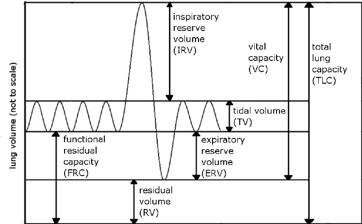
Exhalation, like inhalation, includes the conventional tidal volume as well as the possibility of an expiratory reserve volume (ERV). The ERV may be defined as the amount of air pushed out following a typical breath if you exhale as much as possible. Another popular phrase is vital capacity (VC). VC = TV + expiratory reserve volume + inspiratory reserve volume. VC is a measure of the amount of air that could flow into and out of the lungs if they were strained to their maximum capacity.
Residual volume and total lung capacity are two more terms for lung capacity. Residual volume refers to any lung volume that does not help breathing or gas exchange, such as the dead space between the alveoli in the lungs. Although the air may fill these areas, it cannot be used for any medically useful activities. “Total lung capacity” refers to the total of residual volume and vital capacity.
In this post, we will examine lung volumes and capabilities, including how they are measured and how illness impacts them. It is beneficial to categorize the overall space within the lungs into volumes and capacities. This category assesses the mechanical state of the lungs, including musculature, airway resistance, and the efficacy of gas exchange at the alveolar membrane. These may be determined using easy, inexpensive, and non-invasive testing.
Importance of Tidal Volume in Respiratory Physiology
Tidal volume is critical to respiratory physiology since it is essential for maintaining healthy lung function and enabling the body’s exchange of carbon dioxide (CO2) and oxygen (O2). Here’s a thorough justification of why tidal volume matters in this situation:
Ventilation-Perfusion Matching: Tidal volume has a direct relationship with alveolar ventilation, which refers to the tiny air sacs in the lungs where gas exchange occurs. When breathing in, fresh air that is high in oxygen is continuously delivered to the alveoli thanks to an adequate tidal volume. This, in turn, fosters an efficient match between ventilation (airflow) and perfusion (blood flow) in the lungs. When ventilation and perfusion are regulated, oxygen diffuses effectively into the bloodstream, and carbon dioxide is removed.
Oxygen Exchange: Air that contains oxygen is pulled into the lungs during inhalation. The amount of this oxygen-rich air that enters the alveoli is determined by tidal volume. Through the thin alveolar-capillary membrane, oxygen diffuses from the alveoli into the surrounding blood vessels. There, it attaches to hemoglobin and travels throughout the body. A sufficient supply of oxygen to meet the body’s metabolic needs, supporting energy production and overall cellular function, is ensured by an adequate tidal volume.
Carbon Dioxide Removal: Besides oxygen uptake, tidal volume is important for removing carbon dioxide, a byproduct of cell metabolism. When we exhale, tidal volume determines how much air is expelled from our lungs. This air transports CO2 from the bloodstream into the alveoli. From there, it is expelled during exhalation. If the tidal volume is inadequate, carbon dioxide can collect in the circulation, causing respiratory acidosis and other health problems.
Measurement Methods
Simple Spirometry
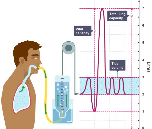
Tidal volume, inspiratory reserve volume, and expiratory reserve volume may all be estimated using simple spirometry techniques. However, it cannot determine residual volume.
Height, age, and gender measurements are all standardized. Height has the largest impact on capacity.
Process: The individual breathes through a closed circuit above the water. The chamber is filled with oxygen, and as they breathe, the gas expands and shrinks the volumes in the circuit. A weight above the chamber adjusts in height with each ventilation, depending on the circuit capacity. The height is measured with a pen to represent the volume inspired or expired over time.
Helium Dilution
Helium dilution is used to calculate total lung capacity. However, it is only accurate when the lungs are not blocked. If there is a point of blockage, helium may not reach all parts of the lung during breathing, resulting in an underestimate because only ventilated lung volumes are assessed.
Process: Following a silent expiration, the individual breathes in a gas containing a known amount of helium. They hold their breath for 10 seconds, enabling the helium to mingle with the air in their lungs, decreasing its concentration. Following expiration, the helium concentration is measured. The volume of ventilated air is then determined based on the degree of helium dilution.
Plethysmography
Plethysmography, often known as body plethysmography, is a technique for measuring tidal volume and lung capacities. It requires placing a patient in a sealed chamber.
Procedure: The patient breathes normally inside the chamber while pressure and volume changes are monitored. Tidal volume is calculated by comparing the chamber’s starting and final volumes.
Ease of Measurement: Although plethysmography is very accurate, it is less typically utilized for regular measurements since it requires specialist equipment and facilities.
Tidal Breathing Flow-Volume Loops
Graphing airflow vs lung volume produces flow-volume loops.
Tidal breathing involves patients breathing into a device that collects their flow and volume data in real time. The area beneath the loop reflects the tidal volume.
Ease of Measurement: Flow-volume loops are often used in research and specialized clinical evaluations, but they are less popular for ordinary measures than spirometry.
Respiratory Inductance Plethysmography (RIP)
Respiratory Inductance Plethysmography (RIP) is a non-invasive procedure for measuring changes in chest and abdominal wall circumferences while breathing.
Tidal volume is calculated using changes in data taken from sensors positioned across the chest and belly.
Ease of Measurement: RIP can be beneficial in some clinical situations, particularly when spirometry is not an option. It is simple to set up and offers continuous tidal volume monitoring.
Nitrogen Washout
A method of assessing serial/anatomical dead space in the conducting airways up to and including the terminal bronchioles (typically 150mL).
Process: The participant breathes in pure oxygen and then exhales via a valve that detects nitrogen levels. Initially, pure oxygen is breathed, reflecting the dead space volume. This is because the exhaled air never entered the alveoli and so never experienced gaseous exchange.
Then, a combination of dead space and alveolar air is expelled. This means that as nitrogen-rich air from the dead zone enters the alveoli, the measured concentration of nitrogen rises. After a few breaths, the lungs are depleted of pure oxygen, indicating that only alveolar air is expelled. The nitrogen levels will match those of alveolar air. Nitrogen levels monitored over time can be used to estimate the anatomical dead space capacity of the lungs.
Minute Ventilation and Tidal Volume
Minute ventilation (VE) is an essential parameter that is linked to tidal volume. It measures the volume of air breathed and exhaled during 60 seconds. Every 60 seconds, an average adult’s VE fluctuates between 4 and 6 liters. You can boost your VE by either taking deeper breaths (which increases Vt) or breathing quicker (which raises your respiratory rate).
Tidal Volume and Minute Ventilation have a direct and fundamental relationship in respiratory physiology. Minute Ventilation is directly dependent on Tidal Volume, as it is calculated by multiplying Tidal Volume by the respiratory rate. This means that any change in Tidal Volume will have a proportional effect on Minute Ventilation, assuming the respiratory rate remains constant. For instance, if Tidal Volume increases while the breathing rate stays the same, Minute Ventilation will increase by the same proportion. Conversely, if Tidal Volume decreases, Minute Ventilation will decrease correspondingly. This direct relationship allows the body to efficiently adjust its overall ventilation to meet changing metabolic demands.
By altering either Tidal Volume or respiratory rate (or both), the body can fine-tune its Minute Ventilation to ensure adequate gas exchange. This flexibility is crucial during activities like exercise, where the body might need to significantly increase its oxygen intake and carbon dioxide output. Understanding this direct relationship is essential in clinical settings, particularly when managing patients on mechanical ventilation, where careful adjustment of Tidal Volume can have immediate and significant impacts on overall Minute Ventilation and, consequently, on the patient’s respiratory status.
Alveolar Ventilation and Tidal Volume
Another important tidal volume indicator is alveolar ventilation (VA). VA determines VE without taking into account airway dead space, which is the volume of air you breathe in the absence of active gas exchange in the lungs.
Dead space is defined as the air that remains above the vocal cords in the upper respiratory system (nasal passages, sinuses, throat, and larynx) and below the vocal cords in the lower respiratory tract. Dead space accounts for around one-third of the air volume moved during casual breathing.
Breathing harder might improve your alveolar ventilation.
Abnormal Tidal Volume Symptoms
Abnormally Low Tidal Volume
Abnormally low tidal volume, also known as hypoventilation, can lead to various symptoms due to inadequate gas exchange in the lungs. Here are the main symptoms and signs associated with abnormally low tidal volume:
- Shortness of breath (dyspnea): Patients may feel as if they are not getting enough air, even when resting.
- Rapid, shallow breathing: To compensate for the low tidal volume, the respiratory rate often increases, resulting in quick, shallow breaths.
- Use of accessory muscles: Patients may use neck and chest muscles to help with breathing, which is visible as increased effort.
- Fatigue: Inadequate oxygenation can cause weariness and weakness.
- Confusion or altered mental status: As carbon dioxide builds up in the blood (hypercapnia), it can affect brain function.
- Headaches: Often caused by elevated blood carbon dioxide levels.
- Cyanosis: A bluish tint to the skin, lips, or nail beds due to poor oxygenation of the blood.
- Anxiety or restlessness: The feeling of not getting enough air can cause significant distress.
- Tachycardia (rapid heart rate): The heart may beat quicker to compensate for low oxygen levels.
- Sleep disturbances: Low tidal volume can worsen during sleep, leading to sleep apnea or frequent awakenings.
- Morning headaches: Often a sign of nocturnal hypoventilation.
- Decreased exercise tolerance: Even mild physical activity may cause excessive breathlessness.
It is crucial to note that the intensity of these symptoms varies and may not be present in all cases. The underlying cause of the low tidal volume (such as neuromuscular disorders, severe obesity, or lung diseases) can also influence the specific symptoms a person experiences.
If someone is experiencing these symptoms, especially persistent shortness of breath or confusion, they should seek medical attention promptly. Abnormally low tidal volume can be a serious condition that requires proper diagnosis and treatment.
Abnormally High Tidal Volume
Abnormally high tidal volume, also known as hyperventilation or overbreathing, occurs when a person breathes deeper or more rapidly than necessary. This condition can lead to various symptoms due to changes in blood gas levels, particularly a decrease in carbon dioxide. Here are the main symptoms and signs associated with abnormally high tidal volume:
- Shortness of breath or a sense of air hunger: Surprisingly, inhaling too much might make you feel like you’re not receiving enough oxygen.
- Rapid breathing: The breathing rate may increase significantly.
- Chest pain or tightness: This might feel comparable to angina or a heart attack in certain situations.
- Lightheadedness or dizziness: Constriction of the blood vessels reduces blood flow to the brain.
- Tingling sensations (paresthesia): Commonly felt in the fingers, toes, and around the mouth due to changes in blood pH.
- Numbness in the extremities: This is often accompanied by tingling feelings.
- Muscle spasms or tremors: Especially in the hands and feet.
- Dry mouth or throat irritation: From increased airflow through the mouth and throat.
- Anxiety or panic feelings: Hyperventilation can trigger anxiety, and anxiety can cause hyperventilation, creating a feedback loop.
- Confusion or disorientation: Changes in blood gas levels can affect cognitive function.
- Fainting (syncope): In severe circumstances, diminished blood supply to the brain can result in a loss of consciousness.
- Carpopedal spasm: In extreme cases, the hands and feet may contort into a characteristic spasm.
- Visual disturbances: Some people may experience blurred vision or tunnel vision.
- Headache: Often described as a tension-type headache.
It’s crucial to remember that these symptoms can be frightening and may resemble more dangerous disorders such as heart attacks or neurological issues. The symptoms of hyperventilation can also differ from person to person and may not be present in all cases.
If someone experiences these symptoms, especially if they’re severe or persistent, they should seek medical attention. While hyperventilation is often not life-threatening, it can be a sign of underlying physical or psychological conditions that need addressing. Additionally, in some cases, what appears to be hyperventilation could be a symptom of a more serious condition requiring immediate medical intervention.
Clinical Applications
Tidal volume is a valuable diagnostic and monitoring tool for a wide range of respiratory ailments, including asthma, chronic obstructive pulmonary disease (COPD), and restrictive lung disease. It is essential for determining lung function and the general health of the respiratory system. In clinical applications, tidal volume is utilized for the following conditions:
Asthma:
Tidal volume measurement is an essential part of the pulmonary function tests used to detect asthma. Asthmatic patients frequently have lower tidal volumes due to increased airway resistance and bronchoconstriction during an episode.
Monitoring: Tidal volume monitoring can measure changes in lung function over time, allowing healthcare practitioners to analyze the efficacy of asthma treatments and interventions. A reduction in tidal volume may indicate deteriorating asthma control, whereas an improvement indicates a good response to therapy.
Chronic Obstructive Pulmonary Disease (COPD):
Diagnosis: Tidal volume measurement, when combined with other lung function tests such as forced expiratory volume in one second (FEV1) and forced vital capacity (FVC), aids in COPD diagnosis. Tidal volume can be normal or increased in COPD, but the ability to exhale air is severely limited.
Tidal volume must be monitored regularly to effectively manage COPD patients. It enables healthcare practitioners to measure disease development and the effectiveness of medications such as bronchodilators and pulmonary rehabilitation. Tidal volume changes may signal deterioration or improvement in lung function.
Restrictive Lung Diseases:
Tidal volume measurements are critical for identifying restrictive lung disorders including pulmonary fibrosis and chest wall abnormalities. In certain situations, the lungs’ capacity to expand and tolerate a normal tidal volume is compromised, resulting in lower tidal volumes.
Monitoring: Tidal volume monitoring is critical for assessing disease progression and therapy response in individuals with restrictive lung illnesses. A rise in tidal volume may indicate improved lung compliance, whereas a consistent decrease shows persisting restrictive pathology.
Assessing Lung Capacity:
Tidal volume is an important component of several lung capacity tests, including vital capacity (VC) and inspiratory capacity (IC). These measures give information on total lung function.
Vital capacity (VC) is the maximum amount of air that may be forcibly evacuated after a maximal inhalation. It covers both tidal volume and extra volumes. Tidal volume changes can influence VC.
Tidal volume is an important component of IC that assesses a person’s ability to take in more air during exercise or deep breathing.
Factors Affecting Tidal Volume
Understanding these variables is critical for evaluating respiratory function and identifying potential problems. Here are the primary elements influencing tidal volume:
- Body Size and Demographics
- Physical Condition
- Respiratory System Health
- Neurological Factors
- Environmental Factors
- Psychological State
- Metabolic Factors
- Mechanical Ventilation
- Pregnancy
- Medications
1. Body Size and Demographics
- Height and Weight: Taller and heavier individuals generally have larger lung capacities and higher tidal volumes.
- Age: Tidal volume tends to decrease with age due to reduced lung elasticity and respiratory muscle strength.
- Sex: Males often have bigger tidal volumes than females due to variations in body and lung size.
2. Physical Condition
- Fitness Level: Well-trained athletes often have higher tidal volumes due to improved lung capacity and respiratory muscle strength.
- Body Position: Tidal volume is typically larger when standing or sitting compared to lying down.
3. Respiratory System Health
- Lung Diseases: Conditions like COPD, asthma, or pulmonary fibrosis can reduce tidal volume.
- Chest Wall Disorders: Conditions affecting the ribcage or diaphragm can limit lung expansion and reduce tidal volume.
4. Neurological Factors
- Central Nervous System Function: Disorders affecting the respiratory centers in the brain can alter tidal volume.
- Neuromuscular Diseases: Conditions affecting the nerves or muscles involved in breathing can reduce tidal volume.
5. Environmental Factors
- Altitude: At high altitudes, tidal volume typically increases to compensate for lower oxygen levels.
- Air Quality: Poor air quality or irritants can lead to changes in breathing patterns and tidal volume.
6. Psychological State
- Stress and Anxiety: Can lead to changes in breathing patterns, often increasing tidal volume.
- Sleep State: Tidal volume usually drops during sleep, particularly REM sleep.
7. Metabolic Factors
- Exercise: To fulfill the increased oxygen needs during physical exercise, tidal volume increases dramatically.
- Fever: Raising body temperature can raise tidal volume and metabolic rate.
8. Mechanical Ventilation
- Tidal volume in ventilator patients may be directly regulated and changed to meet medical demands.
9. Pregnancy
- Tidal volume typically increases during pregnancy, especially in the third trimester, due to hormonal changes and the growing uterus pushing up on the diaphragm.
10. Medications
- Bronchodilators: Can increase tidal volume by opening up airways.
- Sedatives: May decrease tidal volume by depressing respiratory drive.
Understanding these factors is essential for healthcare professionals in interpreting pulmonary function tests, managing respiratory conditions, and optimizing mechanical ventilation strategies.
Conclusion
In conclusion, tidal volume is an important respiratory measure that has broad significance in both clinical and physiological settings. Its relevance in respiratory physiology cannot be emphasized, as it plays a critical role in oxygen exchange, carbon dioxide elimination, and acid-base balance. Tidal volume measurements are essential for diagnosing, monitoring, and treating a variety of respiratory illnesses, including asthma, COPD, and restrictive lung diseases.
In intensive care units and surgical settings, understanding tidal volume is critical for optimizing mechanical ventilation techniques, maintaining lung health, and guaranteeing patient safety. Tidal volume adaptation during exercise and endurance activities is essential for fulfilling the body’s increased oxygen demand during physical effort.
FAQs
What is tidal volume?
Tidal volume is the amount of air that moves in and out of the lungs during normal, relaxed breathing. In a healthy adult, it’s typically about 4-8 mL per kilogram of ideal body weight or approximately 500 mL per breath.
How is tidal volume different from minute ventilation?
Tidal volume is the amount of air moved in a single breath, while minute ventilation is the total volume of air moved in and out of the lungs per minute. Minute ventilation is calculated by multiplying tidal volume by the respiratory rate.
Can tidal volume change?
Yes, tidal volume can change based on various factors including physical activity, stress, altitude, and certain medical conditions. For example, during exercise, tidal volume typically increases to meet the body’s increased oxygen demand.
How is tidal volume measured?
Tidal volume is typically measured using a device called a spirometer. This can be done as part of a pulmonary function test in a clinical setting.
What happens if tidal volume is too low?
If the tidal volume is too low (hypoventilation), it can lead to inadequate gas exchange, potentially resulting in hypoxemia (low blood oxygen) and hypercapnia (high blood CO2). Symptoms may include shortness of breath, fatigue, and in severe cases, confusion or loss of consciousness.
Can tidal volume be too high?
Yes, abnormally high tidal volume (hyperventilation) can occur, often due to anxiety, certain medical conditions, or improper mechanical ventilation. This can lead to respiratory alkalosis (increased blood pH), causing symptoms like lightheadedness, tingling in the extremities, and, in severe cases, muscle spasms.
References
- Hallett, S., Toro, F., & Ashurst, J. V. (2023, May 1). Physiology, Tidal Volume. StatPearls – NCBI Bookshelf. https://www.ncbi.nlm.nih.gov/books/NBK482502/
- Dhameliya, N. (2023, September 3). Tidal Volume. Mobility Physiotherapy Clinic. https://mobilephysiotherapyclinic.net/tidal-volume/#Clinical_Applications
- Hayes, K. (2023, August 11). What Is Tidal Volume? Verywell Health. https://www.verywellhealth.com/tidal-volume-5090250
- Spiegato. (2024, August 6). What Is Tidal Volume? – Spiegato. Spiegato. https://spiegato.com/en/what-is-tidal-volume