Muscle Energy Technique (MET)
What is a Muscle Energy Technique (MET)?
Muscle Energy Technique (MET) is a manual therapy technique commonly used in physical therapy and osteopathic medicine to treat musculoskeletal dysfunctions and improve joint mobility. It involves the active participation of the patient, who engages in specific muscle contractions directed by the therapist.
The goal of MET is to restore normal function to muscles and joints by addressing imbalances, reducing pain, and enhancing overall movement patterns. MET can be applied to various areas of the body and is often utilized in conjunction with other therapeutic modalities to optimize patient outcomes.
Introduction
Fred Mitchell, Sr., D.O. developed the Muscle Energy Technique (MET) in 1948. It’s a kind of manual therapy that osteopaths utilize a lot. By harnessing the natural energy of the muscle through gentle isometric contractions, it lengthens and relaxes muscles.
We refer to this mechanism as reciprocal inhibition or autogenic inhibition. Like static stretching, which is a passive method in which the therapist does all the work, MET is an active technique in which the patient participates actively. MET is based on the principles of autogenic inhibition and reciprocal inhibition.
Autogenic inhibition (MET) occurs when a muscle contracts below its maximum potential and then stretches that same muscle; reciprocal inhibition (MET) occurs when a muscle contracts below its maximum potential and then stretches the opposing muscle.
Anatomy and Physiology
To learn MET, one must have a solid understanding of muscle physiology. There are four types of muscle contractions: isometric, concentric, eccentric, and isolytic. When muscles contract isometrically, it indicates that there is no contact between the muscle’s origin and insertion. Concentrated contractions cause the muscles to shorten.
An eccentric contraction results in the muscle extending. Last but not least, an isolytic contraction is a response to an external stimulus that lengthens a muscular contraction.
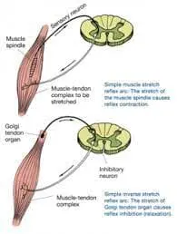
A muscle is composed of several muscle spindles. Every spindle has three to twelve intrafusal muscle fibers, surrounded by a large extrafusal fiber. Every muscle spindle contains both efferent and afferent neural components.
Motor nerve fibers include the gamma motor neurons that innervate the intrafusal fibers and the alpha motor neurons that innervate the extrafusal fibers. The afferent (sensory) components are the Ib fingers, which innervate the Golgi tendon organs at the myotendinous junction, and the Ia and II fibers, which innervate muscle spindles.
Golgi tendon fibers play a significant role in MET through a process known as post-isometric relaxation. Stress activates the fibers in the muscles, producing a negative feedback loop that stops the Ia fibers from contracting anymore.
The thoracic spine, which consists of twelve spinal vertebrae, is particularly significant to osteopathic practitioners because sympathetic nerve fibers are located there. Viscerosomatic roots or biomechanical causes, such as constriction in segments affected by surrounding muscles, maybe the cause of somatic dysfunctions in this area.
Osteopathic care providers need to address and understand viscerosomatic reflexes, even though they are outside the range of this review.
The posterior spinous process, and bilateral transverse processes on both sides and front vertebral body are characteristics of thoracic vertebrae. It is easier to rotate with adjacent segments due to the facet joints of these vertebrae, which are situated above and below. The superior facet joint of the thoracic segment points posteriorly, laterally, and upwards.
Because of the ribs, the thoracic vertebrae additionally include a superior and inferior costal facet near the vertebral body, as well as a transverse costal facet on the transverse process. T12’s joint facet position may be markedly moved from that of the other thoracic vertebrae due to its joint with L1.
Osteopathic schools frequently teach the Rule of Threes as a helpful tool for locating the spinous process about the transverse process. According to this rule, for T1 through T3, the transverse process and the spinous process line up. Segments T4 through T6 have spinous processes located halfway between their transverse processes.
The spinous process (seventh spinous process at T8) is at the level of the successful transverse process from T7 to T10. The transverse process of the eleventh spinous process, which is halfway through, is parallel to that of the twelfth.
There are two types of somatic dysfunctions identified. Extended periods of bad posture are a common source of segment-specific type 1 dysfunctions. Larger, longer supporting muscles in the back, such as the erector spinae, which are ordinarily neutrally positioned but display opposite-directional rotation and side bending, are often affected by these dysfunctions.
On the other hand, Type 2 dysfunctions are more painful and usually include smaller supportive muscles between segments, such as the rotatores, multifidus, interspinal, and intertransversarii muscles. Flexion or extension, side bending, and rotation in the same direction are characteristics of type 2 dysfunctions.
Pathophysiology
An injury may arise from a trauma, accident, hurt, strain sprain, or other incident; not all of them should be treated with muscular energy. These methods work best on the following injury patterns: decreased range of motion due to muscle rigidity, spasticity, hypertonicity, or hypotonicity. Hypertonicity, sometimes brought on by overuse, can result in altered joint posture, increased irritability, and decreased flexibility.
This type of injury often presents with general pain in the affected muscles. Brain damage: The agonist muscles that are attached to a joint or segment are also affected when it becomes dysfunctional. If the antagonistic muscles are not addressed, eventually dysfunction develops in both muscle groups.
What is Autogenic and Reciprocal Inhibition?
When the Golgi tendon organ (GTO) and the muscle spindles are activated, certain muscles are prevented from contracting. This process is known as both autogenic and reciprocal inhibition. These two musculotendinous proprioceptors, which are located in and around the joints and muscles, respond to changes in muscle tension and length to control muscle control and coordination.
The GTO, located between the tendon and the muscular belly, notices an increase in tension as a muscle stretches or contracts. The GTO fires when a muscle contracts. It then contracts the antagonist muscle group in reaction and prevents the original contraction (reflex inhibition). This mechanism is known as autogenic inhibition.
The GTO reaction’s adaptability is essential. When the GTO inhibits the contraction of the (agonist) muscle and facilitates the contraction of the antagonist muscle, the muscle can be stretched farther and more readily. Similar to a low-force, long-duration stretch, static stretching typically causes autogenic inhibition. After seven to ten seconds, muscle tension increases and the GTO response occurs. More muscle stretching is made possible by this temporary inhibition of the muscle spindle in the stretched muscle.
The muscular spindle, which resides inside the muscle belly, stretches together with the muscle. As a result, the muscle spindle is activated, causing the agonist muscle to reflexively contract and the antagonist muscle to relax (this is known as the stretch reflex). This phenomenon is called reciprocal inhibition.
Types of MET:
the reflex neuro-muscular inhibition phenomenon
- Autogenic Inhibition MET
- Post Isometric Relaxation (PIR)
- Post Facilitation Stretching (PFS)
- Reciprocal Inhibition MET
Autogenic Inhibition MET
As mentioned earlier, the concept of autogenic inhibition serves as the foundation for how autogenic inhibition METs work. Based on the concept of autogenic inhibition, post-facilitation stretching (PFS) and post-isometric relaxation (PIR) are the two primary and well-known MET approaches.
Post Isometric Relaxation (PIR)
Later on, Karel Lewitt developed a method known as Post Isometric Relaxation. Post-isometric relaxation (PIR) is the outcome of a reduction in muscular tone in one or more muscles after a brief period of submaximal isometric contraction of the same muscle. The foundation of PIR is the concept of autogenic inhibition.
The PIR approach is applied as follows:
Stretching the hypertonic muscle extends it until the first signs of movement resistance appear, which is typically in the form of pain. The therapist applies resistance in the opposite direction while performing a submaximal (10–20%) contraction of the hypertonic muscle away from the barrier for five to ten seconds. The patient should breathe in with this attempt.
After completing the isometric contraction, the patient is told to relax and stop breathing. Following that, a quick stretch is applied to make up for the lost time before the following challenge. The procedure is restarted at this new barrier two or three times.
Post Facilitation Stretch (PFS)
The Post Facilitation Stretch (PFS) technique was developed by Janda. Although being more aggressive than PIR, this technique also depends on the concept of autogenic inhibition.
The PFS method is applied as follows:
The hypertonic and shortened muscle is in the middle of the stretched and relaxed states.
The therapist will resist the patient’s push for five to ten seconds while the patient is encouraged to make maximum contact with the agonist. The therapist then quickly extends a new barrier while maintaining the position for 10 seconds, after which the patient is told to relax and stop straining themself.
The patient is allowed to relax for around twenty seconds during the three to five repetitions of the process, plus an additional five repetitions. Before each repetition, the muscle is positioned between fully stretched and fully relaxed, as opposed to beginning at a fresh barrier.
Reciprocal Inhibition MET
Reciprocal Inhibition MET is different from PIR and PFS in that it begins with the contraction of one muscle and finishes with the stretching of the opposing muscle since The idea of reciprocal inhibition serves as its foundation.
The Reciprocal Inhibition MET technique is applied as follows:
It is the middle muscle that is injured. The patient’s want towards the restriction or barrier is either completely rejected (isometric) or the therapist allows a movement in that direction (isotonic). After the patient has calmed down and exhaled, the therapist stretches the newly formed barrier passively. Three, five, and even more times are spent doing this process.
Indication
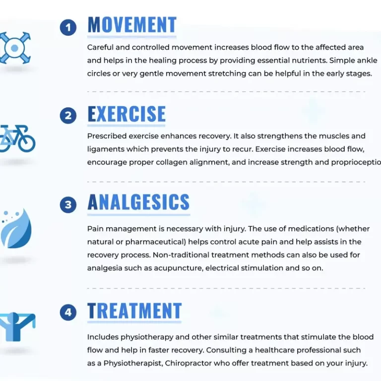
Muscle Energy Techniques can be applied to any condition where the goal is to increase joint range of motion (ROM) and promote muscle relaxation and lengthening. Muscle energy techniques are a safe way to treat almost any joint in the body. Many athletes use MET as a preventative measure to protect their joints and muscles from future damage.
People with limited range of motion (ROM) due to facet joint dysfunction in the neck and back are the main users of it. In addition, it is used to address more general issues like shoulder pain, scoliosis, sciatica, and asymmetrical legs, hips, or arms. It is also used to treat persistent muscle pain, stiffness, or damage.
Contraindications
It is not recommended to utilize MET with post-isometric relaxation in patients who have an acute fracture or dislocation. It may not be the greatest idea to select candidates who have tissue damage in their tendons, ligaments, or muscles.
It is best to attempt this process when the necessary stability has been attained. Methotrexate will not be well received by patients with centrally mediated muscle spasms. Patients have to follow with instructions and work cooperatively for this technique to be successful.
Equipment
MET with post-isometric relaxation is a hands-on osteopathic manipulative treatment that needs a firm, strong platform, preferably an adjustable height cushioned table, for the optimal treatment positioning and comfort of both the patient and the practitioner.
Personnel
A certified provider for MET with post-isometric relaxation is required for muscle energy methods.
Preparation
Informed patient agreement must be acquired for every procedure, including MET and osteopathic manipulative therapy, following a thorough explanation of the risks, advantages, and available treatment options. Before establishing any physical contact, the healthcare provider should explain all of the procedures they would be doing on the patient in detail.
A complete static and dynamic examination of the segments of the thoracic spine is the initial stage of the therapeutic procedure. Clinicians may utilize soft tissue methods as a lead-up to direct approaches like MET with post-isometric relaxation when significant alterations in muscle texture are observed.
Evidence of Muscle Energy Techniques in Physiotherapy
Franke et al. conducted a thorough study comparing the effectiveness of MET with control therapy in the treatment of patients with non-specific low back pain (LBP). It was found that among patient populations with non-specific LBP, the quality of randomized control trials (RCTs) involving MET therapy is low.
This implies that further thorough study is needed to confirm MET’s effectiveness for non-specific LBP. Szulc et al. investigated the efficacy of combining the McKenzie and MET techniques for LBP patients in a randomized control experiment.
The study found that the use of both McKenzie and MET therapies caused improvements in the Oswestry Disability Index, a notable decrease in the extent of the spinal disc herniation, and a marked improvement in the Visual Analogue Scale (VAS) for pain. The combined method is a good treatment for chronic low back pain.
Phadke et al. looked at the effects of static stretching and MET on pain and functional impairment in patients with chronic neck pain in a randomized controlled trial. It was found that MET outperformed the static stretching technique in terms of VAS and Neck Disability Index (NDI) findings.
It was discovered in an RCT by Moore et al. that MET had an immediate effect on the posterior shoulder tightness experienced by basketball players. Both internal rotation and horizontal adduction of the glenohumeral joint’s range of motion were increased.
Examples of Muscle Energy Techniques
Goal StructureStarting PlaceThe therapist’s hand The therapist’s one hand 2. The reaction of the patient The Psoas Major Thomas test position: With your target leg hanging off it and your other leg flexed 90 degrees, lie face down on the plinth with the rest of your body lying on it.
The leg that is hanging off the plinth is flexed just above the knee of the plinth.
Target leg off the plinth, positioned above the superior lower leg on the side The hip abduction extends over the superior lateral thigh. The Hip External RotatorsLeg abduction crook against the therapist with the inferior and lateral mid-thighs over.
Self-Administered MET
Hip Flexion/Hip Extension
The patient sits down and rests their foot on the knee of the opposing side.
knee against foot and foot against knee is pressed.
Hip adduction
Squeezing a ball between their legs, the patient lies down.
Hip external rotation
With their knee and hip flexed to a 90-degree angle, the patient is in the prone position.
The therapist rotates the patient’s knee inside as the patient presses against them.
Scalenes
The patient is in a prone position, with their head and shoulders supported by a pillow.
The therapist shifts in place and opens
By bending and rotating, the patient forces the therapist into a neutral posture.
Osteopathic Manipulative Therapy: Post-Isometric Relaxation Combined With Muscle Energy Technique – Thoracic Vertebrae
Techniques
Muscle energy techniques can be used to treat most body parts. According to one textbook, each approach requires the eight very important procedures listed below:
- Perform and obtain an accurate structural diagnostic.
- Engage the barrier to the highest level possible.
- In an unbreakable counterforce, the patient’s force and the doctor’s force are equal.
- The isometric contraction is performed by the patient for the appropriate duration (5–10 seconds), using the appropriate force and effort that is directed away from the barrier.
- Complete relaxation follows the muscular effort.
The patient is moved into the new limiting barrier as many times as practical. - About three to five times, or until there is no more progress in the range of motion, repeat steps 3-6.
The targetted element of MET treatment for the thoracic spine must be separate and targeted. In the upper thoracic spine, using the head and neck as a lever is common (T1-T4). By altering the patient’s trunk position, the lower thoracic spine segments (T5-T12) can be located.
During treatment, pay attention to mobility in the posterior transverse process and use palpation to identify the difficult region. Post-isometric relaxation is the type of MET that is most commonly used. When inside the barrier, the patient is advised to resist moving in the direction of freedom.
Muscle Energy Technique With Post-Isometric Relaxation
A representation of an osteopathic diagnosis on the right side is a sidebent, rotated, and flexed T3.
Step 1: The patient may be treated in a seated or flat position. In a sitting method, the doctor sits behind the patient while the patient is seated. Given that this is an upper thoracic segment, the head will function as a lever to affect the motion on T3. The right hand will keep an eye on T3’s transverse process.
Step 2: The patient’s head is extended, bent to the side, and rotated to the left by the doctor to contact the barrier until motion is felt at T3. The patient has to be positioned to engage the articular barrier and tighten the fascia.
Step 3: The patient is asked to softly but firmly attempt to bring his head back to an upright, neutral posture as the therapist applies an equal counterforce. The patient is instructed to relax and remain in this position for three to five seconds after taking it. It takes three to five seconds for the patient to become comfortable before engaging a new obstacle.
Step 4: Conduct a passive stretch into the restrictive barrier without involving the patient, and repeat steps 3–5 three times following the last session of therapy. Return the patient to a neutral position in a passive manner.
Step 6: Palpate T3’s transverse processes in the given example to see if the symmetry of the treated segment has improved.
Technical Pearls
When treating the lower thoracic region, it will be simpler to move the patient if the doctor crosses her arms and side-bends her, pressing on the same shoulder with the axilla. When treating type 1 somatic dysfunctions (e.g., T3-6 neutral, side bent right, and rotated left), the peak of the curve is treated first.
It is challenging to cure because the facets above and below T12 face different orientations. The thoracic facets of the vertebrae face posteriorly, upward, and laterally. In contrast, the lumbar facet joints (L1) face posteriorly, upward, and medially. Accurate location is essential while using MET.
How does MET help?
The principle of reciprocal inhibition, which says that when one muscle contracts, the adjacent muscle at that joint relaxes, forms the basis of MET. Put another way, the opposite muscle is stretched when the first one contracts against your physical therapist.
A person can move a joint to its full range of motion and fully extend and contract their muscles using MET. You can achieve greater muscle strength and flexibility by doing this repeatedly.
Who benefits from MET?
MET can help adults in good health maintain the flexibility of their muscles and prevent injuries. Additionally, MET is utilized to treat ailments such
- Pain inhibited movement of the neck, shoulders, and back
- Contractures
- Leg length discrepancy
- Muscle injury
- Muscle tightness
- Sciatica
- Scoliosis
Complications
Patients who use post-isometric relaxation therapy in addition to methadone therapy (MET) should be warned that following the procedure, they may have muscle soreness and fatigue. Following treatment, the doctor could suggest drinking extra water.
Excessive use of force can result in tendon rupture or rib fractures. To decrease the force during post-isometric relaxation, the patient must be encouraged to resist just enough to engage the treated segment.
Conclusion
Muscle Energy Technique (MET) is a hands-on technique used by osteopathic and physical therapists to improve joint mobility and potentially reduce pain. This is an overview:
Goal: Expand the range of motion in your joints and relax your stiff muscles.
Method: The therapist gently inhibits the patient’s movement in the opposite direction while the patient actively contracts specific muscles.
Benefits: may help in improving mobility and reducing pain, especially pain in the lower back.
FAQ
What is the muscle energy technique?
The Muscle Energy Technique (MET) was created by Fred Mitchell, Sr., D.O. in 1948. It is a type of manual therapy that is frequently used in osteopathy. It lengthens and relaxes muscles by using the natural energy of the muscle through mild isometric contractions. This process is known as autogenic or reciprocal inhibition.
What is the difference between muscle energy technique and PNF?
It has been highlighted that MET is a useful method for increasing muscle flexibility, particularly in the hamstrings. One such method that is employed in practice but whose ability to lessen hamstring muscle stiffness has not received as much research is the PNF stretch.
Who is the father of the muscle energy technique?
Fred Mitchell, Sr., DO, created the muscle energy method (MET), a subset of osteopathic manipulative medicine (OMM). The kinematic motion of the pelvis was initially described by Dr. Mitchell in 1948.
What is the OMT technique of muscle energy?
An Osteopathic Manipulative Treatment (OMT) method called “Muscle Energy” involves the patient voluntarily contracting their muscles in opposition to the physician’s counterforce. The doctor applies a counterforce to the confining barrier as the patient tries to move a bodily part toward freedom of mobility.
What is the muscle energy technique in physiotherapy?
Fred Mitchell, Sr., DO, created the muscle energy method (MET), a subset of osteopathic manipulative medicine (OMM). The kinematic motion of the pelvis was initially described by Dr. Mitchell in 1948. Based on this idea and with inspiration from the research of neurophysiologist Charles Sherrington, Dr.
What is the muscle energy technique for reducing pain?
Physical therapists commonly employ the muscular energy technique (MET) as a type of manual therapy to enhance musculoskeletal function and reduce pain. To increase joint mobility and restore normal muscle length, MET entails stretching exercises performed after active muscular contraction and relaxation.
What is myofascial release and muscle energy technique?
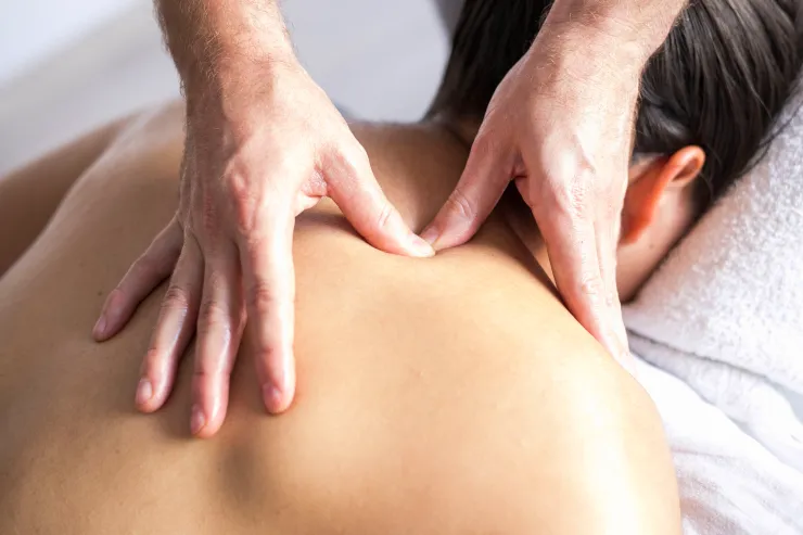
To target the neck muscles, a patient performing a modified exercise therapy (MET) would contract the affected area against resistance, such as pushing their head against the therapist’s palm. After holding this contraction for five to ten seconds, the therapist will stretch the affected muscle.
Why use the muscle energy technique?
In MET, the patient deliberately contracts a muscle in a carefully regulated direction in opposition to the counterforce applied by the therapist. Reducing pain, tightening muscles, strengthening weak muscles, improving local circulation, and releasing joint limitations are some of its therapeutic effects.
What are the risks of the muscle energy technique?
Muscle Energy Technique side effects for the lumbar spine include radicular pain, low back stiffness, increased muscle spasms, and restricted range of motion. Most of the time, these issues are temporary and go away in a day or two.
Is PNF stretching a muscle energy technique?
Many practitioners consider the muscle energy approach and proprioceptive neuromuscular facilitation (PNF) to be nearly equivalent.
References
- Muscle Energy Technique. (n.d.). Physiopedia. https://www.physio-pedia.com/Muscle_Energy_Technique
- Muscle energy technique. (2023, February 20). Wikipedia. https://en.wikipedia.org/wiki/Muscle_energy_technique
- Muscle Energy Techniques – Manual Therapy – Physiotherapy – Treatments – Physio.co.uk. (n.d.). https://www.physio.co.uk/treatments/physiotherapy/manual-therapy/muscle-energy-techniques.php
- Talley, J. T., & Goto, K. K. (2023, September 11). Osteopathic Manipulative Treatment: Muscle Energy Procedure With Post-Isometric Relaxation – Thoracic Vertebrae. StatPearls – NCBI Bookshelf. https://www.ncbi.nlm.nih.gov/books/NBK560895/