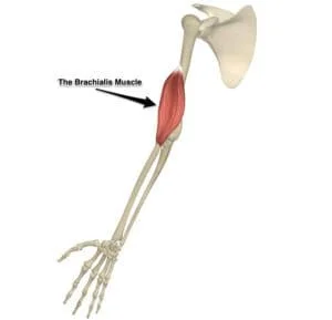
Spring Ligament Tear
| | | |

Spring Ligament Tear

Introduction: A spring ligament tear refers to damage to the plantar calcaneonavicular ligament, a key structure in the foot that supports the arch and stabilizes the midfoot. This ligament connects the heel bone (calcaneus) to the navicular bone. A tear can result from trauma or chronic stress and often leads to pain, swelling, and flattening…

Thorax muscles
| |

Thorax Muscles

Introduction The thorax muscles are a group of muscles located in the chest region that play a key role in respiration and upper body movement. The thoracic wall comprises the subcostalis, transversus thoracis, innermost intercostal muscles, external intercostal muscles, and internal intercostal muscles. During breathing, these muscles are principally in charge of altering the thoracic…

Ischiofemoral ligament
| | |

Ischiofemoral Ligament

Introduction The ischiofemoral ligament is one of the three main ligaments that stabilize the hip joint. It originates from the ischium (part of the pelvis) and attaches to the femur. This ligament helps limit internal rotation and extension of the hip, contributing to joint stability, especially during movement. It attaches to the base of the…

Radial Collateral Ligament (RCL) Injury
| | | |

Radial Collateral Ligament (RCL) Injury

What is a Radial Collateral Ligament (RCL) Injury? Radial Collateral Ligament (RCL) injury involves damage to the ligament on the outer (lateral) side of the elbow that stabilizes the joint against varus stress. It often results from trauma, overuse, or repetitive strain—common in athletes like tennis players. Symptoms typically include lateral elbow pain, instability, and…

Scapholunate Ligament Injury
| | | |

Scapholunate Ligament Injury

What is a Scapholunate Ligament Injury? A scapholunate ligament injury involves damage to the ligament connecting the scaphoid and lunate bones in the wrist. This ligament plays a key role in stabilizing the carpal bones and maintaining proper wrist mechanics. Injury typically results from a fall on an outstretched hand or repetitive strain, leading to…

Perineal nerve
|

Perineal Nerve

Introduction The perineal nerve is a big terminal branch of the pudendal nerve that normally begins during its passage through the pudendal canal or shortly after it exits the canal. The pudendal nerve travels to the perineum before splitting into two terminal branches: the superficial perineal nerve (superficial perineal branch) and the deep perineal nerve….

Pronator quadratus muscle
| |

Pronator Quadratus Muscle

Introduction The pronator quadratus is a quadrangular, thin, short, and flat muscle located in the anterior compartment of the forearm. It is a member of the deep group of forearm flexors, together with flexor digitorum profundus and pollicis longus. The superficial group of forearm flexors overlays these three muscles. The pronator quadratus connects the distal…