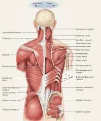
Back muscles
Introduction
The Back muscles are a group of strong, paired muscles located on the trunk’s posterior aspect. They provide spinal movement, trunk stability, and coordination of limb and trunk movements.
Back muscles are divided into two major groups:
Extrinsic (superficial) back muscles are those that are closest to the skin on the back. These muscles are also known as immigrant muscles because they originated in the upper limb and migrated to the back during foetal development. These muscles are subdivided into superficial and intermediate.
The intrinsic (deep) back muscles are also known as true back muscles. The thoracolumbar fascia divides them from the extrinsic muscles, where they are situated deep within. Their primary function is to cause movements in the vertebral column. These muscles are divided into three layers: superficial, deep, and deepest.
Extrinsic back muscles
the extrinsic muscles of the back function similarly to the upper-limb muscles but are located superficially on the posterior trunk. They are separated into:
Superficial extrinsic muscles:
- trapezius
- latissimus dorsi
- rhomboid major
- rhomboid minor
- levator scapulae
Trapezius
- Origin: the spinous processes of C7–T12, the nuchal ligament, the external occipital protuberance, and the superior nuchal line.
- Insertion: the acromion, spine of the scapula, and lateral third of the clavicle
- Innervation: the accessory nerve (12th cranial nerve).
Letissimus dorsi
- Origin: iliac crest, sacrum, thoracolumbar fascia, spinous processes of T7–L5, and 10th–12th ribs.
- Insertion: the intertubercular groove of the humerus
- Innervation: the thoracodorsal nerve.
Rhomboid major
- Origin: the nuchal ligament and spinous processes of C7-T1.
- Insertion: the line connecting the superior angle and the trigonum scapula, which is the superior portion of the scapula’s medial border.
- Innervation: the dorsal scapular nerve.
Rhomboid minor
- Origin: spinous processes of T2-T5.
- Insertion: the line connecting the inferior angle and the trigonum scapula, which is the inferior portion of the scapula’s medial border.
- Innervation: the dorsal scapular nerve.
Levator scapulae
- Origin: the cervical vertebrae’s first four transverse processes.
- Insertion: the superior angle of the scapula.
- Innervation: the dorsal scapular nerve.
Intermediate extrinsic muscles
- Serratus posterior superior and inferior
Serratus posterior superior
- Origin: the spinous processes of the T11-L3 vertebrae.
- Insertion: the inferior border of the 9th–12th ribs.
- Innervation: the T9-T12 spinal nerves
Serratus posterior inferior
- Origin: the C7-T3 vertebrae’s ligamentum nuchae and spinous processes.
- Insertion: the superior aspect of the 2nd-5th ribs.
- Innervation: the anterior rami of the T1-T4 spinal nerves.
Intrinsic back muscles
The true, intrinsic back muscles are the deepest layer of muscles connected to the vertebral column. The muscles of the thoracic area are deep into the thoracolumbar fascia, whereas the muscles of the lumbar area are between the superficial and middle layers of the fascia. Almost all of them get their nerve supply from the posterior (dorsal) rami of spinal nerves, and they’re known as the intrinsic group because they only affect the vertebrae. The numerous muscles in this group are divided into three layers:
Superficial layer:
- splenius muscle
- erector spinae muscle
Splenius muscle
The splenius muscle group includes two muscles:
Splenius Capitis.
- Origin: Spinous processes of C7 vertebrae, T1-T3 (or T4) vertebrae, and supraspinous ligaments.
- Insertion: Mastoid process, a lateral third of the superior nuchal line.
- Innervation: The lateral branches of the C2–C3 dorsal rami.
Splenius Cervicis.
- Origin: T3-T6 spinous processes
- Insertion: atlas and axis transverse processes, posterior tubercle of C3 vertebra.
- Innervation: Lateral branches of the lower cervical dorsal rami.
Erector Spinae Muscles
The erector spinae is a massive muscle group that consists of three muscular columns on each side of the spine. The erector spinae group’s muscles are arranged from medial to lateral:
Spinalis muscle is classified into three regions: spinalis capitis, spinalis cervicis (colli), and spinalis thoracis. They attach to the spinous processes of the vertebrae in their respective regions.
Spinalis Thoracis
- Origin: The most medial erector spinae in the thoracic region originate from the spinous processes of T11-L2.
- Insertion: Upper thoracic vertebrate spinous processes
- Laterally blends with the longissimus thoracis muscle.
Spinalis cervicis and capitis are vaguely defined and underdeveloped. These fibres may not be present in all people.
Spinalis cervicalis
- Origin: ligamentum nuchae and the C7 spinous process.
- Insertion: Spinous processes C3–C4 and axis.
Spinalis Capitis
Instead of the customary insertions on the thoracic transverse processes, a small number of semispinalis capitis fibres are usually inserted into the spinous processes of C7 and T1.
Longissimus muscle is further classified into longissimus capitis, longissimus cervicis (colli), and longissimus thoracis. They attach to the transverse processes of the vertebrae in their respective regions.
Longissimus capitis
- origin: C4-T4 transverse processes.
- Insertion: The posterior edge of the mastoid process.
Longissimus Cervicis
- Origin: T1-T4 transverse processes.
- Insertion: The posterior tubercle of the C2-C6 transverse processes.
Longissimus Thoracis
It includes lumbar and thoracic sections.
The largest erector spinae group
- Origin: Transverse process at the inferior vertebral levels.
- Insertion: Mastoid process and transverse process at superior vertebral level
Iliocostalis muscle
There are three different types of iliocostalis muscle: iliocostalis lumborum, iliocostalis thoracis, and iliocostalis cervicis (colli). They extend between the rib angles and the transverse processes of the corresponding regional vertebra.
Iliocostalis Cervicis
- Origin: Angle of ribs 3 to 6.
- Insertion: posterior tubercle of the transverse process of C4-6.
Iliocostalis Thoracis
- Narrow, fusiform shape.
- Origin: Angle of the lower six ribs.
- Insertion: C7’s transverse process and upper six rib angles.
Iliocostalis Lumborum
- It includes lumbar and thoracic sections.
- Origin: the iliac crest’s dorsal segment and medial end.
- Insertion: angle of ribs 4–12, thoracolumbar fascia, and L1–L4 lumbar transverse processes.
Deep layer:
- transversospinales (semispinalis, multifidus, rotatores)
Transversal Spinalis
This group of muscles connects a spinous process to the transverse process of a vertebra below.
Organised by length and region covered.
Rotatores are the deepest and shortest.
- Span 1-2 segments.
- Eleven pairs between T1 and T12.
- The rotator brevis connects the transverse process of the lower vertebra to the lateral lamina of the upper vertebra directly above.
- The rotator longus connects the transverse process of the lower vertebra to the base of the upper vertebra’s spinous process two levels up.
Multifidus can have two to four segments.
- Covers the lamina of vertebrae.
- Origin: Sacrum and ilium, transverse processes T1-L5, and articular processes C4-C7.
- Insertion: Spinous processes two or four segments above the origin
The semispinalis has four to six segments.
- Origin: Thoracic and cervical transverse processes
- Insertion: Occipital bone and spinous processes in the thoracic and cervical regions. 4-6 segments above the origin.
Deepest layer:
- Interspinales muscle
- Intertransversarii muscle
These muscles work together to maintain the body’s posture and move the vertebral column.
Interspinales Muscles
The interspinales are short muscles that connect the adjacent spinous processes of the vertebrae.
They are classified as interspinales cervicalis, interspinales thoracis, and interspinales lumborum. However, only the cervical and lumbar regions are fully developed, while the thoracic is frequently absent or rudimentary. The cervical and lumbar spines can extend because of the interspinales muscles.
Intertransversarii muscle
The intertransversarii connect the adjacent transverse processes of vertebrae. They are most prominent in the cervical and lumbar spines but are rarely present in the thoracic region.
The anterior and posterior groups of intertransversarii colli have a joint function that aids in stabilising the cervical spine and lateral flexion.
The intertransversarii lumborum is made up of medial and lateral slips that help the spine flex laterally.
Function of the back muscles
The back muscles provide the primary structural support for your trunk (torso). These muscles help you move your entire body, including your head, neck, shoulders, arms, and legs. Bending, twisting, turning your head, and extending your back are all possible because your back muscles work together.
You can sit and stand straighter with the aid of these muscles. They are essential for supporting your spine and allowing you to breathe. Their jobs include:
Superficial muscles: These muscles enable you to maintain a straight back, move your arms, and shrug your shoulders. Superficial muscles include:
- The latissimus dorsi (lats) muscle helps you extend and rotate your shoulder and arm.
- Levator scapulae raise the scapula (shoulder blade).
- Rhomboids are two muscles (rhomboid major and minor) that pull the scapula inward towards the spine.
- Trapezius (traps) muscles help you move your body, raise your arms, and maintain proper posture.
Intermediate muscles. The second extrinsic group consists of two muscles. They connect to the ribs and spine and play an important role in breathing. These two sets of intermediate muscles support your rib cage as your lungs expand and depress during breathing:
- Serratus posterior superior—helps you breathe in
- Serratus posterior inferior—helps you breathe out.
Superficial and intermediate muscles are also known as immigrant muscles. This is because they were originally muscles of the arms and legs but were transferred to the back during foetal development.
Intrinsic muscles. Intrinsic muscles are regarded as the only true back muscles. They are well-developed, large muscles that connect to the bones that support your spine. Intrinsic muscles allow you to maintain your posture, bend, rotate, and flex your back.
Intrinsic muscles are classified into three types, with many smaller, interconnected muscles. The prominent intrinsic muscle groups are:
- Erector spinae are a large group of muscles arranged in three columns around the spine.
- Splenius muscles—located between your upper back and neck
- Transversospinal muscles line the upper spine.
Embryology
In human development, there are three germ layers: the ectoderm, mesoderm, and endoderm. The paraxial mesoderm, which forms the skin’s dermis, also produces the majority of the body’s skeletal muscles and axial skeleton. The back skin’s epidermis is derived from the ectoderm. The spinal cord is derived from an ectodermal structure known as the neural plate. The neural plate develops bilateral neural folds that rise, meet, and fuse to form the neural tube. By day 27, the tube has fused completely and no longer communicates with the amniotic cavity. Failure of this fusion can result in anencephaly.
The term “extrinsic” refers to the back’s superficial and intermediate muscles, which arise from hypaxial myotomes during embryogenesis. Epaxial myotomes form the basis for intrinsic back muscles.
Skeletal muscle develops through epitheliomesenchymal transformation from the somatic mesoderm. Epaxial myotomes are responsible for the development of the vertebral column’s extensor muscles. Because it is challenging to determine the direction of the muscle bundles using current preparation techniques, studying the embryological development of the back muscles has proven to be a challenging subject.
Blood supply
The dorsal branches of the posterior intercostal arteries provide the majority of blood supply to the back’s muscles and skin. These arteries originate from the intercostal arteries or, in some cases, directly from the descending aorta. The intercostal arteries form a groove with the intercostal vein and nerve caudal to the ribs.
The thoracic aorta is anterior to the vertebral column and slightly lateral on the left side. The azygos and hemizygous veins may also be found anterior to the spinal cord. The location and developmental stage of the spinal cord determine which blood supply it receives.
The anterior spinal artery, posterior spinal arteries, and Adamkiewicz artery provide vascular supply to the spinal cord. The anterior and posterior intercostal veins supply venous blood to the back.
The lumbar, posterior intercostal, subcostal, and deep cervical arteries provide blood to every muscle group in the back. Arterial supply varies from person to person.
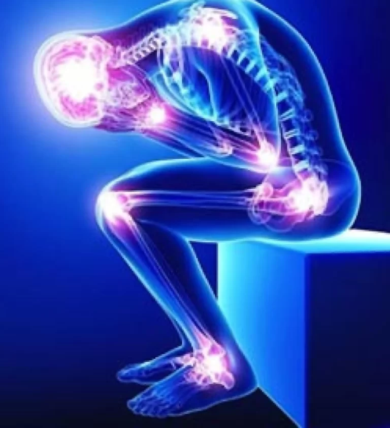
Back muscles injury
In most cases, injuries affect the way the back muscles work. Problems with the back include:
- Muscle strains: The muscles in the back can stretch and tear. These common injuries are usually caused by lifting a heavy object (or lifting incorrectly), exercise, overuse, or an accident. Back strains can cause muscle cramps or spasms. In severe injuries, the back muscles can become paralysed.
- Pain, tightness, and stiffness: Chronic back pain is very common. Pain can cause stiffness and decreased mobility (difficult movement). Tense muscles and back pain can also be caused by depression, stress, and anxiety. Pain in the neck and upper back can cause headaches.
- Ruptured discs: Discs cushion each vertebra in the spine. If the disc ruptures, it puts more pressure on a nerve, causing back pain.
- Bulging discs: Similar to ruptured discs, a bulging disc can increase pressure on a nerve.
- Sciatica is a sharp, shooting pain that travels through the buttocks and down the back of the leg. This can happen when a bulging or herniated disc presses on a nerve, or when a muscle specifically pushes on the sciatic nerve.
- Arthritis: Osteoarthritis can cause problems with the hips, lower back, and other joints in the body. The area around the spinal cord constricts in certain situations. Experts refer to this as spinal stenosis.
- Back pain can result from unusual spine curvature. The condition referred to as scoliosis results in a skewed spine.
- Osteoporosis occurs when bones, including the vertebrae of the spine, become brittle and porous, increasing the likelihood of compression fractures.
Signs and symptoms of back muscle injury
Different things can cause your back muscles to ache. They can sprain (or twist) if you pull them in an awkward motion or exercise. Muscles can also strain or tear when they are subjected to excessive pressure or stretched too far.
A common cause of severe muscle strain is a traumatic injury, such as one sustained while playing sports or in a car accident. Other causes include not getting enough rest between activities, attempting to recover quickly from a sudden movement (such as a loss of balance), or lifting heavy objects.
Age, weight, and fitness level are all factors that can contribute to different types of back injuries. Signs that something may be wrong with your back muscles are:
- Bruising of the muscles
- Decreased mobility
- Limited range of motion.
- Numbness on your back
- Pain and stiffness in the back
- Spasms (often extremely painful).
- Symptoms include swelling and muscle weakness.
Ninety percent of adults will at some point in their lives experience back pain. In some cases, the pain is caused by a mild strain and will subside after a few days of rest. If you are experiencing persistent back or muscle pain symptoms, consult your doctor immediately to learn about treatment options.
Back muscles exercises
Knee-to-chest stretch
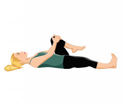
using both feet flat on the ground and bent knees, lie on your back. Using both hands, raise one knee and press it against your chest. Tense your abdominal muscles and press your spine to the floor. Hold for 5 seconds. Return to the starting position, then repeat with the other leg. Return to the starting position. Then repeat with both legs simultaneously. Repeat each stretch 2–3 times. Perform the entire routine once in the morning and once in the evening, if possible.
Lower back rotational stretch.
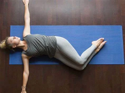
using both feet flat on the ground and bent knees, lie on your back. Keep your shoulders firmly planted on the floor and gradually roll your bent knees to one side. Hold for 5-10 seconds. Slowly return to the starting position. Repeat on the opposite side. Repeat each stretch 2–3 times. Perform the entire routine once in the morning and once in the evening, if possible.
Bridge exercise.
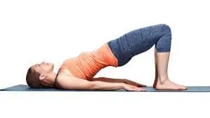
Through the knees bent and your feet flat on the ground, lie on your back. Keep your shoulders and head relaxed on the floor while tightening the muscles in your belly and buttocks. Then raise your hips to create a straight line between your knees and your shoulders. Try to remain in that position long enough to take three deep breaths. Return to where you started and repeat. Begin by doing five repetitions per day and gradually increase to 30.
Cat – Cow exercise
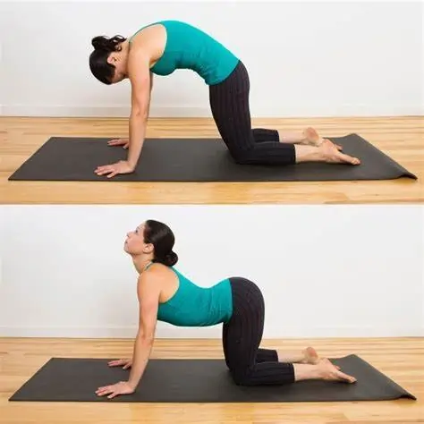
Kneel on your knees and hands. Slowly arch your back, pulling your belly up towards the ceiling as you lower your head. Then, as you lift your head, gradually allow your back and belly to sag towards the floor. Return to where you started. Repeat 3 to 5 times twice daily.
Bird-dog
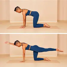
The bird dog exercises the gluteal muscles. It also works the back extensor muscles, which connect to the back of the spine and allow you to stand, bend, and lift objects.
To do the bird-dog exercise, you should follow these steps:
- Begin the exercise on your hands and knees, placing your shoulders directly over your hands and your hips directly above your knees.
- Tense the abdominal muscles and extend the right arm straight in front of the body.
- Maintain the position while remaining balanced.
- Slowly raise the left leg and extend it straight behind the body.
- Hold this position for 15 seconds.
- Repeat on the other side after making a slow return to the starting position.
- Repeat five times.
Abdominal draw-in manoeuvre
Strong abdominal muscles help to support the spine and keep the hips in proper alignment.
To do the ADIM, one should follow these steps:
- Arms by your sides, knees bent, lie on your back.
- Hold the spine in a neutral position and pull the belly button towards it.
- Inhale.
- Exhale while contracting the abdominal muscles and drawing the belly button towards the spine.
- Hold the position for 10 seconds, then release. Pause for 15 seconds.
- Repeat ten times.
Plank
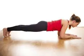
The plank exercise primarily works the abs, but it also engages the arms, shoulders, hip flexors, and feet, making it an excellent full-body stability exercise. This position may also work the back extensor muscles and the quadratus lumborum, the deepest back muscle.
Use the following instructions to execute a plank.
- Lie on your stomach, forearms against the floor, elbows directly in line with the shoulders.
- Tighten your abdominal and gluteal muscles.
- Raise the hips and both knees off the floor.
- Hold the position for 10-30 seconds, not allowing the pelvis to sag towards the floor.
- Repeat five times, taking your time getting back to the starting position.
Side plank
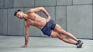
A side plank consists of the following steps:
- Lie on your right side, right leg slightly bent, left leg straight, foot on the floor. Position the right arm directly beneath the right shoulder, with the forearm extended out in front.
- Tighten the abdominal muscles and lift the right hip off the floor.
- Lift the right knee off the floor to straighten the right leg, then stack the feet on top of one another.
- Maintain a straight posture for 10-30 seconds.
- Slowly return to the starting position, then repeat on the other side.
- Repeat the steps outlined above five times.
Summary
The back muscles are a group of paired muscles located on the posterior aspect of the trunk, providing spinal movement, trunk stability, and coordination of limb and trunk movements. They are divided into two major groups: extrinsic (superficial) and intrinsic (deep) back muscles. Extrinsic muscles are located closest to the skin on the back and function similarly to upper-limb muscles but are superficially located on the posterior trunk.
Intrinsic back muscles are the deepest layer connected to the vertebral column and are divided into three layers: superficial, deep, and deepest. The erector spinae muscle group consists of three muscular columns on each side of the spine, classified into three regions: spinalis capitis, spinalis cervicis (colli), and spinalis thoracis. The iliocostalis muscle has three different types and extends between the rib angles and the transverse processes of the corresponding regional vertebra.
Injuries to the back muscles can cause muscle strains, pain, tightness, stiffness, ruptured discs, bulging discs, sciatica, osteoarthritis, unusual spine curvature, and osteoporosis. Ninety percent of adults will at some point in their lives experience back pain. Exercises to strengthen the back muscles include knee-to-chest, lower back rotational, bridge, cat-cow, bird-dog, abdominal draw-in manoeuvre, plank, and side plank.
FAQs
What are the three muscles of the back?
They consist of the longissimus, iliocostalis, and spinalis muscles. Their attachments divide these muscles, and they all share a tendinous origin. They help to move the thoracic cage and flex the upper vertebral column and head. As the back muscles develop, they extend causally.
How do the back muscles divide?
The back muscles are divided into three groups: superficial, intermediate, and deep. Superficial – Associated with shoulder movements. Intermediate – Associated with thoracic cage movements. Deep is associated with vertebral column movements.
Why do back muscles matter?
Your back muscles provide the primary structural support for your trunk (torso). These muscles help you move your entire body, including your head, neck, shoulders, arms, and legs. Bending, twisting, turning your head, and extending your back are all possible because your back muscles work together.
Do you require strong back muscles?
Strong back muscles support the spine, lowering the risk of strains, sprains, and other injuries caused by lifting, bending, or twisting movements. Muscle strains and sprains can be caused by sports, accidents, or daily chores like picking up something from the floor.
Which nerves cause back pain?
The sciatic nerve is a confluence of nerve roots in the lower back. The sciatic nerve runs from the buttocks down each leg. Pain along the sciatic nerve’s path is known as sciatica.
References:
- Overview of the back muscles. (2023, November 3). Kenhub. https://www.kenhub.com/en/library/anatomy/overview-of-back-muscles
- Henson, B., Kadiyala, B., & Edens, M. A. (2023, August 14). Anatomy, Back, Muscles. StatPearls – NCBI Bookshelf. https://www.ncbi.nlm.nih.gov/books/NBK537074/
- Professional, C. C. M. (n.d.). Back Muscles. Cleveland Clinic. https://my.clevelandclinic.org/health/body/21632-back-muscles
- Modes, R. J., & Fahrioglu, S. L. (2023, February 27). Anatomy, Back. StatPearls – NCBI Bookshelf. https://www.ncbi.nlm.nih.gov/books/NBK539746/
- Kashani, S. (2022, August 29). Back Muscles: What to Know. WebMD. https://www.webmd.com/fitness-exercise/back-muscles-what-to-know
- McIntosh, J. (2023, March 17). What is causing my back pain, and how can I remedy it? https://www.medicalnewstoday.com/articles/172943#causes
- Back exercises in 15 minutes a day. (2023, August 15). Mayo Clinic. https://www.mayoclinic.org/healthy-lifestyle/adult-health/in-depth/back-pain/art-20546859
- Cadman, B. (2023, November 30). How to strengthen the lower back. https://www.medicalnewstoday.com/articles/323204#strengthening-exercises
10 Comments