Palatine Bone
Introduction
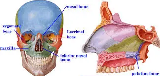
Between the maxillae and the pterygoid process of the sphenoid bone are a pair of bones called the palatine bones. It helps create the orbits, nasal cavity, and oral cavity, the three chambers of the skull. The maxilla, sphenoid, ethmoid, inferior nasal concha, and vomer are the five bones it articulates with to do this. Additionally, the palatine bone supports the skeletal structure of the pterygopalatine, pterygoid, and inferior orbital fissures.
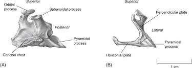
The two horizontal and perpendicular plates that make up the palatine bone are joined to produce the distinctive L-shaped bone. The three processes of the bone are sphenoidal, orbital, and pyramidal.
The palatine bone is located between the maxilla and the pterygoid process of the sphenoid in the rear of the nasal cavity.
It forms two fossae, the pterygopalatine and pterygoid fossæ, and one fissure, the inferior orbital fissure. It helps form the walls of three cavities: the orbit’s floor, the roof of the mouth, and the nasal cavity’s floor and lateral wall.
The palatine bone has a horizontal and vertical portion and three prominent processes—the pyramidal process, which extends backward and laterally from the point where the two parts meet, and the orbital and sphenoidal processes, which rise above the vertical portion and are divided by a deep notch known as the sphenopalatine notch—resembling the letter L in some ways.
Structure
Between the maxilla and the pterygoid process of the sphenoid bone, in the rear of the nasal cavity, are the palatine bones.
They support the walls of three cavities: the roof of the mouth, the floor of the orbits, and the lateral and floor walls of the nasal cavity. They aid in the formation of the inferior orbital fissures, as well as the pterygopalatine and pterygoid fossae.
A horizontal plate, a perpendicular plate, and three projecting processes—the pyramidal process, which is orientated laterally and backward from the junction of the two parts, and the orbital and sphenoidal processes, which rise above the vertical part and are divided by a deep notch known as the sphenopalatine notch—make up each palatine bone, which resembles the letter L in some ways. The two plates connect with the maxillae anteriorly and create the floor of the nasal cavity and the posterior portion of the hard palate. The two horizontal plates articulate with the maxillae more anteriorly at the transverse palatine suture and with each other at the posterior portion of the median palatine suture.
The sphenoid, ethmoid, maxilla, inferior nasal concha, vomer, and opposite palatine are the six bones that make up the human palate.
The larger and smaller palatine foramina are two significant foramina in the palatine bones that provide blood vessels and nerves to this area. In the posterolateral portion of each palatine bone, often near the peak of the maxillary third tooth, lies the bigger greater palatine foramen.
It also possesses the following 3 processes:
- Orbital process.
- Sphenoidal process.
- Pyramidal process.
Horizontal plate
The posterior portion of the hard palate is formed by its medial projection and unification with its opposite-side counterpart.
Perpendicular plate
It is a vertical plate that is attached to the back of the maxilla’s medial outermost layer. The perpendicular plate has two processes at its top border: the orbital process (b) and the sphenoidal process (a). It encloses the sphenopalatine notch between the two processes, which the inferior outermost layer of the sphenoid body transforms into the sphenopalatine foramen. The larger palatine groove is a vertical groove visible on the lateral surface of the perpendicular plate.
Orbital process
In front of the sphenopalatine notch, it protrudes laterally and upward from the anterior portion of the perpendicular plate’s top border. It has two non-articular surfaces and three articular surfaces.
Sphenoid mechanism
It extends medially and upward from the back of the perpendicular plate’s upper edge. It displays three edges and three surfaces.
The Pyramidal Process
From the point where the horizontal and perpendicular plates come together, it extends posterolaterally. The minor palatine canals puncture the pyramidal process. The maxilla and the palatine’s perpendicular plate are separated by the larger palatine canals.
Clinical significance
The Le Fort fractures of the mid-facial skeleton constantly include the perpendicular plates of the palatine bones. The Le Fort I requires the lower one-third of the perpendicular plates of the palatine bones to be included in the Le Fort fractures of the mid-facial skeleton. The bottom third of the perpendicular plates is needed for Le Fort I, whereas the top portions of the palatine plates are needed for Le Fort II and Le Fort III.
Gross anatomy
Between the maxillae and the sphenoid, in the rear of the nasal cavity, are the palatine bones. Each bone is made up of an L-shaped plate that is horizontal and perpendicular. The pyramidal, orbital, and sphenoidal processes are the three types.
They contribute structural elements to the orbital floor, pterygopalatine fossa, nasal cavity, and hard palate. Run in the space between the maxilla and the perpendicular plate of the palatine bone.
Articulations
- with the transverse palatine suture formed anteriorly by the maxillary palatine process
- with the midline of its opposite counterpart
- with the vomer
- utilizing the inferior concha
- with both the sphenoid’s body and medial pterygoid plate
- Using the ethmoid
- Attachments
- Tensor palati to the horizontal plate’s posterior border, uvula muscle to the horizontal plate’s nasal spine
- The medial pterygoid muscle’s superficial head in relation to the pyramidal process
Attachments
- Tensor palati to the rear edge of the horizontal plate.
- Uvula muscle to the horizontal plate’s nasal spine
- The medial pterygoid muscle’s superficial head about the pyramidal process
- Larger foramen of the Palatine
- Transports the pterygopalatine fossa’s larger palatine vessels and nerves to the oral cavity.
- Created by the larger palatine groove and transformed into a canal by maxillary articulation
- Palatine foramen, smaller
- Transmits the vessels and nerves of the smaller palatine
- Foramen sphenopalatine
- Created by the palatine bone’s sphenopalatine notch articulating with the sphenoid body
- Transmits the sphenopalatine artery and vein, the nasopalatine nerve, and the posterior superior nasal nerves, and joins the pterygopalatine fossa to the nasal cavity.
The process of ossification
- Tensor palati to the rear edge of the horizontal plate.
- Uvula muscle to the horizontal plate’s nasal spine
- The medial pterygoid muscle’s superficial head about the pyramidal process
- larger foramen of the Palatine
- Transports the pterygopalatine fossa’s larger palatine vessels and nerves to the oral cavity.
- Created by the larger palatine groove and transformed into a canal by maxillary articulation
- Palatine foramen, smaller
- Transmits the vessels and nerves of the smaller palatine
- Foramen sphenopalatine
- Created by the palatine bone’s sphenopalatine notch articulating with the sphenoid body
- Transmits the sphenopalatine artery and vein, the nasopalatine nerve, and the posterior superior nasal nerves, and joins the pterygopalatine fossa to the nasal cavity.
In the eighth week of pregnancy, intramembranous ossification manifests.
The horizontal and perpendicular plates are the same size at birth.
Processes
Pyramidal process
The intersection of the horizontal and perpendicular plates forms the palatine bone’s pyramidal process. It passes between the lateral and medial pterygoid plates of the sphenoid bone and is orientated posterolaterally.
The inferior surface has the lesser palatine foramina for the passage of the lesser palatine nerves and arteries, while the lateral surface serves as the attachment point for the deep head of the medial pterygoid muscle.
Orbital process
Pictured by Paul Kim, the orbital process of the palatine bone (Processus orbitalis ossis palatini)
- Fissura orbitalis inferior, or inferior orbital fissure; photo by Paul Kim
- Palatine bone orbital process
- Ossis palatini processus orbitalis
The anterior portion of the orbital border of the perpendicular plate is where the orbital process begins. It travels towards the orbit’s floor in a superior and somewhat anterior direction. Three articular and two non-articular surfaces are seen in this process, together with a thin neck.
The maxilla articulates with the anterior (maxillary) surface, which is orientated laterally and anteriorly.
The entrance of an air sinus, which is housed inside this process and connects to the sphenoidal sinus, marks the posterior (sphenoidal) surface, which faces superiorly and posteromedially.
Both anteriorly and medially, the medial (ethmoidal) surface runs. It articulates with the ethmoid bone’s labyrinth. It is this surface, rather than the posterior surface, that has an air sinus opening in certain people. Instead of speaking to the sphenoidal sinus in this instance, the bone speaks to the posterior ethmoidal air cells.
The superior (orbital) and lateral surfaces are the two non-articular surfaces of the orbital process. A notch that forms the medial portion of the inferior border of the inferior orbital fissure divides these two surfaces.
Sphenoidal process
Paul Kim’s image of the palatovaginal canal (Canalis palatovaginalis)
Palatovaginal canalis
Alternatives: Canalis pharyngeus, or pharyngeal channel
From the superior portion of the posterior border of the perpendicular plate, the sphenoidal process extends superomedially.
Its superior surface articulates with the root of the medial pterygoid plate and the sphenoidal concha. A groove that is a portion of the palatovaginal canal also marks this surface.
The nasal cavity’s floor and lateral wall are partially formed by the concave inferomedial surface.
The lateral surface is a component of the pterygopalatine fossa’s medial wall.
There are three boundaries in the sphenoidal process. The medial border articulates with the vomer’s all, while the posterior border articulates with the medial pterygoid plate. The sphenopalatine notch’s posterior boundary is formed by the anterior border.
Horizontal plate of palatine bone
The horizontal plate, which forms the floor of the nasal cavity and the posterior half of the hard palate, is the horizontal component of the palatine bone. There are two surfaces on the palatine bone’s horizontal plate: the nasal surface and the palatine surface.
The horizontal plate’s nasal surface is a component of the inferior nasal meatus and the posterior half of the nasal cavity’s floor.
The bony palate includes the palatine surface, which has the following apertures:
The larger palatine canal is reached by the greater palatine foramen.
The minor palatine canals are reached by the lesser palatine foramina.
The greater palatine nerve exits the greater palatine canal and enters the oral cavity through the greater palatine foramen, a tiny aperture in the palatine bone’s horizontal plate. The greater palatine canal connects the pterygopalatine fossa to the oral cavity by passing through the palatine and sphenoid bones. It conveys the larger palatine nerve, the smaller palatine nerve, and the descending palatine vessels. This canal gives rise to smaller palatine canals.
The smaller palatine canal (-s) runs vertically through the palatine bone’s perpendicular plate as a result of this hole or aperture. The minor palatine vessels and nerves are carried by these canals.
Perpendicular plate of palatine bone
The vertical portion of the palatine bone is called the perpendicular plate. It is a component of the maxillary sinus’s medial wall. The nasal surface and the maxillary surface are the two surfaces of the perpendicular plate. The sphenopalatine notch is another characteristic of this plate.
The nasal surface of the maxilla articulates with the rough, uneven maxillary surface. The super posterior portion of the nasal septum is formed by the nasal surface of the perpendicular plate. It articulates with the ethmoid bone’s cribriform plate superiorly.
Between the palatine bone’s orbital and sphenoidal processes, on the perpendicular plate, is a deep indentation known as the sphenopalatine notch. The notch transforms into the sphenopalatine foramen, which joins the nasal cavity with the pterygopalatine fossa, at the location where the palatine bone articulates with the sphenoid bone’s body.
Palatine Process
A maxillary bone segment: It is not a separate bone, but rather a horizontal projection that extends medially from the maxilla.
Forms the anterior part of the hard palate. 3/4: It joins the palatine bone posteriorly to complete the hard palate.
The structure is quite simple: A ridge separates its palatine (lower) and nasal (upper) flat surfaces.
Articulates with: The palatine bone on the midline, the incisive bone anteriorly, and other parts of the maxilla laterally.
Differences in Anatomy
The most often seen anatomical difference in the palatine bone is the bigger palatine foramen, an opening towards the rear that allows the greater and descending palatine nerves to pass through.
One study found that in almost 73% of the instances, this opening was located across from the third upper molar teeth. Furthermore, about 16% of the time, it was seen between the second and third teeth, and 7% of the time, it was seen across from the second molar.
Function
The sphenopalatine foramen, which connects the sphenoid and orbital processes of the palatine bones, allows branches from the pterygopalatine ganglion and the sphenopalatine artery from the maxillary artery to pass through into the nasal cavity.
The main function of the palatine bone is structural; it helps to carve out important structures inside the skull and defines the lower wall of the cranium.
The bottom portion of the eye sockets (orbits), the nasal and oral cavities, and the roof of the mouth are all formed with the help of this bone.
As was previously explained, they also have apertures through which the palatine nerves can pass. The primary pain-signaling pathways in the mouth and teeth are thus housed by the palatine bones.
The hard palate is created by the combination of the maxillary palatine process and the horizontal plates of the palatine bones. It is the roof of the oral cavity and the floor of the nasal cavity.
The palatine and seven other facial bones make up the orbit bones. The orbit’s floor, or bottom, is composed in part of the maxillary, palatine, and zygomatic bones. Look at the eye sockets below; the places where these separate bones intersect are shown by the many little cracks.
The nasal cavity warms and humidifies the air we breathe while also gathering contaminants and potential pathogens. The cartilaginous septum, the somewhat deflated structure seen in the following photo, separates the nasal canal into two nasal cavities even though humans only have one nose. The horizontal plate of the palatine bone and the palatine process of the maxilla bone comprise the nasal cavity floor.
The os palatinum is also connected to the facial muscles. Four muscles are joined to the palatine bone:
- The medial side of the pterygoid
- Uvula muscle
- Tensor Palitan
- Muscle of the superior pharynx constrictor
Finally, the palatine bone contains three more foramina through which nerves and blood vessels might travel. These nerves supply the hard and soft palate, the nasal cavity, and the motor and sensory pathways.
A group of bones has three foramina:
- The greater palatine foramen is the entry for the larger palatine nerve and blood vessels between the oral cavity and the pterygopalatine fossa.
- The lesser palatine nerve and blood vessels join the oral cavity with the lesser palatine canal through the lesser palatine foramen.
- Between the nasal cavity and the pterygopalatine fossa, the sphenopalatine foramen is the entry to the nasal nerves, nasopalatine nerve, and sphenopalatine artery and vein. The greater palatine foramen is the entry for the greater palatine nerve and blood vessels between the mouth cavity and the pterygopalatine fossa.
- Lesser palatine foramen: the passageway that connects the oral cavity to the lesser palatine canal via the lesser palatine nerve and blood vessels.
- The nasal nerves, nasopalatine nerve, and sphenopalatine artery and vein all enter through the sphenopalatine foramen, which is located between the nasal cavity and the pterygopalatine fossa.
Maxillary bone and details about the palatine bone
The irregular configuration of the confluence of these bones creates space for the pterygopalatine fossa. This fossa is a cavity that accommodates important nerve tissue, such as the maxillary nerve and pterygopalatine ganglion, which innervate different areas of the face, including the skin of the temples, upper lip, and teeth. The pterygopalatine fossa also houses the maxillary artery and several veins. The term “pterygoid” refers to wing-shaped.
The pterygopalatine fossa surrounds nerves and blood vessels.
At the top of the perpendicular plate lies the orbital process, which forms a part of the eye socket. Just below the orbital process is the sphenoidal process. This process not only contributes to the formation of the bony nasal cavity but also connects with the pterygoid plates of the sphenoid bone; it encircles an opening known as the sphenopalatine foramen, linking the nasal cavity with the pterygopalatine fossa.
Other sections of the maxillary surface of the os palatinum help constitute the wall of the maxillary sinus.
The perpendicular plates also contain small holes or foramina. The greater palatine foramen leads into a groove that becomes a canal when the maxilla rests upon it. These open into the greater palatine canal, which safeguards the greater palatine nerve along with its blood supply. The lesser palatine canals contain the lesser palatine nerves. Both the lesser and greater palatine nerves provide sensation to the hard and soft palates, tonsils, and nasal cavities.
One pair of foramina for the greater and lesser palatine nerves is highlighted in pink (not red) below.
Inside the interior of the skull, the fossa often features grooves.
The interior surface of the skull is seldom smooth.
As its name implies, the nasal surface of the perpendicular plate faces the nasal cavity. This portion of the perpendicular plate of the palatine bone adds support at the rear of the nasal passages.
At the junction of the horizontal and perpendicular plates lies the pyramidal process of the palatine bone. This structure fits between two slim plates in the sphenoid bone, termed the pterygoid plates, creating the pterygoid fossa (not to be confused with the pterygopalatine fossa). The pterygoid fossa accommodates the muscles required for chewing.
In the image, the bases of the horizontal plates of the palatine bones can be seen, extending across the pyramidal processes and down along each pterygoid fossa.
Pterygoid fossa – please observe the thin horizontal plates of the palatine bones.
The horizontal plate can be felt towards the back of the hard palate in the oral cavity. It also contributes to the base of the nasal cavity. Similar to the perpendicular plate, this plate possesses two surfaces, which are the nasal and palatine (or palate) surfaces.
The junction where each horizontal plate of the palatine bone meets its counterpart is slightly broader than the rest, forming the nasal crest and nasal spine.
The horizontal plate concludes at its connection with the pyramidal process of the perpendicular plate of the palatine bone; note that the pyramidal process belongs to the perpendicular, not the horizontal, plate.
There is a straightforward distinction between the palatine process and the palatine bone – the palatine bones do not have any palatine processes. Palatine processes are features of the adjacent maxilla bones and articulate with the palatine bones.
The palatine process of the maxilla bone is illustrated in red.
The palatine bone has three distinct processes. When mentioned, these processes should always be identified by the bone from which they originate (in this instance, the palatine bone) to prevent any misunderstanding. The three processes include:
- The orbital process of the palatine bone (which forms part of the orbital structure)
- The pyramidal process of the palatine bone (which is part of the pterygoid fossa)
- The sphenoid process of the palatine bone (which contributes to the pterygopalatine fossa)
When you encounter the term “process” in anatomical references regarding facial bones, it signifies an area or projection that meets the bone it is named after.
The inclusion of “of the,” as in the orbital process of the palatine bone, indicates that this process is a component of the palatine bone and extends to meet the orbital bone. The frontal bone also features two orbital processes, making it vital to specify the bone of origin.
Therefore, the palatine process of the maxilla bone indicates that there is a projection from the maxilla contributing to the structural or functional aspects of the os palatinum.
Function of Palatine Bones
The palatine bone has three purposes. First of all, each bone offers a portion of the orbital bones, nasal cavity, and hard palate of the roof of the mouth.
The horizontal plates of the palatine bones and the maxillary palatine process combine to produce the hard palate. It serves as both the nasal cavity’s floor and the oral cavity’s roof.
The seven facial bones (including the palatine) make up the orbit bones. Parts of the zygomatic, maxillary, and palatine bones make up the orbit’s floor, or bottom. Examine the eye sockets below; the numerous tiny fissures show the points where these disparate bones join.
The orbits resemble intricate jigsaw puzzles.
In addition to trapping pollutants and possible infections, the nasal cavity warms and humidifies the air we breathe. The cartilaginous septum, which is the fairly compressed structure in the accompanying image, separates the nasal canal into two nasal cavities even though humans only have one nose. The nasal cavity floor is made up of the horizontal plate of the palatine bone and the palatine process of the maxilla bone.
The muscles of the face can also adhere to the os palatinum. The palatine bone is attached to four muscles:
- Muscle of the medial pterygoid (to the pyramidal process)
- Constrictor muscle of the superior pharynx (to the horizontal plate)
- Tensor palati (to the plate that is horizontal)
- (To the horizontal plate) Uvula muscle
Lastly, the palatine bone of the face includes three foramina through which blood vessels and nerves can travel. These nerves supply the nasal cavity and hard and soft palate with sensory and motor pathways.
- Skull in an inferior view: hard palate foramen
- Hard palate inferior view of the skull
- There are three foramina on each bone:
- The greater palatine foramen serves as a passageway between the oral cavity and the pterygopalatine fossa for the greater palatine nerve and blood vessels.
- Between the oral cavity and the lesser palatine canal, the lesser palatine foramen serves as a passageway for the lesser palatine nerve and blood vessels.
- Between the pterygopalatine fossa and the nasal cavity, the sphenopalatine foramen serves as the entrance for the sphenopalatine artery and vein, nasopalatine nerve, and nasal nerves.
Palatine Process vs Palatine Bone
When it comes to palatine processes vs palatine bones, the answer is simple: palatine bones do not have palatine processes. The nearby maxilla bones have features called palatine processes that connect with the palatine bones.
The maxillary bone’s palatine process in the upper jaw and nasal cavity
The red palatine process of the maxillary bone
There are three processes in the palatine bone. To prevent misunderstanding, the name of the bone from which these processes originate—in this example, the palatine bone—should always be included when they are mentioned. These three processes are:
The palatine bone’s orbital process, which is a component of the orbital bone
Part of the pterygoid fossa is the pyramidal process of the palatine bone.
The palatine bone’s sphenoidal process, which is a component of the pterygopalatine fossa
When the word “process” appears in literature on the anatomy of the facial bones, you are aware that it refers to a region or projection that meets the bone for which it is called.
The inclusion of “of the” indicates that the process is a part of the palatine bone and extends to meet the orbital bone, as in the case of the palatine bone’s orbital process. It is crucial to indicate the bone of origin because the frontal bone also includes two orbital processes.
Therefore, the maxilla bone’s palatine process indicates that the os platinum’s structure or function is influenced by a projection from the maxilla.
Palatine Process
- The maxilla bone features a section known as the palatine process: It is a horizontal extension that projects medially from the maxilla and is not classified as a separate bone.
- It constitutes the anterior three-quarters of the hard palate: The palatine bone unites with it posteriorly to complete the hard palate.
- The structure is comparatively straightforward: A ridge divides its nasal (upper) and palatine (lower) flat surfaces.
- It articulates with: Other areas of the maxilla laterally, the incisive bone at the front, and the palatine bone in the center.
- The palatine process is a horizontal projection extending medially from the maxilla, rather than being a separate bone.
- It constitutes the anterior three-quarters of the hard palate, joining posteriorly with the palatine bone to complete the hard palate.
- This structure is relatively straightforward, with its nasal (upper) and palatine (lower) flat surfaces divided by a ridge.
- It connects with other parts of the maxilla laterally, the incisive bone anteriorly, and the palatine bone at the midline.
Associated Conditions
Numerous conditions are linked to the palatine bone. In addition to bone outgrowths and congenital malformations, it is possible to suffer fractures and nerve damage.
Damage to Nerves
In dentistry, the palatine bone is most often considered because of the renowned sensitivity of the bigger and smaller palatine nerves. To remove the upper molars and premolars, dentists must anesthetize (number) these nerves.
Because the syringe has the potential to puncture the larger palatine foramen, the injection sites, which are typically situated around 1 centimeter (cm) from the gingival margin, or the “height” of the gums, need to be continuously monitored.
Fractures
Palatine bones can be broken in falls or accidents. Most of these very rare palatal fractures occur in adult males. For medical professionals, their position in the face presents a difficult problem.
There are six primary types of palatine fractures, depending on the location of the bone break:
Sagittal fractures
- parasagittal fractures
- Fractures of the anterior and posterior alveoli
- Transverse fractures
- intricate fractures
- Fractures of the para alveoli
- These issues can lead to misalignment of the teeth and bite in addition to pain and edema in the surrounding tissues.
Cleft Palate
Cleft palates are congenital malformations that are often detected at birth. It happens when the two halves of the palatine bone do not fuse during fetal development. Between one in five hundred and seven hundred newborns are born with a cleft lip or palate.
In addition to dental problems, those who are born with a cleft palate may also have trouble speaking, hearing, eating, and drinking. They could also get colds and ear infections a lot.
Palatine Torus
The formation of palatine bone outgrowths, which are frequently painless and benign, is referred to as “torus palatinus.” They often begin in the middle of the plate and might show up on one or both sides of the palate.
While the majority of cases are asymptomatic and often go undetected, some do lead to:
- Pain
- Oral ulcers
- Abnormal chewing
- Distorted speech
- Usually, those who suffer from torus palatinus are in the
In conclusion
The paired palatine bone is a vital part of the orbitals, or eye sockets, nose, and skull, and it connects to other bones, such as the sphenoid. It protects and permits the passage of critical blood vessels and nerves.
The palatine nerve is required for some tooth extraction operations. Fractures of the palatine bone may also require surgery. Cleft palate is one of the most common palatine disorders; if identified at or before birth, it is typically effectively corrected.
The palatine bone’s main function is structural. To get a correct diagnosis, go to your healthcare provider if you have any troubling symptoms or believe you may have been hurt. erning symptoms or suspect an injury, consult your healthcare provider for a proper evaluation.
FAQs
What is the function of the palatine bone?
The palate splits into a posterior movable soft palate devoid of bone and an anterior immobile hard bony segment throughout development, undergoing intricate morphological modifications to reach its ultimate shape.
What are the eight facial bones?
The facial bones include:
Maxilla (2)
Zygomatic (2)
Mandible (1)
Nasal (2)
Palatine (2)
Inferior nasal concha (2)
Lacrimal (2)
Vomer (1)
What are the palatine and pterygoid bones?
Behind the palatine bones, the pterygoid is a pair of bones that make up the palate of many animals. It is a thin, flat lamina that is joined to the palatine bone’s perpendicular lamina and the medial side of the sphenoid bone’s pterygoid process.
References
- Palatine Bone – Earth’s Lab. Earth’s Lab.
- https://www.earthslab.com/anatomy/palatine-bone/
- Palatine bone – the key to your speech and swallowing.
- Mobile Physiotherapy Clinic. https://mobilephysiotherapyclinic.in/palatine-bone/
- Palatine bone. Radiopaedia.org.
- https://doi.org/10.53347/rid-54775
- Wikipedia contributors. (2024, June 14). Palatine bone. Wikipedia.
- https://en.wikipedia.org/wiki/Palatine_bone
- Palatine bone. (2023, November 3). Kenhub.
- https://www.kenhub.com/en/library/anatomy/the-palatine-bone