Atlanto-Occipital Joint
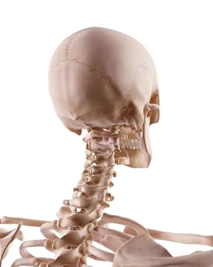
The atlanto-occipital joint is the articulation between the atlas (C1 vertebra) and the occipital bone of the skull. It is a synovial joint that allows for nodding movements of the head, such as flexion (“yes” motion) and slight lateral tilting.
This joint is stabilized by ligaments, including the anterior and posterior atlanto-occipital membranes, and plays a crucial role in supporting and facilitating head movements while maintaining stability.
Introduction
A paired, symmetrical articulation between the base of the skull and the cervical spine is called the atlantooccipital joint (also called the C0-C1 joint).The craniovertebral joints are a collection of joints that includes the atlantoaxial joint.
At the atlantooccipital joint, flexion-extension is the primary movement. This motion allows the head to nod, as is done when expressing acceptance (the “yes” motion). From a functional standpoint, these two ellipsoid (condyloid) joints can be regarded as a single joint because they operate concurrently.
Although stability is sacrificed, the upper cervical spine region is made to provide a great deal of motion. For this reason, the fibrous capsules, ligaments, articular surfaces, and surrounding muscles are primarily responsible for maintaining joint stability in the craniocervical region.
Anatomy
Articulations
Each joint is made up of two concave articular surfaces on the superior aspect of the lateral mass of atlas, which articulate with a convex surface on the occipital condyle.The joint is strengthened by fibrous capsules that support each joint.The atlanta facets are inclined medially.
Capsule
The atlantooccipital articulation capsules surround and connect the occipital bone’s condyles to the atlas’ articular processes; they are thin and loose.
Attachments
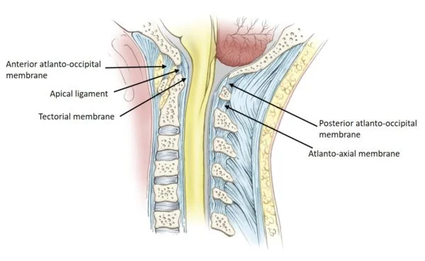
- The anterior atlanto-occipital membrane is a large, dense fibrous structure that connects the top border of the anterior arch of the atlas (C1) to the anterior inferior margin of the foramen magnum. It is a continuation of the anterior longitudinal ligament and helps to prevent excessive neck extension. Laterally, it integrates with the joint capsule, while medially it is reinforced by a strong, rounded cord that joins the basilar occipital bone to the anterior atlantal tubercle.
- The posterior atlanto-occipital membrane is a large but thin fibrous membrane that connects the upper border of the anterior side of the atlas’ posterior arch (C1) to the posterior margin of the foramen magnum. It connects with the posterior atlantoaxial membrane inferiorly (part of the ligamentum flavum) and the ligamentum nuchae posteriorly, and it is located directly posterior to the spinal dura. Suboccipital muscles are located posteriorly. The atlantic (V3) section of the vertebral artery travels anteriorly, piercing the membrane and dura before becoming the dural (V4) portion.
- Tectorial membrane: the posterior longitudinal ligament extends from the dens to the anterior portion of the foramen magnum.
Articular surfaces
The synovial articulation between the occipital bone and the first cervical vertebra (atlas) is known as the atlantooccipital joint. The occipital bone’s convex surfaces articulate with the concave articular facets of the C1 vertebra, which have an oval (elliptical) form and are reciprocally concave-convex. No intervertebral disc separates the occiput from C1. There is hyaline cartilage lining every articular surface. In the first cervical vertebra, the inferior articular facets are located.
These facets can be seen on the superior portion of the lateral mass of the vertebra. They are concave, oval in form, and somewhat slanted medially. In the anteromedial direction, each facet’s two long axes run obliquely, meeting at the midline immediately in front of the atlas. On the inferior part of the occipital bone, near the occipital condyles, are the superior articular facets. Elliptical in shape, these two rounded protuberances are extended and convex on both their long and short axes. The occipital condyles are orientated anteromedially, and are placed immediately lateral to the anterior part of the foramen magnum.
Ligaments & Joint Capsule
The articular capsule that surrounds each atlantooccipital joint is thin and flexible. The synovial membrane lines this fibrous tissue-based capsule. It adheres to the articular facets’ edges. Both the lateral and posterior portions of the capsule exhibit thickenings.
A number of ligaments span the atlantooccipital joint and contribute to its stability. They are the lateral atlantooccipital ligament, anterior atlantooccipital membrane and ligament, posterior atlantooccipital membrane, tectorial membrane, alar ligament, apical ligament, and ligamentum nuchae.Two of these are thought to be the main ligaments of the atlantooccipital joint because they link the occipital bone with the atlas. The following are the:
- Anterior atlantooccipital ligament (and membrane)
- Posterior atlantooccipital membrane
The dense band of fibrous tissue known as the anterior atlantooccipital ligament extends from the top border of the anterior arch of the atlas to the anterior border of the foramen magnum.The anterior longitudinal ligament, which serves as the anterior atlantooccipital membrane, strengthens it medially, and laterally it merges with the atlantooccipital joint capsule.
The posterior part of the atlantooccipital joint is covered by a thin membrane known as the posterior atlantooccipital membrane. It extends inferiorly from the top border of the posterior arch of Atlas to the superior posterior boundary of the foramen magnum. Its lateral edges run the length of the posteromedial joint capsule. It is a significant clinical hallmark that the posterior atlantooccipital membrane is close to the vertebral artery and C1 nerve.
Innervation
The anterior rami of spinal nerve C1 innervate the atlantooccipital joint.
Arterial supply
The deep cervical, occipital, and vertebral arteries anastomose.
Blood supply
An anastomosis between the deep cervical, occipital, and vertebral arteries supplies blood to the atlantooccipital joint.
Function
The following movements are permitted in this joint:
- Flexion and extension around the mediolateral axis, resulting in the typical forward and backward nodding of the head.
- Minor lateral motion, lateroflexion, to one or both sides of the anteroposterior axis.
Flexion is primarily caused by the activity of the longi capitis and recti capitis anteriores, whereas extension is caused by the recti capitis posteriores major and minor, the obliquus capitis superior, the semispinalis capitis, splenius capitis, sternocleidomastoideus, and upper trapezius fibers.
The recti laterales are involved in lateral movement, with the trapezius, splenius capitis, semispinalis capitis, and sternocleidomastoideus on the same side all working together.
Movements
The atlantooccipital joint has two degrees of freedom of motion since it is an ellipsoid joint. Flexion-extension and lateral flexion are two examples. However, flexion and extension are the main movements possible at the atlantooccipital joint. This is due to the atlantal sockets’ form, which is deep enough to keep the occipital condyles from translating too much and enable the atlantooccipital joint to give the head some stability while it balances on the cervical spine.
In the anteroposterior plane, flexion and extension movements take place around a transverse axis. Over the concave facets of the atlas, the convex occipital condyles slide posteriorly and roll forwards concurrently during flexion. Consequently, the occipital bone is moved away from the atlas’s posterior arch. This enables a forward tilting, or downward nod, of the head, like the “yes” movement used to express acceptance. The fibrous structures that surround the joint (joint capsules, posterior atlantooccipital membrane, ligamentum nuchae) and the posterior suboccipital muscles restrict flexion to roughly 5°–10°.
The opposite motions take place in extension. The gap between the occipital bone and the atlas’s posterior arch is closed by the occipital condyles, which roll backward and slide anteriorly on the atlantal facets. The extension range of motion is limited to roughly 10° by the occipital bone, the atlas, and the axis.
The range of motion at the atlantooccipital joint is not significantly affected by lateral flexion.The lateral flexion of the upper cervical spine is actually limited to about 5-8° on each side in cadaveric studies. Additionally, the lateral flexion of the upper cervical spine is a double-joint and linked movement. When lateral flexion and a slight degree of contralateral rotation happen simultaneously, this is referred to as coupled movement. When multiple joints move simultaneously, it’s referred to as double-joint movement.
The combined movements to accomplish lateral flexion of the upper cervical spine are thus as follows: a small quantity of contralateral glide at the occipital condyles (lateral flexion); concurrently, one occipital condyle moves somewhat anteriorly while the other moves posteriorly (rotation); and in addition to these movements, the second vertebra rotates (relatively) against the third cervical vertebra, resulting in an overall range of motion for lateral flexion of the upper cervical spine that is between 5 and 8°.
Muscles acting on the atlantooccipital joint
The cervical spine is made more mobile by the action of the postvertebral and anterior neck muscles on the atlantooccipital joint.
| Flexion from a standing posture | Trapezius, splenius capitis, longissimus capitis, semispinalis capitis, rectus capitis posterior major, rectus capitis posterior minor, obliquus capitis superior |
| From the supine posture, flexion | Sternocleidomastoid, longus capitis, rectus capitis anterior muscles |
| Extending from the standing position | Sternocleidomastoid, longus capitis, rectus capitis anterior muscles |
| Extending from the prone posture | Rectus capitis posterior major, rectus capitis posterior minor, obliquus capitis superior, semispinalis capitis, splenius capitis muscles, cervical part of trapezius |
| Lateral flexion | The splenius capitis, semispinalis capitis, trapezius, rectus capitis lateralis, and sternocleidomastoid |
Muscles acting on the atlantooccipital joint
The rectus capitis anterior and longus capitis muscles are the primary flexors of the head on the neck. The obliquus superior capitis, semispinalis capitis, splenius capitis, trapezius, rectus capitis posterior major, and rectus capitis posterior minor are the primary extensor muscles. Nevertheless, the precise muscles used in these motions may vary based on the head’s starting posture.
Strong motion is required to raise the head and flex it forward on the neck while in a supine position. The muscles of the anterior neck are the primary movers in this situation. These muscles include the rectus capitis anterior, longus capitis, and sternocleidomastoid.
Since the weight of the head can cause it to bend when it is upright, the same strength is not needed. The forward bend movement in this situation is controlled by the posterior muscles of the neck and back. The short suboccipital muscles, the splenius capitis, the longissimus capitis, the semispinalis capitis, and the trapezius are among them.
Similar to this, the anterior neck muscles (such as the sternocleidomastoid and longus capitis) regulate the head’s ability to extend from an upright position by acting against the head’s weight. To raise the head into extension while in the prone position, primary movers are required. These muscles include the cervical portion of the trapezius, the obliquus capitis superior, the semispinalis capitis, the splenius capitis, and the rectus capitis posterior major and minor.
The anterior and posterior neck muscles work together to cause the head to flex laterally. Among these are the muscles of the rectus capitis lateralis, trapezius, splenius capitis, semispinalis capitis, sternocleidomastoid, obliquus capitis superior, and rectus capitis posterior minor. Sternocleidomastoid, rectus capitis posterior minor, obliquus capitis superior, and splenius capitis support the linked movement of rotation.
Clinical significance
Dislocation
The atlanto-occipital joint can be dislocated, particularly in traumatic events like traffic crashes.This can be diagnosed with CT scans or magnetic resonance imaging of the head and neck. Surgery could be utilized to repair the joint and any related bone fractures. Neck movement may be limited for a long time following this injury. Such injuries may also cause hypermobility, which can be identified via radiography. This is especially true if traction is applied during treatment.
Three forms of AOD are distinguished by the occipital dislocation recommendation:
- Anterior displacement
- Posterior displacement
- Longitudinal distraction.
Damage to the related ligaments occurs along with dislocation. The degree of dislocation is the primary determinant of the injury’s severity. Stages I and II show no or very little displacement and sufficient ligament preservation. Stage III is characterized by significant dislocation and extremely unstable damage. Damage to the spinal cord’s cervical area may be linked to stage III.This might be lethal.The neurological abnormalities that survivors of this kind of damage may experience include unilateral or bilateral muscular deficiencies, lower cranial nerve deficits, or even quadriplegia, or paralysis of all four limbs.
FAQs
Is the atlanto-occipital joint an ellipsoid one?
The atlantooccipital joint, which is an ellipsoid, has two degrees of freedom of movement.These include flexion-extension and lateral flexion. However, the primary mobility allowed at the atlantooccipital joint is flexion-extension.
Is the atlanto-occipital joint a pivotal joint?
The atlanto-occipital joint (O-C1) serves as a pivot for the cranium’s flexion/extension motion, with 13 degrees average flexion/extension and 8 degrees lateral bending, enabling just a few degrees of axial rotation.
How stable is the atlanto-occipital joint?
The tectorial membrane and alar ligaments supply the majority of the support for the atlanto-occipital joint, and damage to these ligaments causes instability due to low inherent osseous stability.
What kind of lever is the atlanto-occipital joint?
The Atlanto-Occipital Joint is a First Class Lever.
A first-class lever in the human body is the head and neck during neck extension.The fulcrum (atlanto-occipital joint) connects the load (front of the skull) with the effort (neck extensor muscles).
Is the atlanto-occipital region synovial?
The atlanto-occipital articulation (also called the C0-C1 joint or articulation) is made up of two condyloid synovial joints that connect the occipital bone (C0) to the first cervical vertebra (atlas).
What makes up the atlanto-occipital joint?
The atlanto-occipital joint connects the atlas bone to the occipital bone. It is composed of two condyloid joints. It is a synovial joint.
What are the biomechanical properties of the atlanto-occipital joint?
Atlanto-occipital joint biomechanics. Although the atlanto-occipital joint is capable of flexion, extension, rotation, and lateral bending, cadaveric studies reveal that flexion and extension are its main motions.
Bony components are the primary constraint on this motion (Wolfla, 2006).
Which feature of the atlantooccipital joint is the most distinctive?
This bone’s most distinctive feature is its robust dens, an odontoid process.To put it simply, the atlantooccipital joint is made up of two condyloid joints. The atlantooccipital joints are synovial socket joints, which have shallow sockets when a baby is born and deeper sockets as people age.
References
- Atlantooccipital joint. (2023, August 3). Kenhub. https://www.kenhub.com/en/library/anatomy/atlanto-occipital-joint
- Wikipedia contributors. (2024c, August 24). Atlanto-occipital joint. Wikipedia. https://en.wikipedia.org/wiki/Atlanto-occipital_joint
- Bell, D., & Jarvis, M. (2015). Atlanto-occipital articulation. Radiopaedia.org. https://doi.org/10.53347/rid-35478
- Rupapara, H. (2023, March 29). Atlanto-occipital joint – Anatomy, Ligament, Muscles, Movement. Samarpan Physiotherapy Clinic. https://samarpanphysioclinic.com/atlanto-occipital-joint/

