Cuneiform Bone
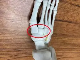
The cuneiform bones, also known as ossa cuneiform in Latin, are a set of three tarsal bones positioned within the first three metatarsals and the proximal navicular bone. The cuboid bone is found with the lateral cuneiform bone on the exterior.
The medial side of each foot contains three cuneiform bones. These cuneiform bones work together to generate the foot’s transverse arch.
What is the Cuneiform Bone?
- Cuneiform bones are sometimes called ossa cuneiform in Latin. The cuneiform bones are located in the middle of the cuboid bone, right below the navicular bone, and among the first, second, and third metatarsals.
- The first, sometimes called the medial cuneiform, the second, sometimes called the middle cuneiform, and the third, often called the lateral cuneiform, are the three cuneiform (“wedge-like structures”) bones that together constitute the human foot.
Structure and Types of the Cuneiform Bone
Cuneiform bones come in three different types:
Medial Cuneiform: One of the three bones, the medial cuneiform (or first cuneiform), is the biggest. In front of the navicular bone, behind the base of the first metatarsal, and on the medial side of the foot, it can be seen.
Intermediate Cuneiform: The intermediate cuneiform, which articulates with the navicular, second cuneiform, and first and second metatarsals, is located laterally to it. medial cuneiform.
Often referred to as the second or middle cuneiform, the intermediate cuneiform’s thin end points downward in a wedge-like structure. The intermediate cuneiform, which connects the medial and lateral cuneiforms, articulates with the navicular posteriorly, the second metatarsal anteriorly, and the other cuneiforms on either side.
Lateral Cuneiform: Often referred to as the third cuneiform or lateral cuneiform, this wedge-shaped bone is within the other two cuneiform bones in size and has a base at the top. It is located in the middle of the front row of tarsal bones, between the third metatarsal in front, the cuboid laterally, the navicular posteriorly, and the intermediate cuneiform medially.
Ligamentous and Muscle attachments
- The medial cuneiform bone is where the peroneus longus (Fibularis) muscle and the tibialis anterior muscle insert.
- The medial cuneiform is where the tibialis posterior muscle inserts and the flexor hallucis brevis muscle begins.
Arterial supply
- The dorsal arterial arch branches transport blood to the cuneiform bones’ dorsal sides, whereas the plantar arterial arch gives blood to the plantar surfaces.
The function of the Cuneiform Bone
- The cuneiform bones’ wedge-shaped structure aids in forming and maintaining the foot’s transverse arch.
Relevant Condition of Cuneiform Bone
- Lisfranc fracture:
- occurs when more than one metatarsal bone separates from the tarsus.
- Cuneiform fracture: Cuneiform breakage Isolated cuneiform fractures are uncommon since ligaments support the midfoot.
- Arthritis: The joints connecting the cuneiform and other foot bones may be impacted by degenerative joint disease.
- Bunions: Although they mostly affect the big toe joint, bunions can occasionally cause the cuneiform bones to become misaligned.
Symptoms of Conditions Associated with Cuneiform Bones:
- Pain in the middle of the foot
- Bruising and Swelling
- Having trouble walking or carrying weight
- Decreased foot range of motion
Diagnosis and Treatment for Relevant Condition of Cuneiform Bone
It’s critical to speak with a medical expert, such as an orthopedic surgeon or podiatrist, if you encounter any of these symptoms. They can identify the root problem and suggest the best course of action, which could involve:
- Ice and Rest
- Physical therapy
- Orthopedic devices
- Medication (anti-inflammatory medications, pain relievers)
- Surgery (in extreme situations)
FAQs
What is causing the cuneiform bone to hurt?
In clinical practice, cuneiform stress fractures cause pain along the foot’s medial or lateral column, which might be confused with a longitudinal arch strain or ligament sprain. There is no usual pattern of presentation for these injuries. They are visible on MRI and can be obscured on conventional radiography.
With a cuneiform fracture, is it possible to walk?
People usually experience severe pain over their dorsal or dorsomedial foot after suffering acute fractures to one or more cuneiform bones. They also struggle to bear weight and move on their toes.
How is a cuneiform bone treated?
Stabilization or total immobilization for a few weeks can be used to treat minor injuries to the cuneiform bones and/or Lisfranc ligament complex so that the damaged components have time to heal. Surgery is used to address severe damage to the ligaments and cuneiform bones.
Which cuneiform is the largest?
The medial (or first) cuneiform is the largest of all of the three cuneiform bones. The word cuneiform is derived from the Latin cuneus (wedge) and forma (likeness), which describes its appearance.
References:
- cuneiform bone. (n.d.). In Merriam-Webster Dictionary. https://www.merriam-webster.com/medical/cuneiform%20bone
- Wikipedia contributors. (2023, December 6). Cuneiform bones. Wikipedia. https://en.wikipedia.org/wiki/Cuneiform_bones
- Next, A. (n.d.). %s | Anatomy.app | Learn anatomy | 3D models, articles, and quizzes. https://anatomy.app/encyclopedia/cuneiform-bones
- Cuneiform bones. (2023, October 30). Kenhub. https://www.kenhub.com/en/library/anatomy/cuneiform-bones

