Calcaneus (Heel Bone)
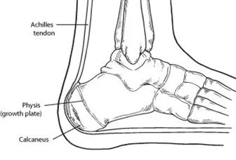
The calcaneus (heel bone) is the largest tarsal bone in the foot. It works as the foundation of the rear part of the foot, forming the heel. The calcaneus plays a crucial role in weight-bearing, shock absorption, and foot mechanics during walking, running, and standing. Structurally, it articulates with the talus above (forming the subtalar joint) and the cuboid in front (forming the calcaneocuboid joint).
Additionally, the calcaneus is a major attachment site for the Achilles tendon, connecting the bone to the calf muscles, and enabling powerful movements like jumping and running.
What is the Calcaneus?
- Human calcaneus belongs to one of the seven connecting bones that collectively make up the foot (tarsus). The calcaneus, the foot’s biggest bone, is located in the hindfoot adjacent to the talus and is sometimes referred to as the heel.
- The calcaneus is attached to a variety of muscles and ligaments that support its function in human bipedal biomechanics.
- It connects with the cuboid anteriorly and the talus superiorly, and it links a joint space to the talocalcaneonavicular joint.
- It is the calcaneus bone that transfers almost all the body weight to the base by using the lower leg.
Anatomy of the Calcaneus
- At the subtalar or talocalcaneal junction, the inferior border of the talus becomes attached to the posterior segment of the calcaneus. The inferior talar surfaces work in conjunction with the anterior and middle calcaneus to produce the talocalcaneonavicular joint.
- The middle talar articular facet is supported on its superior surface by the sustentaculum tali, a shelf-like projection of bone. Conversely, the flexor hallucis longus tendon might go through an aperture on the sustentaculum tali’s inferior surface. The calcaneal sulcus differentiates the center and rear faces. These sulci, along with the talar sulcus above it, make up the tarsal canal, which extends anterolaterally to the tarsal sinus.
- The cuboid bone and calcaneus fuse resulting in the calcaneocuboid joint anteriorly. The calcaneus has a pronounced tuberosity on its posterior side, which is bordered by medial and lateral projections.
Muscle Attachment
- Triceps surae is a tendon that connects the gastrocnemius and soleus to the middle facet of the posterior section of the calcaneus.
- Abductor hallucis develops from the calcaneal tuberosity’s medial process.
- The flexor digitorum brevis expands via the middle processes of plantar aponeurosis as well as calcaneal tuberosity.
- Abductor digit minimi: comes from the medial and lateral processes of calcaneal tuberosity; quadratus plantae: gives rise to the plantar surface of calcaneus. The site of origin of the extensor digitorum breves’s dorsolateral surface
- The tarsal sinus and dorsal surface are the extensor hallucis brevis’s origins.
Ligament Attachment
- Lateral collateral/calcaneofibular ligament in the ankle.
- Inferior: Short plantar ligament (at calcaneal tubercle), long plantar ligament (in front of calcaneal tuberosity), and plantar aponeurosis.
- Superior tarsal sinus ligaments, which include the cervical ligament, and talocalcaneal Interosseous Ligament.
- Bifurcate the ligament.
- The calcaneus’s sustentaculum tali resides ahead of the plantar calcaneonavicular ligament.
Blood Supply
- The posterior tibial artery’s perforating section distributes blood to the middle portion of the calcaneus.
- The lateral calcaneal artery provides blood supply to the calcaneus’s lateral side.
- A watershed, or basin, is formed in the center of the bone by the anastomosis of the medial and lateral intra-osseous arteries.
Palpation
- The Calcaneus bone palpates at the posterior portion of the bone just below the Achilles tendon.
- The inferior element of the calcaneus, or more correctly the calcaneal tuberosity, where the plantar fascia grows, can also be sensation felt.
- By palpating the medial malleolus and moving immediately inferior by 1-2 cm, one can feel the sustentaculum tali on the medial surface.
The function of the Calcaneus
- The calcaneus’s main jobs are to sustain the body’s weight and facilitate mobility in a variety of joints. When the heel strikes the ground while walking, the axial stress of the body travels through the hip, leg, and ankle before arriving at the calcaneus. Ultimately, this tension is supported by the calcaneal tuberosity’s medial and lateral processes. As the body takes a stride, the arches of the foot begin to carry its weight.
- The calcaneus aids in the formation of the two longitudinal arches along the plantar surface of the foot, such as the medial and lateral arches, to absorb shock in a spring-like way when the body’s weight is carried through the step action.
- The calcaneus is additional crucial muscle in plantar flexion. An ankle joint movement known as plantar flexion causes the foot to point downward. The inferior portions of the superficial muscles, including the gastrocnemius, planters, and soleus muscles, make up the calcaneal tendon, also referred to as the Achilles tendon, which is located in the area behind the lower limbs.
- The foot descends from the ankle into plantar flexion when the muscles involved contract, dragging the rear portion of the calcaneus forward. Everyday motions like walking, running, driving, and riding a bicycle all involve this action.
- One of the subtalar joint’s bones, the calcaneus, aids in the eversion and inversion of the foot. The action of the foot external at the ankle joint is called eversion, as opposed to inversion, which is the rotation of the foot upward. The oblique axis of alignment between the talus and calcaneus accommodates these movements.
- Lastly, muscles that originate in the calcaneus enable several joint movements in the foot. The extensor digitorum brevis aids in the extension of the medial four toes at the metatarsophalangeal and interphalangeal joints on the dorsal part of the foot. On the other hand, the great toe can be extended at the metatarsophalangeal joint with the aid of the extensor hallucis brevis.
- The calcaneus is formed by the foot’s abductor hallucis, flexor digitorum brevis, quadratus plantae, and abductor digiti minimi muscles, all of which run down the plantar portion of the foot. The flexor digitorum brevis and quadratus plantae muscles aid in flexing the foot’s lateral four digits, while the abductor hallucis aids in abduction and flexion of the great toe. Finally, the foot’s fifth digit is flexed by the abductor digit minimi.
Relevant Conditions
- Calcaneal fracture
- Calcaneus Talocalcaneal Coalition pseudo-tumor
- Coalition of Calcaneonavicular
- Calcaneal/heel spur
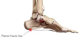
- Plantar fascia discomfort is a disorder characterized by a structural breakdown of the connective tissue that forms the foot’s fascia, or plantar aponeurosis, without any inflammation. Frequently observed near heel spurs.
- A bone exostosis that extends from the posterior superior calcaneus, where the Achilles connects, is known as a Haglund deformity. Although the exact causation of the illness is unknown, high-arched feet, Achilles tendinopathy, and other genetic factors are thought to be contributing factors.
How can fractures of the calcaneus happen?
- Although calcaneal fractures can result from traumas, the majority of heel discomfort is caused by inflammation of the tissues around the calcaneus, such as tendons, bursae, and fasciae.
- Intraarticular fractures, the most widespread kind of calcaneal fracture, take place when multiple fractures extend into the bone’s articulation surface. The subtalar joint is most frequently impacted, and it happens when there is a significant increase in axial strain on the calcaneus, as occurs when someone lands after a jump or falls from a height.
- A comminuted fracture occurs when the calcaneus is compressed as the stress shifts from the talus to the bone, shattering it into several tiny fragments.
- This kind of calcaneal fracture is frequently severe, causes the patient a lot of discomfort, and frequently requires surgery to repair. A pressure dressing can be used to immobilize the foot and ankle before surgery. Long-term arthritis in the afflicted joint may cause chronic pain with weight bearing, inversion, and eversion even after 8–12 weeks of weight-bearing restriction following surgery. Acute compartment syndrome, fracture blisters, edema, and excruciating pain are among the complications that can arise from intraarticular fractures.
- A less frequent kind of calcaneal fracture is an extraarticular fracture, which does not extend into a joint. Most often, falls or joint twisting cause these fractures. Avulsion fractures at tendon or ligament attachment sites are among them, as are fractures restricted to the bone tissue, such as those that occur inside the bone body or across one of the bone’s processes.
- When a force is exerted along the surface of a tendon or ligament, a small fragment of bone is ripped out, resulting in an avulsion fracture.
- Compression dressings, rest, ice application, elevation, and minimizing weight bearing on the injured foot are common initial treatments for extraarticular fractures. While some extraarticular fractures heal in eight to ten weeks, others require a walking boot or cast for four to six weeks.
- Ankle range-of-motion exercises performed at home with increasing weight bearing can assist the person in returning to their regular activities following the removal of a cast or boot. For certain people, more physical treatment could be required.
- Finally, stress fractures of the calcaneus happen when there is a recent increase in activity level, like when runners exercise on hard surfaces, or when there is recurrent loading over time. For milder cases, stress fractures can be treated with decreased activity and heel inserts; for more severe cases, crutches and weight-bearing avoidance are recommended.
Summary
The calcaneus, the largest bone in the foot, supports weight, forms joints with other bones, and serves as the attachment point for several muscle tendons, all of which contribute to stability and walking. Calcaneal fractures can be intraarticular or extraarticular, depending on the state of the joint.
Compared to extraarticular fractures, intraarticular fractures are typically more severe and require a longer recovery period. Some people with intraarticular fractures may develop chronic pain and arthritis, but those with extraarticular fractures typically recover completely with the right care and healing time.
FAQs
What is another term for calcaneus?
A bone of the foot’s tarsus that makes up the heel, the calcaneus (/kaelˈkeɪniʙs/; from the Latin calcaneus or calcaneum, meaning heel; pl.: calcanei or calcanea) or heel bone is found in humans and many other primates. It is the point of the hock in certain other animals.
What makes calcaneum and calcaneus different from one another?
The largest tarsal bone and the main bone in the hindfoot is the calcaneus, also known as the calcaneum (plural: calcanei or calcanea). It articulates with the cuboid anteriorly and the talus superiorly, and it overlaps a joint space with the talonavicular joint, which is more appropriately known as the talocalcaneonavicular joint.
If someone has a calcaneus fracture, can they still walk?
You can resume your regular activities, including walking, after eight weeks. The length of time it takes for a fracture to mend depends on its severity. Within 12 weeks, most people can return to their regular activities.
What is the calcaneus’ primary purpose?
The calcaneus, the biggest bone in the foot, supports weight, forms joints with other bones, and serves as the attachment point for several muscle tendons, all of which contribute to stability and ambulation.
How powerful is the calcaneus?
Typically measuring between 75 and 89 mm from front to back, the calcaneus is the largest and most robust tarsal bone in the foot. The subtalar joint’s three articular surfaces include the posterior facet of the rear dorsal third of the calcaneus, which defines the talus.
References:
- Calcaneus (Heel Bone) Fractures – OrthoInfo – AAOS. (n.d.). https://orthoinfo.aaos.org/en/diseases–conditions/calcaneus-heel-bone-fractures/
- Calcaneus. (2023, October 30). Kenhub. https://www.kenhub.com/en/library/anatomy/calcaneus
- Forsthoefel, C., MD. (n.d.). Calcaneus Fractures – Trauma – Orthobullets. https://www.orthobullets.com/trauma/1051/calcaneus-fractures

One Comment