Facial muscles
Introduction
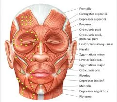
The facial muscles, also known as craniofacial muscles, are a group of approximately twenty flat skeletal muscles that are located beneath the skin of the scalp and face. The majority of them originate in the skull’s bones or fibrous structures before spreading to the skin.
The facial muscles are a collection of striated skeletal muscles supplied by the facial nerve (cranial nerve VII), which, among other things, controls facial expression. Mimetic muscles are another name for these muscles. They are only found in mammals, but they are descended from neural crest cells found in all vertebrates. They are the only muscles that connect with the dermis.
The facial muscles include:
- The occipitofrontalis muscle
- The Procerus muscle
- Nasalis muscle
- Platysma muscle
- Orbicularis Oculi Muscle
- Corrugator supercilii muscle.
- Depressor Septi Nasi.
- Orbicularis Oris.
- Buccinator
- Levator Labii Superioris Muscle
- Depressor labii inferioris muscle.
- Levator Labii Superioris Alaeque Nasi Muscle
- Mentalis Muscle
- Risorius Muscle
- Levator Anguli Oris Muscle
- Depressor Anguli Oris Muscle
- Zygomaticus major.
- Zygomaticus minor.
- Auricular Muscles
The occipitofrontalis muscle
A massive muscle called the occipitofrontalis runs from the brows to the superior nuchal lines of the occipital bones. The muscle is divided into two parts: occipital and frontal, which are connected by a fibrous sheath called the epicranial aponeurosis (galea aponeurotica). Both the occipital and frontal parts have quadrangular muscle heads.
Origin and insertion
Origin: The frontal part is formed by the skin between the brows and the superior parts of the periorbital group of facial muscles.
Insertion: It then curves posterolaterally to meet the epicranial aponeurosis at the level of the skull’s coronal suture.
Origin: The occipital part develops from the lateral two-thirds of the superior nuchal line of the occipital bone.
Insertion: courses superiorly to attach to the epicranial aponeurosis via the lambdoid suture.
Innervation
The facial nerve (CN VII) innervates both parts of the occipitofrontalis. The temporal branches supply the frontal part, whereas the posterior auricular branch of the facial nerve innervates the occipital part.
Blood supply
The ophthalmic and superficial temporal arteries provide blood supply to the frontal part, while the posterior auricular and occipital arteries vascularize the occipital part.
Function
The occipitofrontalis’ function is determined by the part of the muscle that contracts:
- Frontal part: When its forehead attachment is fixed, the frontal part contracts, pulling the scalp forward and wrinkling the forehead, resulting in a frown. If its aponeurotic attachment is fixed, the frontal belly elevates the brows and skin on the forehead, resulting in a surprised expression.
- When its nuchal attachment is secure, the occipital part retracts the scalp. When its aponeurotic attachment is fixed, this part of the muscle moves the scalp forward.
The Procerus muscle
The procerus is a small pyramidal muscle located between the brows.
Origin
It originates from the nasal bone and superior part of the lateral nasal cartilage and extends superiorly in a fan-like fashion.
Insertion
Place on the skin above the glabella and the medial ends of the brows.
Innervation
This muscle is supplied by the temporal, lower zygomatic, and buccal branches of the facial nerve (CN VII).
Blood supply
It receives blood from the angular and lateral nasal branches of the facial artery.
Function
The procerus wrinkles the skin surrounding the glabella and depresses the medial ends of the brows using its superior attachment. This results in a frowning facial expression to convey anger or sorrow, or when exposed to bright light or other eye irritants.
Nasalis muscle
The nasalis is a small muscle located on each side of the dorsum of the nose. According to its origin, it is divided into two parts: alar and transverse.
Origin and insertion
The alar part is located near the nostrils.
- Origin: Originating from the frontal part of the maxilla, superior to the incisive fossa and medial to the transverse part of the nasalis
- Insertion: It extends upwards and anteriorly to insert on the skin of the ala, above the lateral crus of the major alar cartilage.
The transverse part is located over the dorsum of the nose.
- Origin: It develops superolateral to the incisive fossa and laterally to the alar part.
- Insertion: By merging with its counterpart across the nose’s bridge, it passes superomedially to insert at the dorsum of the nose.
Innervation
The nasalis muscle is innervated by the buccal branch of the facial nerve (CN VII)
Blood supply
The superior labial, septal, and lateral nasal branches of the facial artery, as well as the infraorbital branch of the maxillary artery, supply blood to this area.
Function
The nasalis muscle’s transverse part compresses the nasal aperture while the alar part dilates the nostrils. These actions are notable for creating specific facial expressions, such as anger, as well as enhancing deep breathing.
Platysma muscle
The platysma is a muscle that looks like a sheet and is located within the superficial cervical fascia of the anterior neck.
Origin
It grows from the skin and fascia of the superior thoracic and shoulder regions and ascends to the anterolateral sides of the neck. The medial fibres of the platysma attach to the lower border of the mandible and the skin of the lower lip.
Insertion
The lateral fibres of the platysma insert on the skin of the perioral region, where they combine with the various muscles surrounding the mouth to form the modiolus.
Innervation
The platysma gets a nervous supply from the cervical branch of the facial nerve (CN VII).
Blood supply
The blood supply comes from the facial artery’s submental branch and the thyrocervical trunk’s suprascapular branch.
Function
The platysma’s functions vary depending on which part of the muscle contracts. The platysma can help lower the corners of the mouth and lower lip by contracting the lateral fibres that attach to the modiolus, whereas its medial attachment to the mandible can help depress the mandible and open the mouth.
Orbicularis Oculi Muscle
Origin and insertion
The orbitalis muscle is a sphincter-like muscle that surrounds the orbit and periorbital area. It consists of three parts:
The orbital part is the most peripheral part that covers the orbital rim.
- Origin: The medial palpebral ligament, the frontal process of the maxilla, and the nasal portion of the frontal bone are the sources of its development.
- Insertion: Its fibres encircle the orbit and connect to surrounding soft tissue structures.
The palpebral part is the centre of the muscle that forms the eyelids.
- Origin: The medial palpebral ligament is where it starts.
- Insertion: inserts into the lateral palpebral ligament.
The deep palpebral (lacrimal) part is the orbicularis oculi’s deepest point.
- Origin: Located between the medial palpebral ligament and the lacrimal sac.
- Insertion: These fibres pass laterally posterior to the lacrimal sac and attach to the superior and inferior tarsi of the eyelids, as well as the lateral palpebral ligament.
Innervation
The facial nerve’s zygomatic and temporal branches innervate the orbicularis oculi (CN VII).
Blood supply
Blood supply comes from branches of the maxillary, superficial temporal, and facial arteries.
Function
The orbicularis oculi function is determined by the part of the muscle that contracts. Contraction of the orbital part pulls the skin of the forehead and cheeks towards the nose and tightly closes the eyes, typically for protection. In turn, the palpebral part has finer control over the eyelids, closing them gently while blinking or sleeping. In the end, the lacrimal papillae and eyelids are pulled medially by the deep palpebral portion, which also compresses the lacrimal gland and ducts and widens the lacrimal sac. These actions aid in the movement of tears across the lacrimal apparatus.
Corrugator supercilii muscle.
Origin
The corrugator supercilii is a slender muscle located deep within the medial brow. It extends laterally and slightly superiorly, emerging from the medial end of the frontal bone’s superciliary arch.
Insertion
Insert into the skin near the middle of the brow.
Innervation
The facial nerve’s temporal branches innervate the corrugator supercilii (CN VII).
Blood supply
The superficial temporal branch of the external carotid artery and the ophthalmic branch of the internal carotid artery supply it with blood.
Function
A frowning facial expression is produced when the corrugator supercilii contracts, pulling the brows medially and producing vertical wrinkles over the glabella.
Depressor Septi Nasi.
The depressor septi nasi muscle helps the alar part of the nasalis to open the nostrils.
Origin
originate in the maxilla (above the medial incisor tooth).
Insertion
inserts into the nasal septum.
Innervation
Buccal branches of the facial nerve.
Function
Pull the nasal septum inferiorly, widening the nasal opening.
Orbicularis Oris.
The orbicularis oris is the primary sphincter muscle in the lips.
Origin
The maxilla and other cheek muscles form attachments.
Insertion
It attaches to the skin and mucous membrane of the lips.
Innervation
Buccal branches of the facial nerve.
Blood supply
Its blood supply is primarily provided by the superior and inferior labial branches of the facial artery, with support from the mental and infraorbital branches of the maxillary artery and the superficial temporal artery’s transverse facial branch.
Function
The function of the orbicularis oris is to move the lips. A bilateral contraction of the entire muscle pulls the lips together and closes the mouth. Specific muscle groups can contract in isolation to produce a variety of mouth movements, including puckering, twisting, and pouting of the lips. The orbicularis oris facilitates speech and contributes to the production of various facial expressions.
Buccinator
The buccinator muscle is thin and square-shaped. It is located between the mandible and the maxilla, deep within the facial muscles.
Origin
originate in the maxilla and mandible.
Insertion
The fibres run inferomedially, blending with the orbicularis oris muscle and the skin of the lips.
Innervation
Buccal branches of the facial nerve.
Blood supply
The buccal branch of the maxillary artery provides the majority of the vascularization, with contributions from facial artery branches.
Function
The buccinator muscle compresses the cheek against the molar teeth, preventing them from being bitten during mastication. Additionally, it aids in keeping the food bolus in the centre of the mouth cavity and stops it from spilling into the oral vestibule. Furthermore, by compressing the cheeks, the buccinator allows air to be blown from the inflated vestibule, which is necessary for playing wind instruments or whistling.
Levator Labii Superioris Muscle
Origin
The zygomatic process of the maxilla and the maxillary process of the zygomatic bone give rise to the short, triangular levator labii superioris muscle.
Insertion
It runs downward and medially before attaching to the upper lip’s skin and submucosa, where it blends with other facial muscles that are inserted here.
Innervation
The levator labii superioris is innervated by the facial nerve’s zygomatic and buccal branches (CN VII).
Blood supply
It receives blood from the facial artery and the infraorbital branch of the maxillary artery.
Function
It elevates and everts the upper lip, revealing the maxillary teeth and deepening the nasolabial lines. This movement is crucial for producing specific facial expressions, like contempt, a smile, and a grin.
Depressor labii inferioris muscle.
The depressor labii inferioris is a short quadrangular muscle located in the chin region.
Origin
It originates from the oblique line of the mandible and is continuous with the labial part of the platysma.
Insertion
The muscle inserts into the lower lip’s skin and submucosa after travelling superomedially.
Innervation
The mandibular branch of the facial nerve (CN VII) supplies the depressor labii inferioris with nerve fibres.
Blood supply
It is supplied with blood by the inferior labial branch of the facial artery and the inferior alveolar artery’s mental branch.
Function
The depressor labii inferioris is the lower lip’s main tractor, pulling it anteromedially along with the labial part of the platysma.
Levator Labii Superioris Alaeque Nasi Muscle
Origin
The levator labii superioris alaeque nasi is a thin, strap-like muscle located on both sides of the nose. It emerges from the maxilla’s upper frontal process and moves inferolaterally.
Insertion
covering the major alar cartilage of the nose with skin and the perichondrium. A portion of the fibres attach to the levator labii superioris and orbicularis oris when they pass through the lateral region of the upper lip.
Innervation
The buccal and zygomatic nerves supply the levator labii superioris alaeque nasi.
Blood supply
branches of the facial nerve (CN VII). It receives blood from the facial artery and the infraorbital branch of the maxillary artery.
Function
The levator labii superioris alaeque nasi elevates, deepens, and increases the curvature of the nasolabial furrow in addition to lifting and everting the upper lip.
Mentalis Muscle
Origin
The mentalis is a short, conical muscle found in the chin area. It descends inferiorly after emerging from the mandibular incisive fossa.
Insertion
Insert into the chin skin near the mandible’s mento labial sulcus.
Innervation
The mentalis muscle receives nerve supply from the mandibular branch of the facial nerve (CN VII).
Blood supply
It is supplied by the inferior labial branch of the facial artery and the mental branch of the maxillary artery (via the inferior alveolar artery).
Function
The mentalis muscle depresses and everts the base of the lower lip, as well as creating wrinkles on the skin of the chin. When drinking, these gestures help form the lips and produce expressions on the face that suggest doubt, disdain, and melancholy.
Risorius Muscle
Origin
The risorius is a highly variable and inconsistent muscle in the buccolabial region. It has multiple origins, including the fascia of the parotid gland, the fascia of the masseter and platysma muscles, and, on rare occasions, the zygomatic arch.
Insertion
The modiolus is formed by the convergence of the risorius fibres medially and their horizontal movement towards the mouth’s angles, where they unite with other facial muscles.
Innervation
The buccal branch of the facial nerve (CN VII) innervates the risorius muscle.
Blood supply
vascularized by the facial artery’s superior labial branch.
Function
The risorius muscle is commonly referred to as the “smiling muscle” due to its main purpose of pulling the mouth’s angles superiorly and laterally, which causes smiling.
Levator Anguli Oris Muscle
Origin
The maxillary canine fossa is the origin of the levator anguli oris, a thin, sheet-like muscle.
Insertion
It travels almost vertically inferiorly towards the angle of the mouth, attaching to the modiolus and blending with several other facial muscles.
Innervation
The buccal or zygomatic branches of the seventh facial nerve (CN VII) supply the levator anguli oris.
Blood supply
Its blood supply comes from the superior labial branch of the facial artery and the infraorbital branch of the maxillary artery.
Function
The levator anguli oris, like the risorius and zygomaticus major and minor, has the primary function of elevating the angles of the lips, which contributes to the formation of a smile.
Depressor Anguli Oris Muscle
Origin
The depressor anguli oris is a triangular muscle located lateral to the chin on both sides of the face. It originates from the oblique line and mental tubercle of the mandible.
Insertion
courses almost vertically upwards to connect with the modiolus.
Innervation
The marginal mandibular and buccal branches of the facial nerve (CN VII) provide nerve supply to the depressor anguli oris.
Blood supply
It is supplied by the maxillary artery’s mental branch as well as the facial artery’s inferior labial branch.
Function
The depressor anguli oris works to reduce the angle of the mouth, which aids in expressing feelings of sadness or anger. Furthermore, this muscle facilitates opening the mouth for eating or speaking.
Zygomaticus major.
Origin
The lateral surface of the zygomatic bone gives rise to the slender muscle known as the zygomaticus major.
Insertion
reaches diagonally to the angle of the mouth. It interlaces with several other facial muscles to help form the modiolus.
Innervation
The facial nerve (CN VII) innervates the zygomaticus major through the zygomatic and buccal branches.
Blood supply
Its blood supply comes from the superior labial branch of the facial artery.
Function
The zygomaticus major muscle elevates and elevates the angle of the mouth superolateral, producing a smile in conjunction with other muscles.
Zygomaticus minor.
Origin
The zygomaticus minor, like its major counterpart, grows from the lateral surface of the zygomatic bone and extends diagonally towards the lips.
Insertion
It inserts medially into the zygomaticus major and the upper lip’s skin.
Innervation
The zygomatic and buccal branches of the facial nerve (CN VII) innervate the zygomaticus minor.
Blood supply
The superior labial branch of the facial artery provides its vascular supply.
Function
The zygomaticus minor works with other upper lip tractors to elevate and evert the upper lip, allowing for a variety of facial expressions such as smiling, frowning, and grimacing.
Auricular Muscles
The auricular muscles are thin, fan-shaped muscles that connect the auricle to the scalp and move it to a limited extent. These muscles include:
- The auricularis anterior originates from the lateral border of the epicranial aponeurosis and connects to the spine of the auricle helix.
- The auricularis posterior emerges from the temporal bone’s mastoid process and enters the ponticulus on the eminentia conchae.
- The epicranial aponeurosis is the source of the auricular superior, which converges into a thin, flat tendon that joins the upper part of the auricle.
Innervation
The facial nerve branches innervate all of the auricular muscles; the temporal branches supply the auricularis anterior and superior, while the posterior auricular branch supplies the auricularis posterior.
Blood supply
The posterior auricular artery provides the majority of the blood supply to the auricular muscles.
Function
In humans, the auricular muscles serve little purpose because they are so primitive. The majority of ear movements produced by these muscles occur during smiling and yawning, with the auricle being pulled anteriorly, posteriorly, or superiorly.
Embryology
The facial muscles begin to develop in the fourth week from the first and second branchial (pharyngeal) arches. The arches depict the branchial apparatus’ mesodermal components. Branchial clefts contain ectoderm, which develops into skin, whereas branchial pouches contain endoderm, which forms the mucosal lining. The Meckel cartilage, which later forms the maxilla, mandible, malleus, and incus, is located within the first arch. The muscles that develop from this arch are masticatory and controlled by CN V3. Other muscles controlled by this cranial nerve include the tensor veli palatini, which tenses the soft palate, and the tensor tympani, which regulates malleus movement during sound transmission.
The mimetic muscles originate in the second branchial arch, which contains the Reichert cartilage. This structure eventually gives rise to the temporal bone’s styloid process, the stylohyoid ligament, and the hyoid bone’s lesser cornu. The mandibular depressors, stylohyoid and posterior digastric, as well as the facial expression muscles, are all under the control of CN VII, the second branchial arch nerve.
Clinical significance of facial muscles
- Autoimmune diseases, such as Guillain-Barré syndrome or multiple sclerosis, can lead to facial palsy over time.
- Bell’s palsy occurs when swelling compresses the facial nerve, resulting in facial weakness or paralysis on one or both sides of the face. It almost always results in a total inability to wrinkle your forehead. Bell’s palsy develops suddenly but is usually temporary.
- Head and neck cancer: A growing tumour can impair facial muscle function over time.
- Infection: A bacterial or viral infection can cause facial nerve inflammation as well as muscle problems. Examples include ear infections, Lyme disease, and Ramsay-Hunt syndrome.
- Injury to the head or face: Facial trauma, such as a blow to the head or a car accident, can cause nerve and muscle damage.
- Whenever a blood vessel in the brain ruptures or gets blocked, a stroke happens. It can result in sudden facial weakness or paralysis. Other symptoms could include paralysis on one side of the body, confusion, memory loss, and difficulty communicating. A person who has had a stroke can usually still wrinkle their brow, unlike Bell’s palsy.
- Facial paralysis.: This condition can result from local or systemic pathologies affecting the central or peripheral nervous systems. Bell palsy is the most commonly reported cause of facial paralysis. This disorder has a good prognosis, with 70-90% of patients eventually regaining premorbid function. Patients who do not regain normal mimetic function may experience long-term complications such as persistent corneal exposure, difficulty eating and speaking, and psychosocial issues.
Exercises for facial muscles
Here are some practice tips:
- Sit in front of a mirror.
- Do these exercises slowly and carefully.
- To avoid unwanted movement, gently move muscles with your fingers.
Ideally, you may stay faithful to the following general guidelines:
- Perform 5–10 repetitions of each exercise.
- Hold each position for 5-10 seconds, then relax and rest briefly before repeating.
- When appropriate, repeat on the other side.
- Aim to do facial exercises at least three or four times per day.
- As you gain confidence, gradually increase the number of repetitions, duration, and intensity.
Eyebrow Exercises
Eyebrow exercises work the frontalis muscle, which raises the brows. It also facilitates emotional expression and nonverbal communication.
Strengthening this muscle can improve facial symmetry and overall muscle control, making it easier to express shock and surprise.
To exercise this muscle:
- Frown and draw your brows together and downward.
- Gently raise your brows while keeping your eyes open. Use your fingers to help if necessary.
- Press your fingertips firmly into your brow and gradually slide them towards your hairline.
- Gently press the back of your index finger against your eyelid. Then use your opposite hand to gently stretch your brow upward.
Eye exercises.
These exercises work the muscles around the eyes, including the orbicularis oculi muscle, which is responsible for blinking and closing the eyes.
To exercise these muscles:
- Close your eyelids tightly to form wrinkles at the outer corners of your eyes.
- Squint your eyes as if you want to see something far away.
- Widen your eyes without moving your brows. If necessary, hold your brows in place with your fingers.
- Close your eyes and gently press your fingertips against the top and bottom eyelids.
- Wink one eye slowly.
- Maintain a straight neck and slowly gaze downward.
Nose exercises.
These exercises work on strengthening the muscles around the nose and upper lip, such as the nasalis muscle, which flares the nostrils, and the levator labii superioris muscle, which lifts the top lip.
To exercise these muscles:
- Inhale deeply, then sniffle.
- Wrinkle and flare your nostrils as much as possible.
- Flare your nostrils and take a deep breath through them.
- Use your index finger to push the skin down while flaring your nostrils, resulting in wrinkles at the base of your nose.
Tongue exercises
Tongue exercises improve strength, mobility, and control.
To exercise this muscle:
- Press the tongue strongly against the roof of the mouth.
- Slide your tongue back towards your molars, keeping the tip against the back of your top front teeth.
- Extend your tongue as far as possible, then curl the tip towards your nose.
Mouth exercises.
These exercises target the lips, cheeks, and jaw muscles.
To exercise these muscles:
- Slowly pucker your lips and push them forward.
- Gently draw the corners of your mouth down, using your index fingers if necessary.
- Puff your cheeks as much as possible.
- Press your cheeks firmly against your teeth, then pull back the corners of your mouth while drawing your chin back.
- Move your jaw slowly to one side while keeping your teeth together.
- Stretch your lips into a wide smile, revealing your teeth.
Chin and Neck Exercises
These exercises primarily target the platysma muscle, which controls mouth, jaw, and neck movements.
To exercise these muscles:
- Tuck your chin towards your neck.
- Tighten the chin and neck muscles.
- Jut out (extend) your chin.
- Tilt your head slightly back and to the side.
- Tilt your head back, bring the corners of your mouth down, and move your lower jaw up and down.
Summary
Facial muscles, also known as craniofacial muscles, are a group of approximately twenty flat skeletal muscles located beneath the skin of the scalp and face. They originate from the skull’s bones or fibrous structures before spreading to the skin. These muscles are supplied by the facial nerve (cranial nerve VII), which controls facial expression. Mimetic muscles, also known as mimetic muscles, are only found in mammals but are descended from neural crest cells found in all vertebrates.
Facial muscles are categorized into four main types: corrugator supercilii, depressor septi nasi, orbicularis oris, buccinator, levator labii superioris, depressor labii inferioris, levator labii superioris alaeque nasi, mentalis, risorius, and levator anguli oris. Corrugator supercilii is a slender muscle located deep within the medial brow, while depressor septi nasi helps open the nasalis and is found in the maxilla.
The muscle known as the buccinator, which is situated between the mandible and the maxilla and has a thin, square shape, presses the cheek against the molar teeth. Levator labii superioris and depressor labii inferioris work to elevate and evert the upper lip, exposing the maxillary teeth and deepening the nasolabial lines.
Auricular muscles are thin, fan-shaped muscles that connect the auricle to the scalp and move it to a limited extent. Facial muscles develop in the fourth week from the first and second branchial arches, and conditions such as Bell’s palsy, head and neck cancer, infection, facial trauma, stroke, and facial paralysis can impair facial muscle function.
Facial muscles can be strengthened through various exercises, such as eyebrow exercises, eye exercises, nose exercises, tongue exercises, mouth exercises, and chin and neck exercises.
FAQs
What are the most common uses of facial muscles?
Two main functions are carried out by your facial muscles. Chewing (also known as masticating). Make facial expressions like smiling, pouting, or raising your brow in surprise.
What type of muscle is responsible for facial expression?
The facial muscles are striated muscles that connect the facial skin to the skull bone, allowing for important daily functions like mastication and emotional expression. The facial muscles perform a variety of movements but are commonly divided into facial expression (mimetic) and mastication muscles.
What nerve controls facial muscles?
The seventh cranial nerve (CN VII) contains the facial nerve. It originates from the brain stem and runs posteriorly to the abducens nerve and anteriorly to the vestibulocochlear nerve.
What other names do facial muscles go by?
Located beneath the skin of the face and scalp, the facial muscles, also referred to as craniofacial muscles, are a group of about 20 flat skeletal muscles.
Which muscle closes the eye?
The orbicularis oculi muscle
The muscle known as the orbicularis oculi helps to pump tears from the eye into the nasolacrimal duct system and closes the eyelids. The orbital section of the orbicularis oculi is more involved in voluntary eyelid closure, such as winking or forced squeezing.
How do I work my facial muscles?
Purse your lips together, then smile, causing the cheek muscles to rise. Place your fingers on the corners of your mouth and slide them up to the top of your cheeks, then hold for 20 seconds.
References:
- Facial muscles. (2023, November 21). Kenhub. https://www.kenhub.com/en/library/anatomy/the-facial-muscles
- Facial muscles. (2024, February 15). Wikipedia. https://en.wikipedia.org/wiki/Facial_muscles
- Westbrook, K. E., Nessel, T. A., Hohman, M. H., & Varacallo, M. (2024, April 20). Anatomy, Head and Neck: Facial Muscles. StatPearls – NCBI Bookshelf. https://www.ncbi.nlm.nih.gov/books/NBK493209/
- Professional, C. C. M. (n.d.-j). Facial Muscles. Cleveland Clinic. https://my.clevelandclinic.org/health/body/21672-facial-muscles
- Cronkleton, E. (2023, April 10). 6 Types of Facial Exercises for Bell’s Palsy. Healthline. https://www.healthline.com/health/bells-palsy-exercises

One Comment