Types of Nerve Injury
Nerve injury is nerve impairment caused by different processes, including compression, stretching, laceration, or burning. It can be classed based on whether the nerve fibers are severed or intact, which affects the prognosis and the requirement for medical treatment.
What is Nerve?
- Nerve structures are similar to wires, their function is to transmit electrical signals from your brain to the various parts of your body. You may move your muscles and experience sensations thanks to these impulses. Additionally, they keep up several autonomic processes like breathing, perspiration, and food digestion.
- Neurons are another name for nerve cells. Your body is made up of neurons, particularly in the brain and spinal cord. Nerves form the basis of your nervous system, together with your brain and spinal cord. The portion of your nervous system that is not connected to your brain and spinal cord is typically referred to as a “nerve” by medical professionals. Your peripheral nervous system is the term for this.
Types of Nerve
You have two major types of nerves:
- Sensory nerves send information to the brain to facilitate touch, taste, smell, and vision.
- Motor nerves send information to muscles and glands, facilitating movement and function.
Two primary types of nerves branch out from your brain and spinal cord:
- Cranial nerves are 12 pairs that begin in the brain and extend through the face, head, and neck. Cranial nerves can provide sensory tasks, motor functions, or both. For example, cranial nerves assist you in making facial emotions, moving your eyes, and processing odors.
- Spinal nerves are 31 pairs that branch out from the spinal cord. These nerves can perform either sensory or motor functions. For example, spinal nerves can transmit feelings from your joints and muscles to your spinal cord. Spinal nerves also govern certain reflexes or instinctive responses, such as moving your hand away from a hot stove.
Structure of the nerve
Your nerves are composed of:
- Axons are identical to cords, bunches of fibers located in the center of the neuron.
- Dendrites are branches that transmit electrical impulses.
- The endoneurium is a connective tissue barrier that surrounds axons.
- The perineurium is a connective tissue layer that surrounds fascicles, which are axons.
- The epineurium is a layer of connective tissue that surrounds the outer surface of the nerve.
In your brain, oligodendrocytes surround axons. Outside of the central nervous system (brain and spinal cord), Schwann cells are nerve cells that surround the axons. Oligodendrocytes and Schwann cells both contain myelin, a type of fatty tissue.
The axons are multilayer sheaths made of myelin. The myelin sheath functions similarly to electrical wire insulation. If it is injured, your nerves are unable to convey electrical messages as quickly. They occasionally stop sending electrical signals completely.
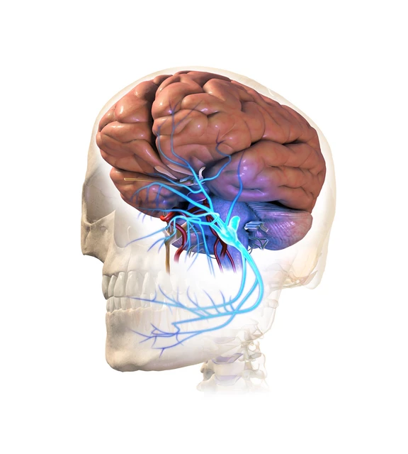
Where are your nerves located?
Your spinal cord is the beginning of several nerves, some of which originate in your brain. They extend throughout your body, especially in
- Arms, containing the ulnar, median, radial, and axillary nerves.
- The chest and belly, include the vagus and phrenic nerves.
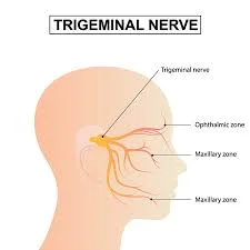
- The facial region is home to the trigeminal, optic, and facial nerves.
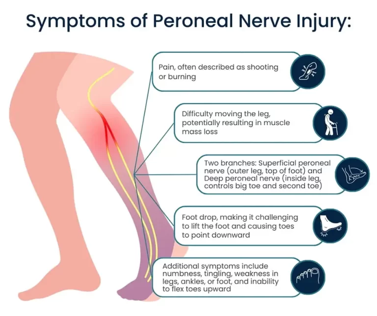
- The sciatic, femoral, tibial, obturator, and sural nerves are located in the legs.
- The pelvis, containing the Pudendal Nerve
The function of the nerve
Nerves carry electrical information from one region of the body to another. These impulses regulate you.
- A voluntary movement.
- Sensory functions such as excitement, pain, temperature, vibration, hearing, balance, taste, smell, and vision.
- Blood pressure.
- Breathing.
- Digestion.
- Heart rate.
- Stress reaction.
Your nerves allow the two sections of your nervous system to communicate with one another.
- The peripheral nervous system connects nerves throughout the body to the spinal cord, which is part of the central nervous system.
- The brain and spinal cord are the elements of the central nervous system. It receives and analyzes nerve signals from the peripheral nervous system. These signals, or inputs, are combined by your brain to control all of your actions, including your movements, emotions, behaviors, and thoughts. Some reactions are reflexive, occurring below the level of consciousness, such as removing your hand from the flames of the stove.
What is the Nerve injury?
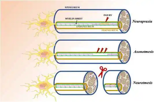
Classifying nerve injury helps predict prognosis and treatment options. Seddon first described the categorization in 1943, followed by Sunderland in 1951. Neurapraxia occurs when the nerve remains intact but the signaling ability is compromised. In the second degree, axonotmesis occurs when the axon is injured but the connective tissue remains intact. Neurotmesis is the final degree of damage, affecting both the axon and connective tissue.
Seddon’s Classification
Neurapraxia (Class I)
Neurapraxia is a transient interruption of conduction that does not result in loss of axonal continuity. Neurapraxia is a physiological blockage of nerve conduction in the affected axons.
Other characteristics:
- The mildest sort of nerve damage.
- Sensory-motor disorders present distal to the site of damage.
- The endoneurium, perineum, and epineurium are all intact.
- Wallerian degeneration is not present.
- The distal and proximal portions have intact conductivity, but there is no conduction across the damage.
- Full nerve conduction recovery takes days to weeks.
- Absence of fibrillation potentials (FP) and positive, sharp ECG waves.
Causes: This type of injury is generally caused by compression or mild trauma that affects nerve conduction but does not sever or significantly injure the nerve fibers. Common reasons include repetitive stress injuries or direct nerve pressure, which can result from a prolonged position or trauma.
Symptoms: Symptoms usually include temporary numbness, tingling, or weakening in the area served by the damaged nerve. For example, in cases of carpal tunnel syndrome, patients may have finger numbness due to median nerve compression.
Axonotmesis (Class II)
Axonotmesis results in the loss of the myelin sheath and axon continuity, although the connective tissue framework (encapsulating tissue, epineurium, and perineum) is preserved.
Other characteristics:
- Distal Wallerian degeneration.
- There are distal sensory and motor impairments.
- Nerve conduction distal to the injury site is nonexistent (3 to 4 days after the damage).
- Fibrillation potentials (FP) and positive, sharp EMG waves (2 to 3 weeks after the injury).
- Surgery is not usually necessary for axonal regeneration and healing, but it may be needed for scar tissue.
Causes: This injury is frequently caused by more severe trauma or crush injuries, which injure the axon while leaving the nerve’s physical structure intact. Axonotmesis can occur as a result of a deep laceration or a severe contusion.
Symptoms: Axonotmesis symptoms are comparable to neurapraxia, however they are more severe and persistent. These can contain significant motor and sensory deficits in the nerve-innervated area.
Neurotmesis (Class III)
Neurotmesis is the entire severance or damage of a nerve fiber. Axon, endoneurium, perineurium, and epineurium are all connected. Neurotmesis can be partial or total.
Other characteristics:
- Distal Wallerian degeneration.
- Connective tissue lesions can be partial or full.
- Severe sensory-motor issues and an autonomic function impairment.
- Nerve conduction distal to the site of injury is not present (3 to 4 days after the lesion).
- There is no distal conduction (EMG or NCV, nerve conduction velocity).
- Surgical intervention is required to restore function.
Causes: Neurotmesis can be caused by high-energy trauma, such as a car accident or serious cuts. Complete nerve rupture demands surgical intervention to heal and restore function.
Symptoms: Symptoms include complete loss of motor and sensory function along the damaged nerve’s spread. Patients may experience entire paralysis of the nerve-innervated muscles as well as substantial sensory impairments.
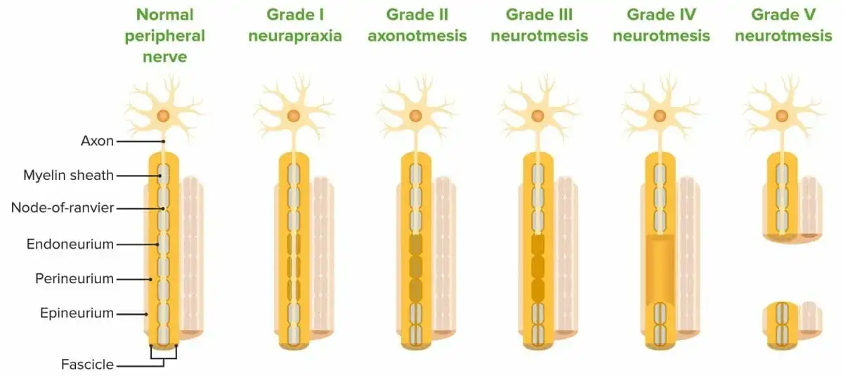
Sunderland’s classification
In 1951, Sunderland increased Seddon’s categorization to five degrees. The first two are identical to Seddon’s. Seddon’s axonogenesis is connected with Sunderland’s third and fourth grades. Sunderland’s third degree is nerve fiber disruption. The epineurium as well as perineurium is undamaged, yet, there is an endoneurium defect.
- Grade I: Neurapraxia – Similar to Seddon’s definition, it causes transient loss of function due to myelin sheath injury but no axonal damage. Full recovery is expected.
- Grade II: Axonotmesis – Axonal rupture with preservation of the connective tissue framework. Nerve fibers can regenerate, leading to eventual recovery, even if this may be partial.
- Grade III: Axonotmesis with Endoneurial Disruption – Damage has spread to the endoneurium. Recovery may be more difficult than in Grade II.
- Grade IV: Axonotmesis with Perineurial Disruption – Causes damage to the perineurium. Recovery is possible, although it may be unpredictable.
- Grade V: Neurotmesis – The nerves, including the endoneurium, perineurium, and epineurium (the outermost layer of connective tissue), are completely severed. Surgical intervention is frequently required, and the outcomes vary greatly.
What illnesses and disorders affect the nervous system?
Certain disorders have an impact on how well your nerves transmit messages. You may develop a neurological disorder if damage or injury disrupts nerve signals.
Common nerve-related conditions include:
- Peripheral neuropathy refers to nerve damage in the peripheral nervous system, separate from the brain and spinal cord.
- Sciatica is discomfort caused by the nerve roots in the lower part of the back either the sciatic nerve, which runs along the backs of both legs.
Clinical significances
Understanding the nature and severity of nerve damage is critical for establishing the best treatment and prognosis. Nerve injuries can have serious consequences, impacting motor and sensory functioning as well as one’s general quality of life.
Symptoms:
- Depending on the nerve involved (e.g., median, ulnar, or radial), symptoms may include pain, numbness, tingling, weakness, or loss of hand and wrist function.
Diagnosis:
Diagnosis usually involves a combination of clinical examination and diagnostic tests. Neurological examinations, electromyography (EMG), and nerve conduction studies (NCS) assist in determining the amount and form of the injury. Imaging techniques such as MRI and ultrasound can be used to detect structural damage.
Treatment:
Treatment techniques are different depending on the degree of the injury.
- Conservative treatment for neurapraxia, such as rest, immobilization, and physical therapy, is often sufficient. It is possible to manage pain and inflammation with medication.
- Axonotmesis may necessitate more intense rehabilitation, including physical therapy, to improve nerve function and strength. In some circumstances, surgical intervention may be required if recovery is partial or delayed.
- Neurotmesis: Surgical repair is usually required. Direct suturing, nerve grafting, and nerve transfer are some of the techniques used. Post-surgical rehabilitation is crucial for maximizing functional recovery.
Pain Management
- This includes both pharmaceutical and non-pharmacological techniques to manage pain and discomfort caused by nerve damage.
- Rest, immobilization, and physical therapy are frequently adequate forms of conservative treatment. Medication is a useful tool for treating pain and inflammation.
Physical Therapy
One essential element of preventing nerve injury is physical therapy. It aims to restore function, relieve pain, and improve overall quality of life.
- Range of Motion Exercise: Exercises may aim to improve the range of motion and avoid muscular atrophy.
- Strengthening Exercise: Aims to regain strength, flexibility, and coordination.
- Electrical stimulation (ES) is the considerable often used nonsurgical treatment. Most research used low-frequency ES to enhance nerve regeneration. Regardless, the mechanism and frequency range of ES requires to be standardized because high-frequency ES can cause nerve damage.
Prognosis
- The prognosis for nerve injuries is determined by various factors, including the nature and severity of the damage, the timing and efficacy of treatment, and individual patient characteristics. Early intervention often results in better outcomes, with many patients seeing considerable improvements with the right therapy.
How do you maintain your nerves healthy?
- To maintain a healthy nervous system, adopt healthy habits such as avoiding tobacco and quitting smoking.
- A nutritious diet includes entire grains, healthy fats, lean protein, fruits, and vegetables.
- Limit your alcohol intake.
- Manage nerve-related health issues, including diabetes.
- Reduce stress with healthy coping skills like meditation and exercise.
- Grab around seven to eight hours of sleep per night.
- Stay hydrated by consuming plenty of fluids.
Summary
Nerve injuries, particularly those affecting the wrist and upper extremities, are difficult to diagnose, treat, and recover from. Understanding the different types of nerve injuries and their impact on life is critical for successful care and recovery. With advances in medical science and technology, there is the possibility of better results and a higher quality of life for people suffering from these disorders.
FAQs
What are the three types of nerve injuries?
Classification of nerve injuries. Seddon2 divided nerve damage into three major categories: neurapraxia, axonotmesis, and neurotmesis.
What are the three types of nerves?
The body’s nerves are classified into three types: Autonomic nerves, Motor nerves, and Sensory nerves.
Is it possible to repair nerves?
Nerve cells can repair and grow again at a rate of roughly an inch per month, although the process is often imperfect and delayed. This is an entire nerve injury, meaning there has been a rupture in the nerve sheath and underlying neurons. If there is an open cut, a neurosurgeon can see and repair the damaged nerve endings during surgery.
What is the best vitamin for nerve repair?
B vitamins, including B12, B6, B3, and B1, are needed for nerve function. These vitamins, known as ‘neurotropic’ vitamins, can aid in nerve repair and relieve symptoms such as numbness and tingling.
Is it possible to remove damaged nerves?
If a portion of an injured nerve is fully cut or destroyed beyond repair. A surgeon can remove the damaged part and reconnect the healthy nerve endings. This is referred to as nerve repair. As an alternative, to close the gap between the nerves, the surgeon could insert a segment of the nerve from another area of the body.
Can damage to the nerves be repaired by exercise?
Treatment approaches often center on relieving pain and addressing the underlying problem. However, research indicates that exercise can successfully keep their nerves functioning and promote nerve regeneration.
What is the quickest way to heal from nerve damage?
If your nerve is simply damaged, it may heal on its own without the need for surgery. Nerves heal slowly, sometimes taking several months. Nonsurgical treatment options for minor nerve injuries include medication, physical therapy, and massage therapy. Peripheral nerve surgery can rebuild or repair damaged nerves.
References:
- Classification. (n.d.). https://nerveclinic.co.uk/nerve-injuries/classification
- Moscony, A. M. (2014). Peripheral Nerve Problems. Elsevier eBooks, 272–311. https://doi.org/10.1016/b978-0-323-09104-6.00024-9
- Murphy, A., & Goel, A. (2016). Nerve injury classification. Radiopaedia.org. https://doi.org/10.53347/rid-47058
- Nerve injury classification. (2024, April 28). Wikipedia. https://en.wikipedia.org/wiki/Nerve_injury_classification
- Professional, C. C. M. (n.d.). Nerves. Cleveland Clinic. https://my.clevelandclinic.org/health/body/22584-nerves