Patellar Dislocation
What is a Patellar Dislocation?
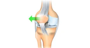
Patellar dislocation is caused by a lateral movement of the patella, which leaves the trochlea groove of the femoral condyle. This usually results in a rupture of the medial patellofemoral ligament.
Patellar (kneecap) dislocations are common, particularly in younger athletes, and the majority of them occur laterally (outside). When this occurs, they are accompanied by severe pain and swelling. Following a patellar dislocation, the initial step is to reposition the kneecap in the trochlear groove.
This frequently occurs spontaneously when the individual extends the knee, either on the field of play or in an emergency department or training room as the knee is stretched for assessment. When relocation occurs before examination, its occurrence must be solved by discovering related issues.
What is the Patellar Dislocation?
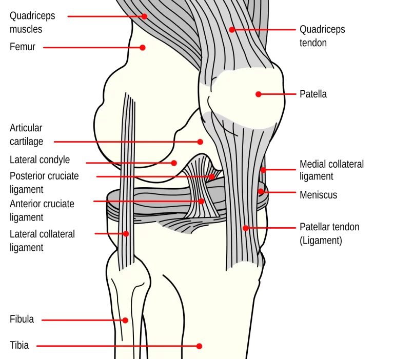
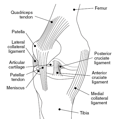
- A patella dislocation is the separation of the kneecap (the patella) from its groove at the knee joint. The knee joint connects three bones: the thighbone, the shinbone, and the kneecap in the center.
- When you bend and straighten your leg, the kneecap typically moves up and down a vertical groove between the bottom end of the thighbone and the top end of the shinbone (the trochlear groove). A network of tendons and ligaments holds the kneecap in place inside the groove, allowing it to flex while moving.
- When the patella dislocates, it is pushed out of the trochlear groove and cannot move up and down. This locks the knee and forces the ligaments out of position, frequently ripping them. Most of the time (93%), the kneecap pops out laterally, to the side of the groove.
- Patellar dislocation is often an acute injury induced by a collision or a sharp spin and twist. It is painful and debilitating, just like any other dislocation, unless repaired. However, the displaced kneecap can occasionally fix itself.
Anatomy of The Patellofemoral Joint
The patellofemoral joint forms part of the knee joint. The patellofemoral joint, sometimes called the kneecap joint, is a complicated and important structure in the knee.
It is generated when the posterior side of the patella (kneecap), the body’s biggest sesamoid bone, articulates with the trochlear groove of the distal femur (lower end of the thighbone).
Here’s an overview of the main anatomical aspects:
Bones:
Patella: A triangular-shaped bone with a smooth, articular surface on the posterior side.
The trochlear groove on the distal anterior femur creates a shallow groove for the patella to glide in.
Cartilage:
Articular cartilage forms a smooth, low-friction contact between the patella and femur, facilitating joint mobility.
Ligaments:
- The medial patellofemoral ligament (MPFL) is the major stabilizer of the patella, preventing lateral movement.
- The lateral patellofemoral ligament (LPFL) provides additional stability against medial movement.
- The quadriceps tendon connects the quadriceps muscle to the patella, delivering force during knee extension.
- The patellar tendon connects the patella to the tibia (shinbone), conveying force during knee extension.
Other structures:
- The retinaculum is a sleeve of tissue that encircles the patella and serves to maintain it in place.
The synovial membrane lines the joint capsule and generates synovial fluid, which lubricates the joint. - The patellofemoral joint permits the patella to glide and track along the trochlear groove during knee flexion and extension.
- This gliding action helps disperse stresses throughout the joint and improves quadriceps muscle activity during knee extension.
Function
The patellofemoral joint is essential for several tasks, including:
- Knee extension is primarily driven by the quadriceps muscle, which acts through the patella and patellar tendon.
- Joint stability: The patella and its ligaments serve to stabilize the knee joint and prevent abnormal patellar movement.
- Joint protection: The articular cartilage absorbs stress and distributes forces throughout the joint, cushioning the bone beneath.
Causes of the Patellar Dislocation
Causes of patellar dislocation may include:
- A direct hit on the knee
- An abrupt rotation or twist of the knee.
- Participating in activities that require plenty of turning or leaping, such as basketball, volleyball, or skiing.
- Having loose ligaments or other anatomical problems in your knee.
Types of the Patellar Dislocation
There are two major forms of patellar dislocations:
- Acute dislocation is the most frequent variety, and it happens when the kneecap is rapidly pulled out of position, usually as a result of a fall, twist, or direct hit to the knee.
- Chronic dislocation occurs when the kneecap frequently dislocates, which is generally caused by underlying structural issues in the knee joint.
What’s the difference between patella dislocation and subluxation?
Some people may believe they have a patella dislocation when they have a patella subluxation. A subluxation is a partial dislocation. It indicates that the bone is unstable in the joint and may have moved slightly out of its correct position, but it has not popped out. When you have a patella subluxation, your kneecap remains in the groove and you may walk, but it may feel unpleasant or unsteady, and you may hear a popping noise when it moves. Patellar subluxation can be caused by an injury or general joint looseness (patellar instability).
What’s the difference between a patellar dislocation and a knee dislocation?
A patella dislocation is the dislocation of the kneecap. A “dislocated knee” refers to the other two bones that make up the knee joint: the femur (thighbone) and the tibia (shinbone). When your knee is dislocated, the femur and tibia do not connect at the knee joint. One of the bones has been pushed back or forward compared to the other. A dislocated knee (tibiofemoral dislocation) is less common and more harmful than a dislocated kneecap due to the force necessary to misalign the leg bones and the ligament damage it causes.
Symptoms of the Patellar Dislocation
Symptoms of patellar dislocation may include
- Severe discomfort in the knee.
- An audible pop.
- Swelling and bruises around the knee.
- Having difficulty bending or straightening the knee(Locking of the knee.)
- Inability to walk.
- The kneecap feels out of position.
- Numbness or tingling in your knee. Buckling of the knee
- Bruising on the kneecap.
Who is affected by the Patellar Dislocation?
Anyone can dislocate their patella due to injury. However, certain people are more vulnerable, including:
- Athletes, particularly in high-impact sports.
- Dancers who do rapid pivots.
- Teenagers have looser joints and ligaments due to their continual growth.
- Women’s larger hips and looser ligaments cause additional lateral stress on the knee.
- Big and tall guys, whose joints are under additional stress.
- People with patellar instability, especially if they have previously dislocated their patella.
Doctors are unsure what causes congenital patella dislocation, but the increased occurrence among family members implies a hereditary relationship.
Other congenital diseases connected with the following conditions:
- Larson Syndrome.
- Arthrogryposis.
- Diastrophic Dysplasia.
- Nail-patella Syndrome.
- Down syndrome.
- Ellis-van Creveld syndrome.
Diagnosis of the Patellar Dislocation
Medical history and Physical examination
During the medical history and physical examination, the doctor will inquire about your symptoms, the cause of the injury, and any prior knee issues.
They will next do a physical assessment of your knee, looking for:
- Swelling and bruises
- Tenderness
- Deformity (kneecap may appear out of place)
- Range of motion (difficulty bending and straightening the knee)
- Instability (the patella may feel lax or easily displaced)
Imaging tests:
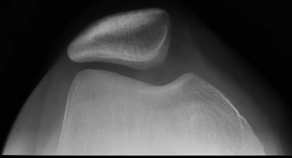
- X-rays are often performed to confirm dislocation and rule out fractures.
- Other imaging examinations, such as An MRI scan produce comprehensive pictures of soft tissues such as ligaments and cartilage, which aids in the identification of ligament rips or cartilage damage.
- CT scans provide a more thorough image of the bones, particularly if X-rays are uncertain.
Special Test:
The doctor may do specialized tests to evaluate patellar stability, including:
- The patellar apprehension test: In this test gently apply pressure to the kneecap to examine if it dislocates easily.
- Tilt test: Detects unusual patellar tracking during knee flexion.
Differential Diagnosis of Patellar Dislocation
The doctor might be required to check out other medical conditions with comparable symptoms, such as:
- Meniscus tear.
- ligament sprain.
- Osteochondral fracture.
- Patellofemoral Pain Syndrome.
Treatment of the Patellar Dislocation
If you have dislocated your patella, you should seek medical assistance immediately. A doctor will be able to reposition the kneecap and offer therapy to avoid further dislocation. Treatment for a patellar dislocation might include:
- Manual reduction: The doctor carefully moves the kneecap back into its groove.
Immobilization: The knee will be immobilized in a splint or brace for a while to allow the healing process to start. - Rehabilitation: For the first several days of rehabilitation, you will be given pain relievers and a splint to wear. Periodically elevating and cooling the joint might help reduce swelling. You’ll start walking again gradually, using crutches and a brace to keep the joint stable.
- Physical Therapy: Physical treatment is essential for strengthening the muscles and reducing the range of motion until the joint is stabilized. A displaced patella often takes six weeks to three months to fully heal.
- Surgery: In some circumstances, surgery may be required to repair ligament damage or other structures in the knee. surgency may also be required if you have recurring patellar dislocations or persistent patellar instability. Repairing and strengthening the cartilage and ligaments is a prophylactic approach to stabilize the knee. Patellar dislocation is congenital, and the joint can only be corrected surgically.
Strengthening Exercises to Prevent Patellar Dislocation
- Quadriceps strengthening: These are the primary muscles that extend your knee and stabilize the patella. Squats, lunges, leg extensions, and stair climbing are all exercises that can be beneficial.
- Hamstring strengthening: Strong hamstrings serve to balance the quadriceps and avoid excessive patellar tracking. Exercises like hamstring curls and bridges are good.
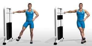
- Strengthening both the inner and outer thighs: These muscles aid in controlling the medial and lateral mobility of the kneecap. Exercises like side leg lifts, hip abduction/adduction, and clamshells are suggested.
Squats
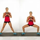
- Wall squats: Stand with your back to a wall and your feet shoulder-width apart. Slowly slide down the wall as if you were going to sit in a chair, maintaining your back straight and your core active. Return to your starting position by sliding up.
- Chair squats: Stand in front of a chair and gently lower yourself until your glutes just touch the chair. Then rise back up to your starting posture.
- Single-leg squats: Stand on one leg and gently lower yourself down as if you were about to sit in a chair. Elevate your other leg straight in front of you.
- Return to the First position, then repeat with the opposite leg.
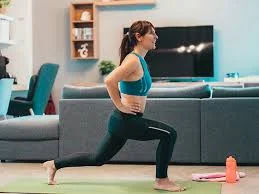
Lunges
- Put your hands on your forward knee. Maintain a comfortable posture with your shoulders down, hips even, chest open, and gaze straight ahead.
- Press your hands down and press your hips forward until you feel a stretch on your left side from the front of your hip, groin, and thigh.
- For around twenty to thirty seconds, hold the stretch.
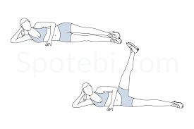
Leg Extensions
- Holding onto a wall or chair for balance, stand on one leg.
- With your toes pointing front, straighten and extend the other leg back.
- At the peak of the action, hold for a brief moment before lowering your leg back down gradually.
- After doing the required number of repetitions, swap legs.
Hamstring Curls
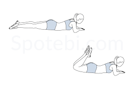
- With your hands flat on the ground beneath your shoulders or grabbed behind your back, assume a prone position.
- Stretch your legs hip-width apart and point your toes upward with your feet flexed.
- Squeeze your belly button against your spine and bring your hips down a little towards the floor to activate your core and glutes.
- With your lower back flat and your core tight, raise your legs off the floor until they are straight.
- At the peak of the exercise, tighten your hamstrings and hold the position for a moment.
- With control, slowly return your legs to the beginning position. Continue for however many times you wish.
Bridges
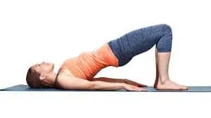
- Lay flat on your back, feet hip-width apart, knees bent.
- Pull your belly button in the direction of your spine to activate your core.
- Raise your hips off the floor by applying pressure with your heels until your body forms a straight line from your shoulders to your knees.
- After a few moments of gluteal contraction, carefully bring your hips back down. Continue for the appropriate amount of times.
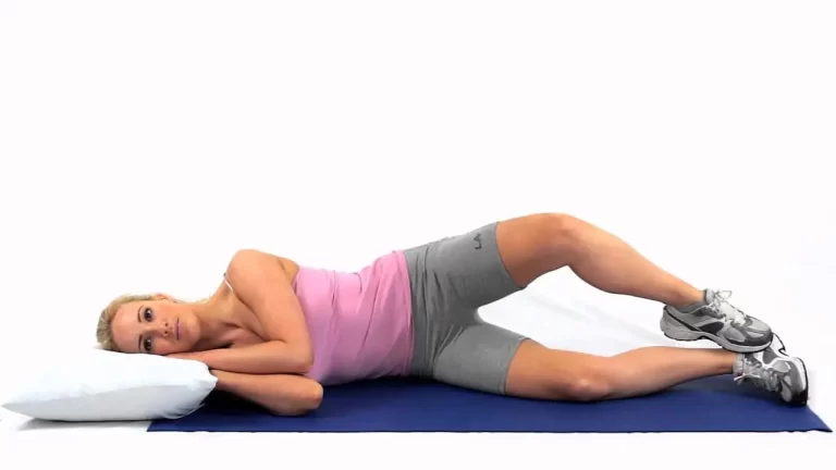
Side leg lifts
- Stretch and stack your legs as you lie on your side.
- As high as you can raise your upper leg off the ground, then gradually bring it back down.
- Continue with the opposite leg.
Hip adduction
- Stand on one leg and lift your other leg out to the side while maintaining it straight.
- Hold for a few seconds before slowly lowering it back down. Repeat on the opposite side.
- This workout tests your balance and engages your inner thighs.
Clamshells
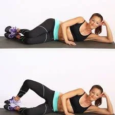
- Bend and stack your knees while lying on your side.
- Feel the engagement in your inner thigh as you raise your upper knee off the ground while maintaining your feet together.
- On the other side, repeat (10–15 repetitions on each side, two–three sets).
Stair Climbing
- Maintain a straight back, engaged core, and relaxed shoulders.
- Instead of relying on momentum, propel your steps using your leg muscles.
- To be balanced and attentive, avoid gazing at your feet.
- Take slower, more deliberate steps down to reduce the impact on your knees.
Prevention of the Patellar Dislocation
- Maintaining a healthy weight can lessen stress on your knee joints.
- Warm up thoroughly before engaging in physical exercise and cool down afterward.
- Avoid activities that repeatedly place your knee in susceptible situations.
- Listen to your body and take pauses when you feel pain or discomfort.
- For those who have previously dislocated their kneecap, physical treatment can help recover strength, flexibility, and normal movement patterns, reducing the chance of recurrence.
- A healthcare practitioner can provide personalized recommendations based on your specific risk factors and preventative tactics.
- For high-risk activities, wear a knee brace to support and stabilize the patella.
- Proper athletic technique can reduce stress on the knee joint. Consult a coach or trainer for advice.
- Exercises that stress balance and proprioception can improve reaction time and avoid knee twisting. Yoga, tai chi, or single-leg balancing exercises might be beneficial.
- Strengthening exercises include the quadriceps, which help expand and stabilize the kneecap.
- Strengthening the hamstrings helps to balance the quadriceps and prevents excessive patellar tracking.
- Strengthening the inner and outer thighs: These muscles assist regulate the kneecap’s medial and lateral movements.
Summary
A dislocated patella can be frightening and painful, but it is less dangerous than other dislocation injuries. The patella dislocates with less power than other bones, reducing the risk of collateral injury to blood vessels or nerves. It also moves more readily, sometimes by itself.
If your kneecap dislocates, the first thing to consider is reattaching it. the qualified expert can fix it on-site, and administer medicine to alleviate the discomfort. Following that, rest and therapy should have you back on your knee within six weeks.
FAQs
How do you treat a dislocated patella?
A displaced kneecap requires immediate emergency attention. After administering pain medication, the doctor or other health care professional will carefully slip the kneecap back into place while straightening the leg. This rapid maneuver is known as a reduction. Then they will order an X-ray to look for fractures.
Is patellar dislocation serious?
Kneecap dislocation harms the knee joint. It can cause cartilage damage and raise the chance of getting osteoarthritis at a young age. Repeated dislocations exacerbate the problem and make it more difficult to cure
How long does it take to heal from a patella dislocation?
How long does it take to heal from a patella dislocation?
A dislocated kneecap can take 6 to 8 weeks to fully recover, although you should be able to walk normally within a few days. You may be given a knee brace to wear for two weeks to help it mend. If walking is unpleasant, a crutch can assist.
Is surgery necessary for patellar dislocation?
If you dislocate your kneecap for the first time, conservative therapy is usually recommended. If it occurs again or repeatedly, surgery is typically recommended.
Should I bend my knee after having a patellar dislocation?
The majority of patients have considerable pain reduction when they perform muscle-strengthening exercises and avoid activities that bend the knee beyond 90 degrees. Physical therapy can help train the muscles around the kneecap to pull uniformly in all directions, allowing your kneecap to remain in proper alignment.
References
- Professional, C. C. M. (n.d.-j). Patellar dislocations. Cleveland Clinic. https://my.clevelandclinic.org/health/diseases/21633-patellar-dislocations
- Patellar dislocation. (n.d.). Physiopedia. https://www.physio-pedia.com/Patellar_dislocation
- Hayat, Z. (2023, July 4). Patella Dislocation. StatPearls – NCBI Bookshelf. https://www.ncbi.nlm.nih.gov/books/NBK538288/
- Knee dislocation and instability in children – OrthoInfo – AAOS. (n.d.). https://orthoinfo.aaos.org/en/diseases–conditions/patellar-dislocation-and-instability-in-children-unstable-kneecap/
- Aglietti, P., Ciardullo, A., & Cuomo, P. (2006b). Patellofemoral dysfunction. In Elsevier eBooks (pp. 155–180). https://doi.org/10.1016/b978-0-7216-0331-5.50021-5