Blog

  • Thenar Muscle Pain

    Thenar Muscle Pain

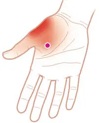
    Thenar muscle pain refers to discomfort or soreness in the muscles of the thenar eminence, the fleshy area at the base of the thumb. It can result from repetitive strain, overuse, poor ergonomics, or conditions like carpal tunnel syndrome.

    Symptoms may include aching, weakness, or difficulty gripping objects. Proper rest, stretching, strengthening exercises, and ergonomic adjustments can help alleviate pain and prevent further strain.

    What is a Thenar Muscle Pain?

    Pain in the thenar prominence, a soft region near the base of the thumb, is referred to as “thenar muscle pain.” The thenar muscles are subject to strain, overuse, and nerve-related problems since they are essential for thumb movement and grip strength. Injuries, underlying medical disorders like arthritis, repeated activities, and nerve compression (as in carpal tunnel syndrome) can all cause this kind of pain. Maintaining hand function and avoiding increasing symptoms require accurate diagnosis and treatment.

    The thumb’s mobility and grip strength are controlled by the thenar muscles, a trio of muscles at the thumb’s base. Several factors, such as overuse, strain, nerve compression, or underlying medical disorders, can produce the soreness, hurting, or pain that is commonly referred to as “thenar muscle pain.”

    The soft, fleshy spot at the base of your thumb is known as your thenar eminence. Your thumb is opposable thanks to these four muscles. In other words, they enable your thumb to grasp and retain little items like a spoon, sewing needle, or pencil. You can also handle heavy bags, grasp and turn a doorknob, and text on your phone with an opposable thumb.

    Many of your everyday activities are done using your thumb. These repetitive movements have the potential to overstress the muscles controlling your thumb over time, leading to pain and inflammation. To find out how to diagnose, treat, and prevent thenar eminence pain, continue reading.

    What is the thenar muscle?

    • Both hands have the thenar eminence, which is often called the “thumb pad.” The abductor pollicis brevis, flexor pollicis brevis, and opponens pollicis are the three muscles that make up the thenar eminence.
    • The thumb joint is stabilized by the tension in all three muscles; however, prolonged active tension in any one of these muscles can result in thenar eminence muscular pain.

    Common Causes of Thenar Muscle Pain:

    Overuse & Repetitive Motion: Prolonged holding of things, texting, or typing can all cause the thenar muscles to become strained.

    • Carpal Tunnel Syndrome (CTS): Pain, weakness, and tingling in the thenar area can result from compression of the median nerve.
    • Trauma or Injury: Excessive force, falls, or direct collision can all result in strained or damaged muscles.
    • Arthritis: The thumb joint may be affected by osteoarthritis or rheumatoid arthritis, which can cause pain in the thenar region.
    • De Quervain’s Tenosynovitis: Pain that radiates to the thenar muscles can be caused by inflammation of the tendons close to the thumb.
    • Nerve Disorders: Pain and weakness may be caused by conditions that impact the nerves, such as peripheral neuropathy.

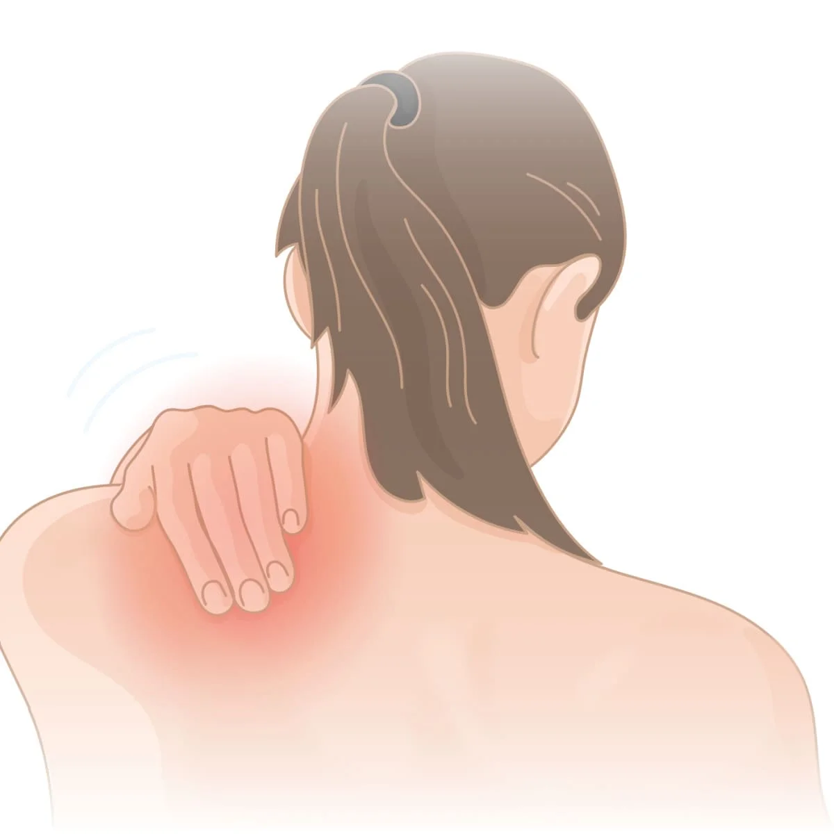
    Symptoms of Thenar Muscle Pain:

    • Aching, soreness, or a sharp ache at the base of the thumb are examples of pain and tenderness.
    • Weak Grip Strength: The inability to hold or grip something securely.
    • Swelling or Inflammation: The thenar area may appear swollen or sensitive.
    • Pins-and-needles sensations, such as numbness or tingling, are frequently caused by nerve involvement (e.g., carpal tunnel syndrome).
    • Thumb Movement Issues: Having trouble grasping, pinching, or using fine motor skills.
    • Thumb mobility is restricted due to stiffness or limited range of motion, particularly after rest.
    • pain that radiates past the thenar eminence to the hand or wrist.
    • For an accurate diagnosis and course of therapy, it is advised to seek a medical assessment if these symptoms intensify or continue.

    Diagnosis of Thenar Muscle Pain

    A combination of a physical examination, medical history, and diagnostic tests are used to identify the underlying reason for thenar muscle pain.

    Medical History & Symptom Assessment

    • The length, severity, and type of the pain
    • Any past medical issues, trauma, or repeated hand movements (e.g., arthritis, carpal tunnel syndrome)
    • Related symptoms such as weakness, tingling, or numbness

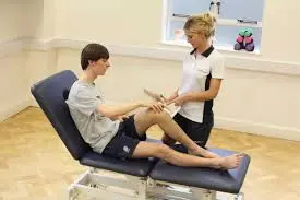
    Physical Examination

    • Palpation: Examining the thenar area for soreness, edema, or muscular atrophy
    • The Range of Motion (ROM) Test measures the stiffness and mobility of the thumb. The Grip & Pinch Strength Test evaluates the thumb and hand function.
    • Examining for nerve compression, such as carpal tunnel syndrome, using Tinel’s Sign and Phalen’s Test

    Diagnostic Tests

    • Electromyography (EMG) and nerve conduction studies (NCS) are used to evaluate nerve function and identify disorders such as carpal tunnel syndrome.
    • X-ray – Recognizes problems with bones, like arthritis or fractures
    • Examine soft tissues, muscles, and tendons for signs of inflammation, rips, or nerve entrapment using ultrasound and magnetic resonance imaging.
    • Blood tests are used to look for autoimmune or inflammatory diseases such as rheumatoid arthritis.
    • A correct diagnosis aids in choosing the most effective course of treatment, guaranteeing efficient pain control and recuperation. It is imperative to seek medical attention if symptoms intensify or continue.

    Treatment of thenar muscle pain:

    It normally gets better if you can quit the activity that’s generating the pain and irritation. Because it is a work activity, this is frequently not feasible. You might not want to give it up if it’s because of a sport or pastime.

    Even if the offending behavior is not entirely stopped, medical interventions and home remedies can be beneficial. Usually, the best results come from combining the two groups.

    Medical Treatment:

    Therar eminence pain is frequently treated with a thumb splint. Your thumb becomes immobile as a result, preventing misuse of the muscles. Your muscles will have more time to recover and the pain will be lessened.

    If the splint affects your ability to do your job, you may not be able to wear it all the time, but you should wear it whenever you can.

    Additional medical interventions consist of:

    • Using kinesiology tape to immobilize your thumb and nonsteroidal anti-inflammatory medications like naproxen and ibuprofen
    • injections of steroids
    • Dry needling, acupressure, or acupuncture
    • Apply cold therapy for more recent pain and warm therapy for more chronic pain. Ice the region for ten minutes, three to four times a day.
    • Stretch your hands and thumbs and massage the area.

    Home remedies for thenar muscle pain:
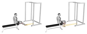
    Rest

    Avoid spending a lot of time on activities that cause your thenar eminence pain, such as sewing, weeding, or texting. To prevent these prolonged postures that are aggravating your pain, take rests in between tasks. For chores like texting, you might try switching hands.

    Heat therapy

    Put some heat on your thenar eminence. You can also soak your hands in a warm water bath or wash them in warm water. The heat promotes blood circulation in the area and relaxes your muscles, facilitating recovery. To prevent irritating the skin or giving this area superficial burns, use caution while applying excessive heat. Applying heat for 10 to 15 minutes at a time is often advised.
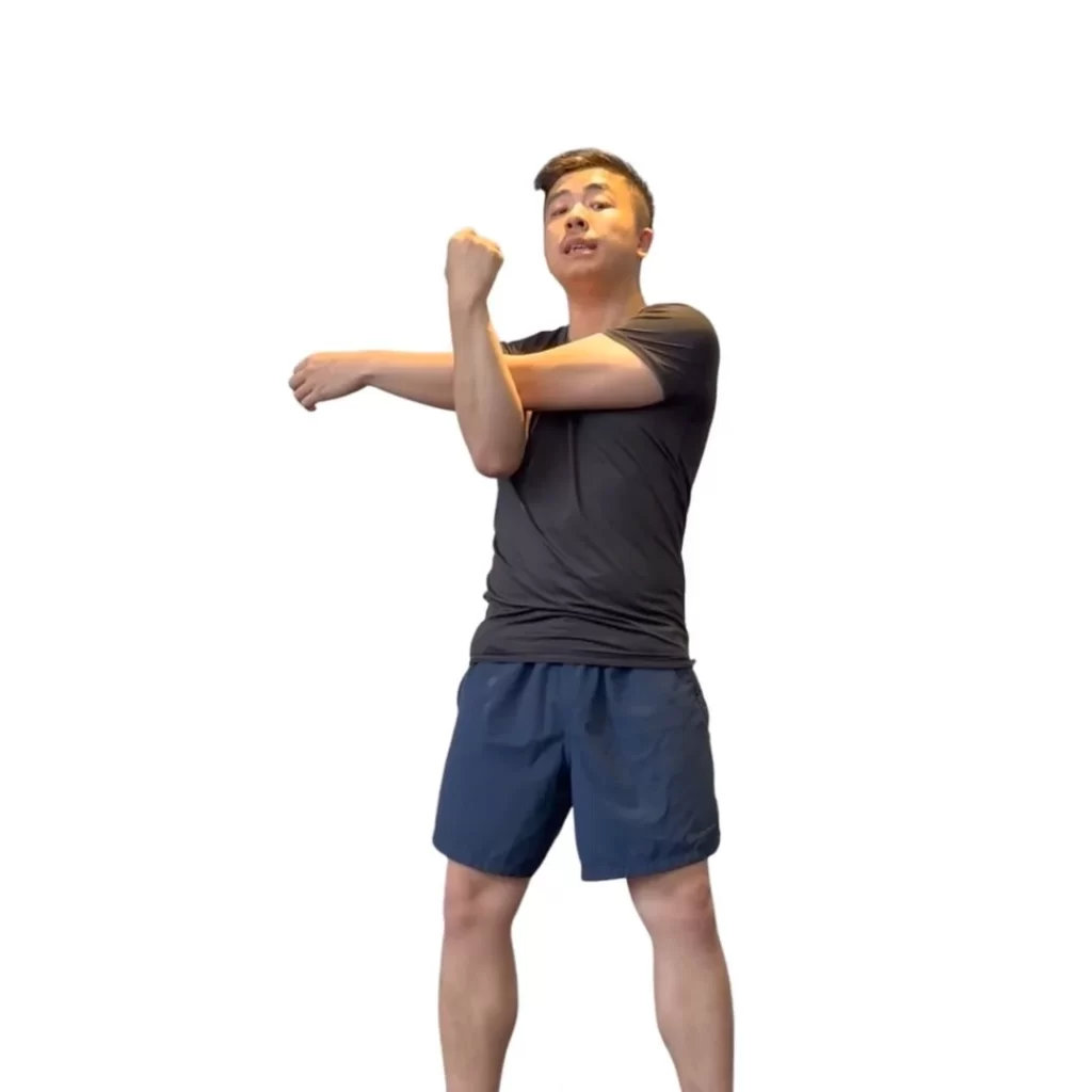
    Stretches

    Press your palms down and spread your thumb and index finger as widely apart as you can against a level surface. A diamond (or L and reverse L) should form between your two hands as a result of this stretch. You may also perform this stretch at the bottom of a warm water sink. The muscles will further relax if you use warm water.

    With your palm facing up, use your other hand to grasp your thumb and gradually drag it to the ground. After 30 seconds, switch to the other hand and hold this stretch.

    How to prevent thenar muscle pain:

    • Avoiding repetitive thumb movement activities is the greatest approach to prevent thenar eminence pain from developing or returning.
    • Sometimes you are unable to cease these actions because they are essential for your job or because you wish to carry on with the activity that is causing them. In this situation, you should relax the muscles that control your thumb by taking frequent rests.
    • There are other ways to complete the task that don’t need to use your thumb excessively.
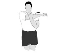
    • Another way to keep your hand and thumb muscles from being stiff is to stretch them. The following stretches are beneficial for your thenar eminence:
    • Spread your other fingers apart and gently slide your thumb back toward your forearm.
      Keeping your thumb and index finger as far apart as possible, press your palm down against a level surface.
      With your palm facing up, place your hand on a level surface. Then, use your elbow to gently lean into your thenar eminence and move it around the area.

    FAQs

    What’s causing my thenar pain?

    Overuse syndrome, which is caused by repetitive thumb movements, is typically the cause of thenar eminence pain. Usually, a mix of home remedies and medical therapies helps it get well. Avoiding activities that involve repetitive thumb movements can help prevent thenar eminence soreness.

    What makes my thenar muscle so large?

    Pseudohypertrophy or genuine hypertrophy associated with spontaneous activity, increased effort of remaining fibers, and fiber stretching 2, 3 can cause thenar hypertrophy. The probability of real thenar hypertrophy is increased by spontaneous activity on needle EMG and chronic denervation.

    Why do thenar muscles deteriorate?

    The median nerve innervates the thenar muscles, which might exhibit atrophy in Carpel Tunnel Syndrome (CTS), if median nerve impairment occurs. Other CTS tests, such as the Tinel’s test and the Phalen’s sign, should be performed if indicated.

    How is the thenar stretched?

    The muscles will relax even more if you use warm water. Hold onto your thumb with your other hand and gradually pull it to the floor, palm facing up. After 30 seconds of holding this stretch, switch to the other hand.

    For thenar pain, what is the differential diagnosis?

    In conclusion, for baseball players who need manual power to hold a baseball and grip a bat, thenar pain can be a major functional constraint. When a baseball player experiences thenar pain, osteocartilaginous, ligamentous, vascular, and musculotendinous reasons are among the potential diagnoses.

    What is the function of the thenar muscles?

    The thumb’s intrinsic muscles are composed of the four thenar muscles. These consist of the flexor pollicis brevis, opponens pollicis, adductor pollicis, and abductor pollicis. These intrinsic muscles work together to provide thumb mobility.

    How is the thenar muscle massaged?

    Work your way outward from the palm’s center, beginning with the palmar fascia and moving layer by layer deeper until you reach the thenar eminence.

    How can the thenar muscle be stretched?

    Wrap your hand with a rubber band so that it rests at the base of your finger joints. Move your thumb as far away from your other fingers as you can with gentleness. After 30 to 60 seconds of holding this position, let go. With each hand, repeat ten to fifteen times.

    What is causing the thenar muscle pain?

    Overuse syndrome, which is caused by repetitive thumb movements, is typically the cause of thenar eminence pain. Usually, a mix of home remedies and medical therapies helps it get well. Avoiding activities that involve repetitive thumb movements can help prevent thenar eminence soreness.

    References

    • The Healthline Editorial Team. (2024, January 27). How to Diagnose, treat, and prevent thenar Eminence Pain. Healthline. https://www.healthline.com/health/thenar-eminence-pain
    • Posts, V. M. (2020, November 18). 4 Tips for thenar muscle pain relief. Urban Health HQ. https://urbanhealthhq.com.au/2020/11/18/4-tips-for-thenar-muscle-pain-relief/
    • Seladi-Schulman, J., PhD. (2019, May 28). Thenar Eminence overview. Healthline. https://www.healthline.com/health/thenar-eminence#nerves

  • Pectineus Muscle Pain

    Pectineus Muscle Pain

    Pectineus muscle pain is suggested when you have tingling, weakness, or numbness in the groin or leg along with groin or upper thigh pain.
    The pop/snapping sound is also audible with this muscular soreness.

    What is the pectineus muscle pain?

    Pectineus Muscle Pain occurs due to strain, overuse, or sudden movements affecting the upper inner thigh. It can cause groin discomfort, stiffness, and restricted hip mobility, often impacting activities like running, cycling, or kicking. Proper rest, stretching, and strengthening exercises help in recovery.

    • The flat, quadrangular pectineus muscle, which is situated in the center of the thigh, aids in flexing and moving the leg in the direction of the body.
    • Excruciating injuries can result from strains and pulls on the pectineus muscle caused by activities like running, skating, kicking a soccer ball, playing basketball, and other activities that use fatigued muscles.
    • The injury causes the patient’s legs to become weak and numb.
    • The rice concept, physical therapy, and medication are utilized to relieve this problem.

    Anatomy of the pectineus muscle?

    • The pectineal line of the pubic bone, commonly referred to as the pectin pubis, is where the pectineus muscle begins.
    • The pectineus muscle then integrates into the pectineal line of the thigh bone after continuing downhill and posterolaterally behind the side.
    • One of the five major muscle groups in the medial (middle) thigh is the pectineus, a hip adductor muscle.The obturator nerve innervates the pectineus muscle.
    • The pectineus muscle is responsible for flexion, adduction, and external rotation, which is the hip joint’s lateral movement.

    Cause of the pectineus muscle pain?

    • The pectineus muscle is harmed when one or both legs are overextended to the side and front of the body.
    • Injury to the pectineus muscle is caused by quick motions and repetitive exercises like running and jumping.
    • weak muscles in the adductors
    • If you alter your course too rapidly while running
    • when you spend too much time sitting with one leg crossed.
    • in the event that a history of groin or hip injuries exists.

    The effect of this pain is strain and pull in the groin:

    There are three strain levels of this groin strain.

    Grade 1 strain:

    • Torn and overstretched muscles can cause damage to up to 5% of muscle fibers.
    • This grade one strain does not create pain when walking, but it does when jumping, jogging, and stretching.

    Grade 2 strain:

    • The muscle fibers in this strain sustain more severe damage when the muscle tears.
    • The patient experiences pain when walking.
    • The patient needs crutches to walk at this grade.

    Grade 3 strain:

    • Due to the tearing of much or all of the muscle and tendon, this strain results in rapid, severe pain, swelling, and bruising.
    • You can feel the gap where the muscle fibers have been ripped as a result of this serious injury.
    • In this situation, it’s crucial to refrain from running or leaping as these activities could exacerbate the injury and to get medical help right once.

    Symptoms of the pectineus muscle pain?

    • The patient is in pain.
    • There is redness in the skin.
    • Where the pain is, there is also swelling.
    • The hurting area has trigger points and some point soreness.
    • Feel the stiffness in the muscle.
    • A pop and snapping sound has also been heard by you.

    Which disease requires you to call a doctor?

    • if you get excruciating groin pain and edema.
    • If the groin and upper thighs’ colors shift
    • if you feel weak, tingly, or numb in the leg and groin region.
    • if the leg is immobile.
    • if the weight cannot be supported by the leg.

    A diagnosis made by the physician:

    • Since the symptoms of a groin strain are always identical to those of hip strain, bursitis, and stress fracture, the doctor suggests magnetic resonance imaging, or MRI, to determine the extent of the muscle damage.
    • The area and degree of the damage are assessed at regular intervals throughout the recuperation phase with the aid of musculoskeletal ultrasonography, which aids in further visualizing the muscles, ligaments, nerves, and tendon and bone attachment sites.

    Treatment of pectineus muscle pain?

    RICE principle:

    The first phases of pain treatment involve the application of the RICE concept.

    • R-rest = keep away of actions that could exacerbate pain and injury when it occurs. Therefore, either take a short break or limit activities that employ the pectineus muscle, such as jogging and walking.
    • To alleviate pain and swelling, I-ice is applied to the affected area and left there for 20 minutes. Ice packs and frozen peas can also be used.
    • C-compression = Apply C-compression to the compression bandage to reduce muscles and edema.
    • E-elevation: The patient is elevated to the affected leg using a pillow in order to lessen edema.

    Pain medication :

    • NSAIDs (nonsteroidal anti-inflammatory drugs), such as naproxen and ibuprofen, are among the treatments that patients might use when they have muscle pain.
    • In addition, the patient can apply pain gel or patches to reduce pain in the affected area.

    Groin strain treatment:

    • The grade determines how to treat a groin strain.
    • When groin strain pain first appears, it is usually treated with acetaminophen and non-steroidal anti-inflammatory medications.
    • Pain management can also be approached using the RICE principle.
    • It takes longer to heal from mild strains and requires three to six weeks of rest.
    • A full tear denotes three degrees of strain, requiring surgery and up to three months of recovery time.

    Heating therapy:

    • After the patient has had muscle soreness for two to three days, use a heating pad and a warm water bottle to provide heat until the patient’s groin area is comfortable.
    • But remember this all the time. Sleeping with a heating pad on your skin increases the risk of burns.

    Physical therapy treatment for pectineus muscle pain?

    The physical therapy treatment for muscle soreness includes electrotherapy, stretching, and exercise.

    Electrotherapy for the pectineus muscle pain:

    • This electrotherapy treatment includes the US machine, which lowers swelling and pain, TENS, IFT, and SWD.
    • Ultrasound therapy: A US machine is utilized to target the trigger and tender sites in order to reduce edema.
    • The affected area is subjected to short-wave diathermy, or SWD, to relieve muscle pain.
    • The affected area is treated with TENS and IFT equipment to reduce pain and edema.

    Stretching for the pectineus muscle pain:

    Muscle soreness is caused by tension in the groin, which this stretching helps to relieve.

    • Standing Groin Stretch
    • Seated Groin Stretch
    • Squatting Groin Stretch
    • Hip Opener & Groin Stretch

    Standing Groin Stretch:

    • The patient is placed both legs wide apart as you stand.
    • Next, move the weight to the left.
    • To get the left knee over the left foot, let it bend.
    • The patient’s right groin is feeling stretched.
    • Surely Keep your feet planted firmly so that they face front.
    • After that, stretch the other side as well.
    • On each side, this stretching is done three times.

    Seated Groin Stretch:

    GROIN BUTTERFLY STRETCH
    Seated Groin Stretch
    • The butterfly stretch is another name for this stretch.
    • The stretch is performed while the patient is seated.
    • Bring the soles of the feet together and bend the knee joint.
    • Place your elbow joint on your knees and use your hands to hold your feet.
    • Avoid slouching by allowing the knee joint to drop toward the floor while maintaining a straight back.
    • Next, gently press on the knees with the elbow joint to apply pressure to the inner thigh.
    • The patient experiences mild groin strain and tugging.
    • Avoid bouncing and applying excessive pressure.
    • For 20 to 30 seconds, hold the stretching position.
    • Next, In a single session, release and repeat three times.
    • To make the foot more flexible and closer to the groin.

    Squatting Groin Stretch:

    • The patient is Place your feet wide apart and point your toes outwards as you stand.
    • Next, bend to a 90-degree angle and squat down gradually until the knee joints are directly above the ankles.
    • To open the hip joint, place the hands on top of the inner thighs and gradually press outward.
    • Both legs’ groin muscles are stretched, according to the patient.
    • After holding this stretch for 20 to 30 seconds, release it and do it three times.

    Hip Opener & Groin Stretch :

    Hip-Opener-and-Groin-Stretch
    Hip Opener & Groin Stretch
    • Drop the left knee to the floor and begin in the forward lunge stance.
    • On the inside of the right knee, position the elbow joint.
    • Next, shift your torso to the left and gently press your right elbow against your right knee.
    • Reach behind the left arm until the patient’s right groin and lower back begin to gently stretch.
    • After holding this stretch for 20 to 30 seconds, let go and repeat with the other leg.

    Exercises for the pectineus muscle pain:

    You can strengthen your muscles and lessen your pain with this activity.

    • Standing hip flexion exercise
    • Lateral Lunges
    • Side-Lying Hip Adduction

    Standing hip flexion exercise:

    • The patient faces a bench and other raised surfaces, such as a stable and fastened chair, while standing.
    • Keep your distance from it at around the leg’s length.
    • One foot should be placed on the bench and an elevated surface while the hands are on the hip joint.
    • While maintaining a straight body, slowly bend the lifted leg until it lunges forward.
    • Hold this stretching position for at least two seconds while pushing the hip joint slightly forward; as strength improves, the patient can hold it for longer.
    • After completing ten repetitions on one leg, switch to the other leg to resume the workout.

    Lateral Lunges:

    side lunge
    Lateral Lunges
    • To begin, place your feet slightly less than shoulder-width apart while standing.
    • To aid with balance, place the hands on the hip joint out to the side.
    • Step out to the right and squat down on one side with your right foot.
    • The patient Doesn’t go below the point where the thigh is parallel to the floor but goes as far down as is comfortable.
    • The right foot should then be gently pushed back into the starting position to help you stand up again.
    • Surely During this workout, the abdominal muscles were contracted to maintain the stability of the core muscle.
    • As your strength increases, increase the number of repetitions and sets after performing these lateral lunge exercises four times on each side.

    Side-Lying Hip Adduction:

    • The mat and padded floor surface are used for this workout.
    • The patient’s legs are straight and piled on top of one another while they are on their side.
    • To liberate the bottom leg, slide the top leg slightly backward. Then, pull the bottom leg from the hip joint and raise it a few inches off the ground.
    • To finish the leg repetitions, slowly lower yourself back to the floor and then repeat.

    FAQs

    Is your pectineus muscle palpable?

    This muscle connects to a crease in the femur bone after emerging from the pubis. From the outside of your thigh, move your hand toward your groin. A wrinkle will be felt toward the interior. Your pectineus should be the first muscle you feel there.

    Which movement is controlled by the pectineus?

    Along with four other major big muscles, the Pectineus muscle is situated on the medial thigh and aids in hip adduction and flexion.

    Who is Pectineus’s adversary?

    One of the three buttock muscles that aid in thigh mobility is the gluteus maximus. It is Pectineus’s adversary. The gluteus relaxes when the pectineus contracts. An agonist is a muscle that contracts, whereas an antagonist is a muscle that lengthens or relaxes.

    Which location causes pectineus pain?

    Innervation of the pectineus occurs through the femoral and sometimes the obturator nerves.Referred pain in the leg, primarily medial thigh pain and then anterior thigh pain, is caused by the trigger point, which is situated in the upper thigh close to the pubic bone.

    What are some ways to strengthen my pectineus muscle?

    Hands on hips, place one foot on the bench or other elevated surface. Bend your raised leg slightly to execute a lunge forward while keeping your body straight. Push your hips forward a little bit and hold the stretch for at least two seconds. As your strength increases, you can hold it for longer.

    Which motions are caused by the pectineus?

    One muscle located in the upper medial region of the femur is the pectineus muscle. It helps the thigh rotate medially and is in charge of the adduction and flexion of the thigh.

    How much time does it take to heal a strained pectineus?

    There may be bruising or swelling in the groin or inner thigh area. A significant strain may cause you to hobble as it heals. The muscle can mend with rest and various at-home treatments. It may take three weeks or longer to heal.

    How can I get rid of pain in my pectineus?

    Pectineus pain is covered in this article. It describes the structure of the groin and upper thigh muscles, typical reasons for pectineus pain, and how to maintain the health of your groin muscles.

    What is the heart’s pectineus muscle?

    The pectinate muscles are parallel muscular columns that resemble the “teeth of a comb” and are found on the inside wall of the left and right atria. The pectinate muscles in the left atrium are thin and smooth, whereas those in the right atrium are thick and coarse.

    How do I make Pectineus active?

    In conclusion, the pectineus was moderately recruited during workouts that required rotational hip stabilization in either direction as opposed to internal hip rotation alone, and it was substantially stimulated during hip flexion exercises.

    What causes pain in the pectineus?

    Pectineus pain is most often caused by a damaged pectineus muscle. Damage to the pectineus muscle may result from extending the leg or legs excessively to the front or side of the torso.

    How much time does it take for a pectineus strain to recover?

    It takes a month or two for most people to fully recover from a minor or severe groin strain (grade 1 or grade 2). Chronic strains (repeatedly stressing the same muscle) and severe groin strains (grade 3) can take months to heal.

    The pectineus muscle is supplied by which nerve?

    The femoral nerve innervates the pectineus primarily, but when the auxiliary obturator nerve is present, it may also innervate the pectineus. Obturator nerve innervation is somewhat uncommon.

    How is the pectineus muscle repaired?

    Rehabilitation and Physical Therapy
    Among the methods are stretching exercises, which increase the range of motion and ease tense muscles. Exercises for Strengthening: Hip flexion exercises aid in regaining muscle mass and averting further strains. Manual therapy: Practical methods to reduce pain and increase range of motion.

    How may pain in the pectineus muscles be relieved?

    Every 15 to 20 minutes, ice. Stretching and exercise should be resumed gradually to aid with the pectineus’ recovery. Such stretching or exercising should be halted right away if there is severe pain.

    References

    • Ladva, V. (2023b, August 30). Pectineus muscle pain: Cause, Symptoms, Treatment, Exercise. Samarpan Physiotherapy Clinic. https://samarpanphysioclinic.com/pectineus-muscle-pain/
    • Valand, B. (2025, January 3). Pectineus muscle strain – Samarpan Physiotherapy Clinic. Samarpan Physiotherapy Clinic. https://samarpanphysioclinic.com/pectineus-muscle-strain/
  • Biceps Femoris Muscle Pain

    Biceps Femoris Muscle Pain

    Biceps Femoris Muscle Pain occurs due to strain, overuse, or sudden movements, affecting the back of the thigh. It can cause tenderness, stiffness, and difficulty in knee movement. Proper stretching, strengthening, and rest help in recovery and prevention.

    What is a Biceps Femoris Muscle Pain?

    • When you feel pain in the rear side of the legs, it is considered muscle soreness in the biceps femoris muscle.
    • This muscular soreness arises largely due to the misuse of the biceps femoris muscle.
    • Muscle strains, rips, and tendon rupture are among the major conditions that might occasionally result from this muscle soreness.
    • pain, swelling, and spasms in the affected area accompany pain from the biceps femoris muscle.
    • The RICE concept, pain medication, and physical therapy treatment alleviate the pain in the biceps femoris muscle.

    Anatomy of the biceps femoris muscle?

    • The biceps femoris muscle is the most prevalent portion of the hamstring muscle.
    • This muscle is a muscle of the posterior compartment of the thigh & placed in the posterolateral aspect.
    • Long-head superficial, and short-head deep are the two heads that emerge proximally from this muscle.
    • The biceps femoris muscle’s long head is derived from the ischial tuberosity.
    • The biceps femoris muscle’s short head: linea The femur bone’s aspera and lateral supracondylar line
    • The lateral aspect of the fibular head is equal to the insertion of the biceps femoris muscle. The muscle’s actions include:
    • The long head of the biceps femoris muscle helps in external rotation of the thigh when the hip joint is extended, flexion of the knee joint, extension of the hip joint, and external rotation of the lower leg when the knee joint is slightly flexed.
    • When the knee joint is slightly flexed, the short head of the biceps femoris muscle externally rotates the lower leg.

    What does the muscle known as the biceps femoris contain?

    • Thousands of small, elastic muscular fibers make up the biceps femoris muscle.
    • The biceps femoris muscle fibers aid in the contraction and tightening of the muscles.
    • These are red and white muscular fibers.
    • This biceps femoris muscle, visible beneath the skin, is striated, or striped.

    Causes of biceps femoris muscle pain?

    • Knee joint overuse and overexertion during regular activities are the primary causes of biceps femoris muscle pain.
    • Microtears in the muscle fibers are the cause of this pain.

    Muscle injury:

    • Muscle injuries in the biceps femoris are the primary cause of muscle pain.
    • During everyday activity, the biceps femoris muscle lengthens, contracts, and shortens, resulting in strains and tears.
    • Additionally, during everyday activities, this muscle injury causes the biceps femoris muscle to be overextended.

    Tendinitis:

    • Muscle pain results from tendinitis, a tendon inflammation after daily activity.
    • After the knee joint is repeatedly moved during regular activities, tendonitis develops.
    • As you move, the pain in your muscles from tendinitis gets worse.

    Tendon rupture:

    • Rarely, tendon rupture is the cause of muscle soreness.
    • Large tendon tears occur with this syndrome.
    • Both partial and complete tendon ruptures can occasionally occur.

    Symptoms of biceps femoris muscle pain?

    • A dull, aching sensation is felt across the biceps femoris muscle.
    • The biceps femoris muscle is tearing and ripping, which causes this muscle pain.
    • Bruising is occasionally seen close to the location of muscular pain.
    • In muscle pain, you can also feel the trigger point and tenderness.
    • In the area of your muscles that hurt, you are seen to have swelling and spasms.
    • As a result of this muscle soreness, you have heard popping and clicking sounds when moving your legs.

    When is it necessary to contact a doctor in an emergency?

    • when daily activities do not alleviate the biceps femoris muscle soreness within a day or two of home treatment.
    • as your pain worsens over time.
    • Sometimes, following certain activities, you experience a slow onset of tingling or numbness and weakness in the leg.
    • When a skin rash spreads, it feels warm to the touch, similar to a fever.

    Treatment for biceps femoris muscle pain?

    RICE principle:

    The RICE concept is recommended by a doctor as a primary treatment or at-home remedy for biceps femoris muscle pain.

    • R-rest = When you experience muscle soreness following an activity, your doctor may recommend that you take a break. This can involve complete rest or even the use of crutches and other mobility assistance to support your weight.
    • I-ice = You apply ice to the area of pain for 20 minutes, allowing swelling and muscular pain to subside. To avoid ice burn, always use a cloth between the ice and your skin. You can also use frozen peas and an ice pack for ice treatment.
    • C-compression = around reduce swelling and mobility, think about applying an elastic wrap around the thigh.
    • E-elevation: To reduce swelling, try to keep the leg raised onto the cushion.

    Pain medications:

    • When you have pain from a biceps femoris muscle injury, an over-the-counter medication can assist.
    • Oral NSAIDs are non-steroidal anti-inflammatory medications that are useful for temporary pain relief, such as ibuprofen or another over-the-counter pain reliever like Tylenol.
    • Non-steroidal anti-inflammatory medications, or topical NSAIDs, are used in creams and gels to help reduce pain.
    • It is important to consult a physician before self-medicating if the patient feels seriously hurt.
    • Applying pain-relieving gel and spray, to the area of muscle soreness and swelling is another option.

    Physical therapy treatment for biceps femoris muscle pain?

    • The doctor has recommended physical therapy as a treatment for muscle pain if it does not go away after home remedies and painkillers.
    • You can get relief from pain, edema, spasms, and tightness in your muscles with physical therapy treatment.

    Massage:

    • When you are unable to relieve your muscle pain and soreness after two to three days of adhering to the RICE principle, a massage is administered.

    Electrotherapy treatment:

    • Muscle discomfort can be relieved with electrotherapy if the RICE principle, pain medication, and massage are ineffective.
    • To lessen discomfort, edema, and spasms, the therapist is advised to employ electrotherapy.
    • Many machines are used by the treatment therapist in electrotherapy.
    • In order to alleviate muscle discomfort when trigger and tender points are present, therapists are advised to employ ultrasound treatment, or US.
    • For five to ten minutes, the hurting area is treated with a gel.
    • Pain and edema are lessened with this treatment.
    • Short wave diathermy (SWD), interferential treatment (IFT), and transcutaneous electrical nerve stimulation (TENS) are applied to the afflicted muscle by a pain reduction therapist.
    • One kind of hot therapy used to treat muscle soreness is called short-wave diathermy, or SWD.
    • IFT (interferential therapy) and TENS (transcutaneous electrical nerve stimulation) are applied to the afflicted muscle region using gel and electrodes.
    • Ten minutes are spent applying this therapy to the affected muscle area.

    Exercise treatment for pain in the biceps femoris:

    • The RICE approach, main therapy, and pain medication help you feel better after two to three days.
    • When you are excessively calm and pain-free, the physical therapist recommends exercise to relieve stiffness and weakness in your muscles.
    • Muscle pain exercise therapy includes both strengthening and stretching activities.
    • While stretching exercises help you loosen up tense muscles, strengthening exercises assist you loosen up weak muscles.

    Stretching exercise:

    The physical therapist recommends stretching to relieve muscle tightness after the patient has received follow-up electrotherapy for two to three days to relieve muscle pain.

    • Lying hamstring stretch
    • Sitting hamstring stretch
    • Standing hamstring stretch
    • Strap Stretch
    • Hurdler Hamstring Stretch
    • Biceps Femoris Stretch

    Lying hamstring stretch:

    lying-hamstring-stretch-with-band
    Lying hamstring stretch
    • You lie flat on a mat with your legs spread wide.
    • Grasp the rear of the right knee joint with both hands to extend the right leg.
    • The goal is to draw the leg up to the chest.
    • Gradually extend the knee joint until the muscle stretches.
    • For 30 seconds, maintain the stretching position.
    • Do this stretching exercise three times a day and three times in one sitting.

    Sitting hamstring stretch:

    • With the left foot pointing inward and the left leg bowed at the knee, you sit on the ground.
    • Make an effort to extend the right leg while maintaining a little bend at the knee.
    • Next, bend forward at the waist in a straight back position.
    • 30 seconds should be spent in this stretching position.

    Standing hamstring stretch:

    standing-hamstring-stretch
    Standing hamstring stretch
    • Your spine is in a neutral position and your posture is straight.
    • Place the leg with the foot flexed in front of the body.
    • The heel is pressed into the ground, while the toe points upward.
    • Next, slightly bend the left knee joint.
    • With your hands on the straight leg, try to stoop a little forward.
    • A neutral spine must be maintained.
    • For 30 seconds, maintain this stretching position.
    • Do this stretching exercise three times a day and three times in one sitting.

    Strap Stretch:

    • You have a resistance band wrapped around the bottom of one foot while you lay on your back on the floor.
    • Next, pull the foot in your direction while extending the leg with the resistance band into the air.
    • The rear of the leg feels stretched.
    • By pushing on the band even further, you can also intensify the strain.
    • Hold this exercise position for 30 to 60 seconds.
    • Do this stretching exercise three times a day and three times in one sitting.

    Hurdler Hamstring Stretch:

    hurdler hamstring stretch
    Hurdler Hamstring Stretch
    • One leg is out in front of you while you sit on the floor.
    • Try to bend the second leg at the knee joint by placing its sole against your opposing inner thigh.
    • Then, bending at the waist as comfortably as possible, reach forward over the straight leg with your arms extended.
    • For 30 seconds, maintain this stretching position.

    Biceps Femoris Stretch:

    • The hamstring muscle group includes the biceps femoris, which is situated at the rear of the thigh.
    • A basic method for stretching the biceps femoris is to sit on the floor with one leg outstretched and the other bent at the knee, pressing the sole of the bent leg on the straight leg.
    • Reach for the outstretched leg’s toes while leaning the torso forward.
    • This stretching exercise should be done three times a day and three times in one sitting.

    Strengthening Exercises:

    • The physical therapist recommends massage for two to three days to relieve pain and strengthening activities for weak muscles following electrotherapy.
    • It is often advised to do this strengthening activity when you are at ease and wish to reduce pain.
    • This is an all-strengthening routine that will help strengthen and tone your muscles.
    • Nordic hamstring curl
    • High Kicks
    • Cross-leg Toe Touch
    • Seated Toe Touch
    • Standing knee flexion
    • bridge exercises
    • Seated hamstring curl
    • Good mornings
    • Single-Leg Deadlift
    • Lying Leg Curl

    Nordic hamstring curl:

    Nordic-hamstring-curl
    Nordic hamstring curl
    • Kneeling, you place a mat or pillow in front of yourself.
    • When you’re ready, have someone support your ankles as you carefully descend your upper body.
    • Stretch your arms out in front of you when you’re ready.
    • As you approach the floor, stop yourself.
    • Make an effort to touch the floor.
    • After then, return to your starting point and repeat this practice.
    • Three times a day and ten times in a single session, do this exercise.

    High Kicks:

    • A dynamic warm-up for the biceps femoris workout frequently includes high kicks.
    • To begin, stand up straight.
    • You’re using one leg to execute a high kick.
    • Kicking the leg up to mid-chest height and beyond is the goal of this exercise.
    • To contact the toe at the apex of your kick, you are reaching out with the arm on the same side of your body.
    • You can also perform this exercise by moving forward with a high kick on each leg.

    Cross-leg Toe Touch:

    • begins with your feet together and you standing erect.
    • Bend forward and cross one foot over the other.
    • Make an effort to extend your hands toward your toes.
    • After 30 seconds of maintaining this hunched posture, switch legs.

    Seated Toe Touch:

    • placing both feet out in front of you while seated on the ground.
    • Attempt to touch your toes by extending your hands forward.
    • Avoid bending at the knee.
    • For the duration of the stretch, try to maintain the legs straight.
    • For 30 seconds, maintain this workout stance.
    standing-knee-flexion
    Standing knee flexion

    Standing knee flexion:

    • With only gravity acting as resistance, you stand on one leg while bending the other.
    • This is an early-stage exercise that should be started cautiously and gently.
    • This exercise’s goal is to perform three sets of ten repetitions once a day, gradually increasing to four sets of twenty.
    • To add weight to the ankle joint, this exercise requires an increase in load.

    Bridge exercises:

    • You perform this exercise while lying supine with your back knee bent.
    • Raise the hip joint to engage the biceps femoris and gluteal muscles.
    • To start, try to push up with both feet on the floor.
    • For a short time, hold this workout stance and then lower yourself.
    • Build up to 3 sets of 12 reps from 3 sets of 8 reps.
    • This practice progresses to single-leg bridges.

    Seated hamstring curl:

    resisted-seated-hamstring-curl
    Seated hamstring curl
    • A resistance band’s first end is fastened to a stationary point.
    • A therapist holds a resistance band while the other end is fastened to the foot.
    • Pull your heel into your buttocks if you can.
    • For ten seconds, maintain this exercise position.

    Good mornings:

    • A more complex variation of the single-leg ball pick-up, the Good Morning exercise strengthens the hamstrings or lower back eccentrically, primarily the fibers that run up the top of the thigh.
    • Start by performing the single-leg ball pick-up exercise.
    • Then maintain a straight back while bending forward at the waist.
    • For ten seconds, maintain this exercise position.

    Single-Leg Deadlift:

    Single-leg-deadlift
    Single-Leg Deadlift
    • You are standing on your right leg, bending your knee joint.
    • Hold a kettlebell with your right hand first, keeping your left foot off the ground.
    • Keep your left leg straight back behind you and your chest high.
    • Continue as far as you can until your right hamstring starts to tense.
    • You can either go back to the initial position or stand up straight again and clench your glutes.
    • Before switching to the left leg, finish all of the repetitions on the right leg.
    • Perform eight to ten reps for each leg.

    Lying Leg Curl:

    • Make sure your heels are lying on the roller pad before beginning this exercise.
    • The roller pad is placed on top of your lower calves while you are in the prone posture.
    • Next, grasp the machine’s grips on either side.
    • While keeping your hips on the bench, try to raise your feet and flex your knees or draw your ankles in toward your glutes.
    • After a little pause, carefully drop your leg back to the beginning position.
    • Perform the ten to fifteen repetitions.
    • You are selecting a weight that is sufficiently light to allow for tight form.
    • Reduce the weight if your hip joint is separating from the bench during curling.

    Home exercise for the Biceps Femoris muscle:

    • Do a 90-degree leg lift and strengthen and stretch your biceps femoris muscle?
    • You are in a supine position on your yoga mat, with your back pleasantly flat.
    • Have both legs bowed when you begin this exercise.
    • With the exercise band wrapped around the sole of your foot, try to straighten one leg onto the mat while gradually raising the other leg.
    • The lifting angle is as near to 90 degrees as it can be.
    • Keep your back and opposite leg as flat on the floor as you can.
    • For a maximum of one minute, maintain this workout position.
    • This exercise should be done three times, using the opposite side each time.
    • Avoid straining your head and neck when you lift and release your legs.

    FAQs

    How should a strain of the biceps femoris be treated?

    Avoid engaging in intense activities if the pain and decreased mobility are severe. Additionally, your physical therapist could suggest rest, applying heat or ice, and, if required, taking pain medication. They might also advise you to use straps or braces for support.

    Which workouts are beneficial for pain in the biceps femoris?

    Exercises like kettlebell swings, squat jumps, and burpees are excellent examples. These vigorous motions work the muscles and build strength in a way that promotes recovery.

    Why do I have tight biceps femoris?

    Nerve entrapment is one of the main reasons for hamstring pain. The sciatic nerve can occasionally become trapped behind the bicep femoris muscle, which runs over it.

    What’s causing my femoris muscle pain?

    One of the strong quadriceps muscles is the rectus femoris. Overuse or explosive stresses on the muscle, such as during kicking or sprinting, can cause a rupture of the tendon at the top of the muscle close to the hip. During physical activities that include the usage of this muscle, groin pain is caused by inflammation of the muscle.

    How may an pain to the biceps femoris be healed?

    For four to six weeks, non-operative treatment consists of rest, ice, non-steroid anti-inflammatory medications, mild stretching, and therapeutic exercise. Non-operative treatment is usually used for myotendinous ruptures and isolated biceps femoris tendon ruptures.

    How is pain in the biceps femoris tested?

    Particularly helpful in eliciting these diagnostic symptoms are tests such as the bent-knee stretch test and the Puranen-Orava test. The ‘taking off the shoe’ (TOST) is a self-administered test that shows good sensitivity and specificity in identifying biceps femoris tendon pain.

    How can the bicep femoris be relaxed?

    Turn your leg slightly so that your foot is facing inward. The Bicep Femoris is positioned on the outside rear of your thigh, and this is where you will feel it now. Release after around 30 seconds of holding. Don’t forget to stretch your other leg.

    What is the term for biceps femoris in everyday speech?

    The biceps femoris, semitendinosus, and semimembranosus muscles comprise the hamstring muscle. The popliteal fossa, a triangular area behind the knee, is bounded by the hamstring muscles.

    Which workouts are beneficial for the rehab of the biceps femoris?

    Therefore, the flywheel leg curl and Nordic hamstring curl are great workouts for strengthening the hamstrings in general or for focusing on the biceps femoris short head, semitendinosus, and membranosus. The Russian belt exercise is a great addition.

    Why do pain to the biceps femoris occur most frequently?

    Given that the biceps femoris requires more force to perform a longer stretch in the same amount of time than the semimembranosus or semitendinosus muscles, making it more prone to pain, one such application would be to propose that, if these other hamstring group muscles could.

    What causes soreness in the biceps femoris?

    A common cause of pain in the biceps femoris, a hamstring muscle involved in knee flexion and hip extension, is overuse pain or strain from physical activity. Pain, swelling, and weakness at the back of the leg are signs of biceps femoris pain, which can be mistaken for sciatica.

    How can pain in the bicep femoris be treated?

    Make an appointment for physical therapy right away if you have biceps femoris tendinopathy. In most cases, it does not improve without treatment. In the interim, you can start applying a bag of frozen peas or crushed ice wrapped in a moist cloth to the sore area for 15 to 20 minutes every one to two hours.

    The biceps femoris is controlled by which nerve?

    The sciatic nerve
    The root values of the tibial and common fibular divisions (spinal nerves L5, S1, and S2) are identical.

    What is the recuperation period for biceps femoris?

    Biceps femoris central tendon pain required a much longer recovery period (72 to 91 days). It makes sense that when the central tendon is torn, healing takes longer since tendon blood flow is lower than that of muscle fibers.

    Which workouts are beneficial for tendinitis of the biceps femoris?

    However, alternate activities that incorporate jumping become more appropriate in situations such as biceps femoris tendonitis, where jogging is discouraged during rehabilitation. Exercises like kettlebell swings, squat jumps, and burpees are excellent examples.

    What is biceps femoris calcific tendinitis?

    The rotator cuff tendons are most frequently affected by calcific tendinitis, a prevalent source of periarticular pain. Less frequently, it may affect the gluteus maximus, longus colli, and Achilles tendon. A very uncommon location of involvement that results in lateral knee pain is the distal tendon of the biceps femoris.

    References

    • Ladva, V. (2024g, December 11). Biceps femoris muscle pain : Cause, symptoms, treatment. Samarpan Physiotherapy Clinic. https://samarpanphysioclinic.com/biceps-femoris-muscle-pain/
  • Shoulder Blade Muscles Pain

    Shoulder Blade Muscles Pain

    Shoulder blade muscle pain is suggested when you have upper back pain, typically around the scapula bone, and weakness in the affected arm, especially when doing overhead movements.

    • Additionally, you hear a snapping sound when you attempt to move your shoulder joint.
    • There are numerous causes of this pain, including shoulder blade injuries and muscle strains.
    • This injury is caused by pain in the affected muscle area accompanied by edema and redness.
    • The RICE protocol, pain relieving medicine, and physical therapy are used to relieve this pain.

    What is shoulder blade muscle pain?

    • The muscles between the shoulder blades are frequently a source of pain.
    • Doctors may call this sensation “inter-scapular pain.”
    • You usually experience dull, throbbing, shooting, and sore shoulder blade pain in the space between your shoulder blades.
    • This pain is usually not very severe, but occasionally it has become an indication of a more serious illness.
    • Therefore, you should make an effort to learn about the causes, remedies, and preventative measures for muscular pain.

    What is the anatomy of the shoulder blade?

    • The scapula bone is another name for the shoulder blade.
    • This big, triangular bone is located in the upper back.
    • A sophisticated network of muscles surrounds and supports the scapula bone, enabling you to move your arm.
    • The teres major and rotator cuff muscles are located around the shoulder blade.
    • The muscles of the rotator cuff include the teres minor, subscapularis, infraspinatus, and supraspinatus.
    • Around the shoulder blade are all of these muscles.
    • The shoulder blade muscle serves a variety of purposes. Supporting the shoulder joint’s crucial movements is one of these roles.

    Causes of the shoulder blade muscle pain?

    Muscle strains:

    • occur when a muscle is overstretched and torn, which can happen for a variety of causes, such as lifting a large object, having bad posture, spending a lot of time at a computer, exercising, and sleeping.

    Injuries:

    • Pain between your shoulder blades might also result from injuries to other parts of your body.
    • Among these injuries are fractures of the spine.
    • Dislocation of the shoulder
    • Tears in the rotator cuff
    • If additional injuries, such as trauma, occur.

    Other causes:

    • Additional reasons for this pain include:
    • Disc degeneration and scoliosis
    • arise from a bulging and herniated disc in the spine.
    • When people develop osteoarthritis in the joints surrounding their neck, spine, and ribs
    • Any number of factors can lead to spinal stenosis and spinal cord narrowing.
    • Myofascial pain syndrome
    • Shingles
    • Fibromyalgia
    • Acid reflux
    • Certain cancer conditions like lymphomas, lung cancer, esophageal cancer, liver cancer & all cancers that are spread to bones.

    Some Emergency causes:

    • When unexpected circumstances lead to shoulder pain that necessitates immediate medical attention. These ailments, which cause excruciating pain, include the following:
    • Shoulder blade pain might occasionally be a sign of a heart attack, which primarily affects women.
    • Aortic tears cause sudden, excruciating pain in your upper middle back and are caused by a tear and rupture in the aorta’s inner layer.
    • When a pulmonary embolism occurs, it is also accompanied by shortness of breath and abrupt, acute pain in the shoulder blades.

    Symptoms of shoulder blade muscle Pain?

    You have either dull or severe pain around the scapula bone; you have weakness in the affected arm, which is primarily felt when you try to do overhead movements; restricted range of motion (ROM) that makes it hard to raise your arm above your shoulder joint; you feel as though your posture is skewed on the affected side; you occasionally hear a snapping sound when you move your shoulder joint; and you see the scapula projecting visibly, a condition known as winging of the scapula.

    Diagnosis of shoulder blade muscle pain?

    When you visit the doctor for an injury, the doctor asks you a few questions about your medical history and symptoms.

    The doctor performs a physical examination, checking for weakness, tenderness, and tightness in the scapula and shoulder joint. The doctor then recommends imaging tests for diagnosis, such as an MRI, CT scan, or X-ray. At times, the doctor refers you to a specialist, such as an orthopedist or neurologist, for a more thorough diagnosis and specialized care.

    Under what circumstances is it necessary to call a doctor in an emergency?

    • When your pain becomes excessive, out of the ordinary, and persists beyond a week and the initial treatment, you should see a doctor.

    You should see a doctor if you experience any of the following pain symptoms for several days:

    • Chest pain
    • Excessive sweating
    • Coughing up blood
    • Shortness of breath
    • Rapid or irregular heart rate
    • Lightheadedness
    • Fever
    • Loss of vision & Consciousness
    • Sudden difficulty speaking
    • Paralysis on one side of your body
    • Unexplained weight loss

    Treatment of the shoulder blade muscle pain?

    Depending on the reason and intensity of your shoulder blade pain, treatment will vary from person to person, as will recovery time.

    RICE principle:

    The RICE concept is recommended by the doctor as a primary treatment or at-home remedy when you experience muscle pain.

    • R-rest = You must take a break from activities that cause muscle pain.
    • I-ice = You apply ice to the affected area for 20 minutes to relieve swelling and pain but always wrap the ice with a towel to prevent ice burn. You can also use frozen peas and an ice pack for ice therapy.
    • C-compression = You can also apply compression bandages to relieve swelling and pain.
    • E-elevation = You must be raised to the injured arm with a pillow beneath it to relieve swelling and muscular pain.

    Pain Medications:

    • Certain drugs can aid you with shoulder blade pain and stiffness.
      You can take a variety of anti-inflammatory medications, such as Ketoprofen and Naproxen sodium.
    • For severe pain, the doctor may occasionally recommend steroids, such as pills or injections, to assist you reduce inflammation and pain.
    • Muscle relaxants and some antidepressants are sometimes prescribed by doctors to treat pain.
    • Additionally, you can use pain-relieving gels and sprays, to relieve muscle soreness.

    Hot & cold therapy:

    • pain is reduced when you apply hot and cold compresses between your shoulder blades.
    • This therapy works best when administered every few hours for 15 minutes at a time.

    Diet:

    • Certain meals add to inflammation in your body which may increase your symptoms.
    • So avoid processed foods & eat plenty of fruits & veggies or Select omega-3 fatty acid-rich foods, such as salmon.

    Physical therapy treatment for shoulder blade muscle pain?

    Massage:

    • In order to alleviate muscle pain, the therapist is advised to provide massage therapy when trigger or tender points are discovered in the affected area.
    • After following the RICE principle for two to three days, the massage is performed three times a day at home using oil for five to ten minutes each time.

    Electrotherapy treatment :

    • When massage, painkillers, and the RICE principle fail to ease muscle pain, electrotherapy is used.
    • Your therapist suggests an electrotherapy session to alleviate your pain, swelling, and edema.
    • The treatment therapist uses a lot of machines in electrotherapy.
    • When the trigger & tender points are present clinicians utilize US = ultrasound therapy for the relief of muscle pain.
    • Using a gel, this treatment is applied to the painful area for five to ten minutes.
    • This treatment helps to reduce edema and pain.
    • SWD = Short wave diathermy is heat therapy for release to spams on the location of pain.
    • IFC (interferential current therapy) and TENS (transcutaneous electrical nerve stimulation) are applied to the sore area using gel and electrodes.

    Exercise therapy for Shoulder blade muscle pain :

    • After two to three days of applying the RICE approach at home, getting primary care, and taking pain medication, you feel better.
    • Once you are at ease and your muscle pain has subsided, the physical therapist recommends an exercise regimen to help with muscle weakness and stiffness.
    • One component of exercise treatment for muscle pain is stretching and strengthening activities.
    • Stretching exercises help to relax and strengthen stiff muscles. Exercise helps relieve muscle weakness.

    Stretching exercise:

    After the follow-up Electrotherapy for 2-3 days for release to muscle pain by the physical therapist then the therapist is encouraged to stretch for release to muscle tightness.
    You apply this stretching once your pain has subsided and you are at peace.
    This Stretching exercise helps you remove muscle pain & tightness.

    • Shoulder stretch
    • Ear to shoulder
    • Cross arm stretch
    • Thread the needle
    • Across-the-chest stretch
    • Doorway shoulder stretch
    • Pendulum stretch
    • Cross-body shoulder stretch:
    • Eagle arm stretch
    • Standing wall stretch

    Shoulder stretch:

    Shoulder and Triceps Stretch
    Shoulder stretch
    • You have one arm out in front of you.
    • The elbow joint of the outstretched arm should be pulled toward your chest with your other arm.
    • This stretching position should be maintained for about 30 seconds.
    • Perform this stretches three times a day and ten times in one sitting.

    Ear to shoulder:

    • With a straight spine, you lean your head toward your right shoulder joint.
    • Next, move as far as you can without putting undue strain on your left shoulder joint.
    • Gently bring your head down with your right hand to deepen the stretch.
    • For 30 seconds, maintain this stretching posture.
    • Perform this stretching exercise three times each day and three times in a single session.

    Cross arm stretch:

    Crossover-arm-stretch
    Cross arm stretch
    • Starting with your left arm at chest height, place it across the front of your body.
    • For support, use the elbow joint crease of your right arm with your left arm.
    • Afterward, use your right hand to grip your left arm.
    • Keep your face forward and extend your shoulder joints.
    • For 30 seconds, hold this stretching position.
    • Perform this stretching exercise three times each day and three times in a single session.

    Thread the needle:

    • With your hands just beneath your shoulders and your knee behind your hip, you start this exercise on all fours.
    • With your palm facing up, raise your right hand and slowly move it to your left.
    • With your head turned to the left, rest your body on your right shoulder.
    • Be sure you are not putting too much weight on your shoulders.
    • For 30 seconds, maintain this workout stance.
    • Release slowly and return to the starting position.
    • Three times a day and three times in a single session, perform this stretching exercise.

    Across-the-chest stretch:

    • This exercise starts with your right arm placed over your chest.
    • Support your arm with your left hand while placing it in the crease of your left elbow joint.
    • Maintain these stretches for a maximum of one minute.
    • Next, repeat on the other side.
    • Stretch each side three to five times.
    • To make the stretch more profound, raise your arm to shoulder height.
    • Three times a day and three times in a single session, perform this stretching.

    Doorway shoulder stretch:

    • You stand in a doorway with your elbows and arms at a 90-degree angle.
    • Step forward with your right foot and press your palms against the sides of the door frame.
    • Lean forward & engage your core muscle.
    • This stretching position should be maintained for up to 30 seconds.
    • Stretch again, putting your left foot forward.
    • Three times a day and three times in a single session, perform this stretching exercise.

    Pendulum stretch:

    Pendulum Stretches
    Pendulum stretch
    • Lean forward and look at the floor while standing with your feet hip-width apart.
    • Then, place your right hand on a table or chair for support.
    • Then, let your left arm hang down.
    • Allow gravity to do the majority of the work as you swing your left arm gently in small circular motions for 30 to 1 minute.
    • After that, switch up the motion’s direction. Three times a day and three times in a single session, repeat this stretching exercise.

    Cross-body shoulder stretch:

    • You stand with your feet hip-width apart.
    • Next, attempt to straighten out your right arm.
    • Place the hand on the opposite side of the left leg pointing to the floor as you begin moving the right arm across the torso.
    • At the elbow joint, bend the left arm.
    • Next, use the right arm above the elbow joint for support and hook the left forearm beneath the right arm.
    • Pull the right arm more in and across the body with the left forearm, and attempt to extend the back of the right shoulder.
    • Hold it for 30 seconds, then repeat the stretching on the other side.
    • Three times a day and three times in a single session, perform this stretching exercise.

    Eagle arm stretch:

    Eagle arm stretch
    Eagle arm stretch
    • When you stand, your feet are hip-width apart.
    • Lift your arms to your sides as you take a breath.
    • As you release the breath, swing your arms in toward your body.
    • Next, let the right arm pass beneath the left.
    • In the crook of the right elbow joint, place the left elbow joint.
    • Starting with the palms together will force them to reach for each other.
    • When the palms of your hands are not touching, hold their backs together.
    • Then take a deep breath three or four times.
    • Cross the left arm beneath the right and repeat the stretch on the opposite side.
    • Three times a day and three times in a single session, perform this stretching exercise.
    Bilateral Standing Pectoral and Bicep Stretch
    Standing wall stretch

    Standing wall stretch:

    • Start with placing both hands at a 90-degree angle to your body on a wall. Then, walk your feet back until your arms are bent and straight, at the hip joint.
    • Be careful not to push against the wall or raise your arms too high to prevent shoulder impingement; instead, keep your shoulder blades set back and refrain from tensely hunching your shoulder joint around your neck.
    • Three times a day and three times in a single session, repeat this stretching exercise.

    Strengthening Exercises:

    Following two to three days of electrotherapy and massage to relieve muscle pain, the physical therapist recommends strengthening exercises to relieve muscle weakness. These exercises are always recommended when you feel comfortable and ready to release pain.

    • Shoulder blade squeeze
    • Arm circles
    • Plank
    • Shoulder raises
    • Shoulder rolls
    • Chin retraction
    • Standing arm swings
    • Standing arm lifts
    • Wide-legged standing forward bend
    • Cat cow pose
    • Reverse prayer pose
    • Cow face pose
    • Eagle arms spinal rolls
    • Cross-body arm swings
    • Ragdoll Pose
    • Shoulder rotation
    • T, Y & I movements

    Shoulder blade squeeze:

    Shoulder blade squeeze
    • You’re standing erect.
    • Raise the elbow joint inward and backward.
    • bringing the shoulder blades back and downward.
    • Next, Go back to where you were before.
    • Three times a day and 10 times in one session, perform this exercise.

    Arm circles:

    • With your feet shoulder-width apart and your arms straight down your sides, you are standing upright.
    • Then Move your arms around in huge circles going forward.
    • Make sure your arms are straight.
    • After a few repetitions, change to the opposite way, moving your arms backward in a circle.
    • Three times a day and 10 times in one session, perform this exercise.

    Plank:

    Plank Variations
    Plank
    • With your arms straight down your sides and your feet shoulder-width apart, you are standing upright.
    • Then Move your arms around in huge circles going forward.
    • Make sure your arms are straight.
    • After a few repetitions, change to the opposite way, moving your arms backward in a circle.
    • Perform this exercise three times a day and ten times in one sitting.

    Shoulder raises:

    • You are either sitting or standing.
    • With your arms by your sides and your back straight, begin this exercise.
    • After that, slowly lift your shoulder joint toward your ears.
    • Return to the slow starting position after holding this exercise for ten seconds.
    • Perform this exercise ten times in a single session and three times daily.

    Shoulder rolls:

    Shoulder Rolls
    Shoulder rolls
    • First, maintain a straight posture while sitting or standing.
    • Roll your shoulders down, back, and up after that.
    • Perform this workout motion ten times.
    • After that, roll your shoulder joint up, forward, and down ten times.
    • Perform this exercise three times a day and ten times in one sitting.

    Chin retraction:

    • First, maintain the posture of your head, neck, and spine while standing and sitting.
    • As far as you can without straining, extend your arm and chin in front of you.
    • Pull your chin back into your throat and neck after that.
    • Perform this exercise three times a day and ten times in one sitting.

    Standing arm lifts:

    Arm lifts (standing)
    Standing arm lifts
    • First, clench your hands into fists and place them in front of your hips.
    • Then, as you raise your arms overhead until your hands are joined above your head, take the inhale.
    • Then, lower yourself back to where you were before.
    • Perform this exercise three times a day and ten times in one sitting.

    Wide-legged standing forward bend:

    • You are standing with your toes pointed forward and your feet wider than hip-distance apart.
    • After that, bring your hands together behind your back and open your chest.
    • Maintain a small bend in your knee and engage your leg muscles.
    • To fold forward and bring your arms over your head and toward the floor, hinge at the hip joint.
    • After that, drop your chin slightly toward your chest and let your head hang down.
    • For no more than a minute, maintain this posture.
    • Perform this exercise three times a day and ten times in one sitting.

    Cat cow pose:

    Seated Cat-Cow Pose
    Cat cow pose
    • Initially Put your knee joint beneath your hip joint and your hands beneath your shoulder joint.
    • As you glance up, fill your tummy with air and let it sink.
    • As you exhale, contract your abdominal muscles.
    • Next, attempt to wrap your chin around your spine and into your chest.
    • For a few minutes, keep doing this exercise, paying close attention to your shoulder joint.
    • Perform this exercise three times a day and ten times in one sitting.

    Reverse prayer pose:

    • This stance is performed in the tree pose, sitting, and standing.
    • Your hands should then be behind your back, fingertips down, and the backs of your hands facing each other as you start this exercise.
    • Turn your hands in the opposite direction, fingers facing up, from this position.
    • Next, turn your palms toward one another.
    • Pull your elbows back slightly, open your chest, and place your hands together.
    • Your spine must remain straight.
    • Then, maintain this posture for ten seconds.
    • Perform this exercise three times a day and ten times in one sitting.

    Cow face pose:

    Gomukhasana (cow pose)
    Cow face pose
    • Start this exercise sitting with your hand along your spine. Your left elbow should be raised to the side of your head.
    • Use your right hand to pull your left elbow joint to the right as it moves down your spine.
    • Additionally, if you feel comfortable doing so, you can grip your left hand by bending your right arm and raising your right hand.
    • For a minute, hold the posture.
    • Perform this exercise three times a day and ten times in one sitting.

    Eagle arms spinal rolls:

    • Cross your elbows in front of your body and place your right arm on top.
    • After that, bend your elbow joint and put your hands and forearms back together.
    • Try stretching your right hand around to bring your palms together.
    • Hold this position for 30 seconds.
    • During the exhale, roll your spine as you draw your elbow joint in toward your chest.
    • As you take a breath, raise your arms and spread your chest.
    • Then continue this motion for a minute.
    • Perform this exercise ten times in a single session and three times daily.

    Cross-body arm swings:

    • Standing, place your feet hip-width apart.
    • Lift your arms to your sides and squeeze your shoulder blades together as you take a breath.
    • As you release the breath, gently draw the arms in toward each other.
    • With both arms straight, attempt to cross the right arm under the left.
    • Swing your arms back out to the sides while you inhale and press your shoulder blades together.
    • Return your arms to each other gently as you release the breath.
    • When you cross the left arm under the right, both arms must stay straight.
    • Three times a day and 10 times in one session, perform this exercise.

    Ragdoll Pose:

    ragdoll-pose
    Ragdoll Pose
    • It’s a forward-bend yoga technique that helps relieve shoulder joint strain.
    • You are standing with your feet hip-width apart.
    • The knee joint should be somewhat bent.
    • Bend forward and try touching the toes.
    • Surely To support the lower back, keep the stomach pressed up against the bent knee.
    • Next, place both hands on the elbow joint of the other arm.
    • Try to crown the head in a downward direction.
    • Let go of any tightness in the neck and shoulders and let the head drop.
    • Hold this posture for one minute.
    • Perform this exercise ten times in a single session and three times daily.

    Shoulder rotation:

    shoulder-internal-and-external-rotation
    Shoulder rotation
    • You can bring both elbow joints out to 90 degrees and maintain a neutral shoulder blade position by leaning your back against a wall.
    • Then, without shifting your elbow joint, raise your right arm until the back of it reaches the wall, then lower your left arm till your left palm touches the wall.
    • After that, switch slowly for around 30 seconds while attempting to maintain your arms at a 90-degree angle.
    • Perform this exercise three times a day and ten times in one sitting.

    T, Y & I movements:

    • Start working out. Position yourself with your back to a wall and your palms facing out.
    • Then, keeping your arms and back against the wall, carefully lift your arms into a T form.
    • Then continue to raise your arms in a Y shape, then try to touch your thumbs over your head while making an I shape.
    • but must be kept up. Position your shoulder blades flat on the wall.
    • Perform this exercise three times a day and ten times in each shape.

    Surgical treatment:

    When you no longer have pain following medical and physical therapy treatment, the doctor may recommend surgery.
    Rarely is surgery necessary for shoulder blade pain.

    However, in certain extreme circumstances, surgery is required for:

    • Excruciating arthritis
    • Excruciating pain in the shoulder blades
    • Surgery-treatable injuries
    • Inability to respond to more conservative therapy
    • fracture of the shoulder blade
    • In the upper back or shoulder region, the operation entails tendon restoration and scar tissue removal.
    • The doctor may occasionally recommend shoulder replacement surgery.

    Prevention points for the shoulder blade muscle pain?

    • Maintaining proper posture is essential in day-to-day living.
    • One of the best defenses against all shoulder blade problems is proper posture.
    • Maintain proper posture every day by sitting up straight and avoiding slouching.
    • To help you, you must be accustomed to ergonomic office supplies like seats and pillows.
    • When using a computer or desk, one must sit for brief amounts of time and stretch periodically.
    • Always exercise caution when lifting anything because lifting big objects can potentially result in injury. However, your knee joint bends first when you attempt to lift a big object.
    • Always complete the warm-up first.
    • Always do light stretching as a warm-up before a workout & exercise time.
    • It helps you avoid injury and gets your muscles ready for activities.
    • Keep up a healthy lifestyle at all times.
    • Perform the workout regularly.
    • Always look for strategies to stay happy and handle your everyday stress.

    FAQs

    Which sleeping posture is ideal for people who have shoulder blade pain?

    Generally speaking, the greatest sleeping posture for shoulder pain alleviation is to sleep on your back or non-painful side with a supportive pillow. keep away of sleeping on the sore side of your tummy.

    Which malignancies hurt the shoulder blades?

    Shoulder pain can happen when lung cancer spreads to the shoulder’s surrounding bones or when a lung tumor presses against a neighboring nerve. But it’s crucial to remember that shoulder pain might also be caused by an illness like arthritis that has nothing to do with cancer.

    Can shoulder blade pain be caused by gas?

    Yes, that is the response! Bloating, cramping, and upper back pain between the shoulder blades can all be symptoms of trapped gas in the digestive tract, which puts additional strain on the back.

    Which organ is the source of pain in the right shoulder blade?

    a lung condition, like pneumonia, in which pain may be felt in the neck, armpit, upper chest, upper arm, shoulder blade region, and shoulder. Shoulder pain typically occurs on the same side as the lung tissue. Other ailments include thoracic outlet syndrome, Paget’s disease, or herpes zoster (shingles).

    Can physical therapy help with shoulder pain?

    Pain and inflammation may occur if the muscles, ligaments, or tendons are not effectively coordinating or functioning as a unit. When it comes to treating and preventing shoulder injuries caused by sports, physical therapy is helpful.

    Which tablets help with shoulder pain?

    NSAIDs, or nonsteroidal anti-inflammatory drugs, work by decreasing inflammation in the shoulder joint. Doctors often recommend ibuprofen and naproxen as NSAIDs; if these over-the-counter medications don’t help, your doctor may prescribe a higher dosage for weeks or months.

    What is the duration of shoulder blade pain?

    Mild shoulder pain can take four to six weeks to completely go away. To help relieve shoulder pain, there are a few things you should and shouldn’t do. Before shoulder pain begins to subside, you typically need to do these exercises for two weeks.

    How can shoulder blade pain be relieved while I sleep?

    Try to sleep on your side or back instead of your front, as this will certainly result in pain for all types of shoulder impingement. Shoulder impingement is frequently linked to rotator cuff issues; in this situation, you should take care to prevent further rotator cuff damage.

    How can shoulder blade pain be massaged?

    Using a racquetball or foam roller are two methods for self-massaging the shoulder blade. (A lacrosse ball or tennis ball will also work.) The foam roller or ball can be used for these kinds of massages by positioning it between the back and a wall and then gently rolling it up and down or side to side.

    Which topical creams or gels are the best for shoulder blade pain?

    Products with 10% menthol (Icy Hot, BenGay) or diclofenac (Voltaren) may help relieve pain without the need for pills. If those don’t work, try other over-the-counter pain relievers like naproxen sodium (Aleve), ibuprofen (Advil, Motrin IB, and others), and acetaminophen (Tylenol, among others).

    Is it possible to treat shoulder blade pain?

    Depending on the underlying reason, treatment options for shoulder pain may include: Reducing arm-intensive physical activity. taking drugs to lessen pain and inflammation. seeing a physical therapist or chiropractor to increase your flexibility, stretch, and alignment.

    The number of physical therapy sessions for shoulder pain?

    Although this can be considerably less or much more, four to six sessions are usually needed. A series of stretches and other exercises will be led by your practitioner to help you start moving your shoulder more easily once more.

    How is pain in the shoulder blades treated?

    arm-straight pull at chest level
    Either sit or straighten up. Your hands should be shoulder-width apart as you hold an exercise band.
    Relax your shoulders. …
    Slowly return to your starting position.
    Repeat 8 to 12 times.

    How quickly can shoulder blade pain be resolved?

    Rest: Avoid doing anything that puts a lot of strain on your upper back for at least two days. Ice: Apply an ice pack four to eight times a day for 20 minutes to the sore spot. Compress: Swelling can be lessened by wearing a compression garment. Elevate: Use a pillow to support your shoulder blades so they are higher than your heart.

    Can shoulder blade pain be relieved by a physical therapist?

    In addition to helping you with exercises, physical therapy can aid by easing your pain and gently extending and mobilizing some of the tissues surrounding the joint. To relieve pain and support the sensitive muscles surrounding the shoulder and neck, your physical therapist may also employ acupuncture and specialized taping techniques.

    References

    • Ladva, V. (2024e, December 10). Shoulder blade muscles pain – Cause, Symptoms, Treatment, Exercise. Samarpan Physiotherapy Clinic. https://samarpanphysioclinic.com/shoulder-blade-muscles-pain-treatment/
  • Sternocostal Joints

    Sternocostal Joints

    The sternocostal joints are the articulations between the sternum (breastbone) and the costal cartilages of the ribs. These joints are classified as synovial plane joints, except for the first sternocostal joint, which is a cartilaginous joint (synchondrosis).

    They play a key role in the mechanics of breathing, allowing slight movements of the ribs during inspiration and expiration. Their flexibility and stability are essential for maintaining the rib cage’s structure and function.

    Introduction

    The thoracic costal cartilages (-chondral) and sternum (-sterno-) are joined by synovial plane joints known as chondrosternal or sternocostal joints. As a major cartilaginous joint, the first sternochondral joint is an exception.

    The first sternochondral joint connects to the manubrium of the sternum, the next five attach mostly to its body, and the seventh sternochondral joint attaches to the xiphoid process. This total number of sternochondral joint pairs corresponds to the seven pairs of genuine ribs. Three ligaments—the intraarticular sternochondral, radiate sternochondral, and xiphichondral ligaments—reinforce these joints.

    By allowing the costal cartilage to move with the ribs during inspiration and expiration, the sternochondral joint primarily facilitates mechanical breathing.

    Articular surfaces

    The term “sternochondral joint” refers to the intersection of the corresponding sternal ends of the first seven costal cartilages with the costal notches along the lateral border of the sternum.

    From an anterior view, the sternal costal notches are visible as shallow, concave depressions on the sternum.The superior and inferior costal notches have different surface areas in the sagittal plane. The inferior costal notches eventually become more irregular and rectagular quadrilaterals, whereas the superior ones are spheroid or spherical. With its articular surfaces forming a sharp angle, the seventh costal notch is the deepest of all. The costal grooves’ edges are generally smooth, with a few tiny, uneven areas scattered throughout.

    The costal cartilages’ sternal ends are big, convex, and nearly semiround in the coronal plane. Their size, surface area, and shape in the sagittal plane are comparable to those of the equivalent sternal costal notches. The first and seventh costal cartilages are the only two outliers; the former is somewhat less spherical and only slightly convex than the others, while the latter is pointed. The costal notches and costal cartilages may accommodate one another more easily because to these associated features, which provide a lock-and-key fit.

    There is a difference between the second sternochondral joint and the others. Instead of being one continuous articular surface, the sternal articulation is a demifacet. This results from the existence of the sternum’s manubriosternal joint. The sternum’s manubrium has a superior articular surface that points inferolaterally and is beveled in the frontal plane.

    The inferior articular surface of the sternum is a crescent-shaped circular depression on its body. It points in the frontal plane superolaterally. An intraarticular ligament separates the two compartments of the joint formed by the sternal end of the second costal cartilage, which resembles the demifacet in shape.

    A comparable circumstance occurs at the seventh sternochondral joint. The xiphisternal joint, which has nearly the same articular surface features as the second sternochondral joint, makes it a demifacet as well. However, due to the absence of an intraarticular ligament, the seventh sternochondral joint only has one joint cavity.

    Joint capsule

    Synovial joints make up the second to seventh sternochondral joints. The sternochondral ligaments that surround them support the thin fibrous capsule that envelops them. A synovial membrane that serves as the lining of the fibrous capsule secretes a viscous lubricating fluid.

    From the second to the seventh sternochondral joints, joint cavities are also present. But as people age, the thin cavities—particularly in the inferior sternochondral joints—usually go away. In these situations, intraarticular fibrocartilage takes the place of the destroyed joint cavities. The second sternochondral joint is an exception, with joint cavities that stay open even as people age. In every sternochondral joint, fibrocartilage lines the articular surfaces.

    Instead of being a synovial joint like its neighbors, the first sternochondral joint is categorized as a primary cartilaginous joint (symphysis). As a result, there is no joint cavity between the cartilage and the corresponding costal notch on the sternum, and there is hardly any movement. The inferior sternochondral joints’ propensity to be more cartilaginous can also cause the seventh sternochondral joint to be symphysitic in some people.

    Ligaments

    There are multiple ligaments that support the thin fibrous capsules of the sternochondral joints, including the intraarticular, xiphichondral, and radiating sternochondral ligaments.

    The anterior and posterior radiating sternochondral ligaments are two sets of broad, short, and thin ligaments. The anterior margins of the relevant costal notches of the sternal body and the anterior surface of the sternal ends of the costal cartilage are separated by the anterior ligaments. Their posterior equivalents are identical and are connected by the posterior ligaments.

    Thus, both anteriorly and posteriorly, the radiating sternochondral ligaments directly reinforce the sternochondral joints. Connecting with the ligaments from the other side, the fibers of those ligaments extend over the sternal surfaces. In the process, they weave together with pectoralis major tendinous fibers to form the sternal membrane. All of the sternum’s joints are supported by this thick, fibrous, crisscrossed membrane, which is strongest inferiorly.

    The second sternochondral joint has very different radiating sternochondral ligaments. Both the anterior and posterior ligaments adhere to more than just the sternum’s body; they also attach superiorly to the manubrium, horizontally to the manubriosternal joint’s fibrocartilage, and inferiorly to the sternum’s body.

    Together, these horizontal fibers make up the intraarticular sternochondral ligament, which reaches the second costal cartilage’s sternal end. This intraarticular ligament limits the second sternochondral joint’s range of motion while also giving it additional support. The intraarticular sternochondral ligaments may also be able to attach the third sternochondral joints to the first or second sternochondral joints in some people.

    That seventh sternochondral joint is the sole one reinforced by the xiphichondral ligaments. These ligaments come in two varieties: anterior and posterior. The anterior one is more prominent and connects the anterior margin of the seventh sternal costal notch on the xiphoid process to the anterior surface of the sternal end of the seventh costal cartilage. On the other side, the posterior xiphichondral ligament performs the same function.

    Innervation

    The anterior rami of spinal nerves T1 through T11 are represented by the intercostal nerves, which innervate the sternochondral joints.

    Blood supply

    Branches of the internal thoracic artery, which originates from the subclavian artery, provide blood to the sternochondral joints.

    Movements

    The initial sternochondral joint is categorized as a primary cartilaginous joint, or synchondrosis, both physically and functionally, because it lacks a joint cavity and has intraarticular fibrocartilage.

    The joint is therefore nonaxial and allows for very little movement. During mechanical ventilation, this factor is crucial. This immobile sternochondral joint aids in the automatic upward and outward, or “pump handle,” movement of the sternum when the ribs rise and fall and their anterior ends elevate. Thus, during inspiration, the first sternocostal joint contributes to the thorax’s increased anteroposterior diameter. The opposite occurs when it expires.

    For the remaining six sternochondral joints, the presence of a joint cavity and the articular surfaces make them structurally flat synovial joints. They only allow nonaxial, translational movements in terms of functionality.

    During this uniplanar motion, the articular surfaces of the sternal costal notches and the corresponding sternal ends of the costal cartilages slide or glide linearly. The intraarticular sternochondral ligament restricts the second sternochondral joint’s range of motion even further. During mechanical breathing, the second to seventh sternochondral joints’ articular configuration makes thoracic movements easier.

    The sternal ends of the costal cartilages slide superoinferiorly into the sternal costal grooves when the ribs rise and fall and the sternum revolves upward and outward (a movement known as the “pump handle”). The sixth and seventh ribs’ sternochondral joints allow the movement axis to pass through them, allowing for thoracic expansion as they likewise move laterally and outward (also known as “bucket handle” movement). During expiration, the anteroposterior and transverse thoracic diameters decrease, and the opposite occurs.

    Muscles acting on the sternochondral joints

    There is no direct muscle activation on the sternochondral joints. Their movements instead occur indirectly because of the sternum, costal cartilages, and true ribs.

    Many muscles join to the ribs, but the breathing-related anterolateral trunk muscles are the most crucial. The transverse abdominis, rectus abdominis, quadratus lumborum, transversus thoracis, abdominal oblique (internal, external), and intercostal (internal, innermost) muscles are among them. During inspiration and expiration, these muscles work together to raise or lower the ribs as necessary. The costal cartilage’s sternal end is then moved in relation to the sternal costal notch as a result.

    The deep (intrinsic) and superficial (extrinsic) muscles of the back are the second set of muscles that link to the ribs and move the sternochondral joints. Notable mentions include the iliocostalis, longissimus, levatores costarum, and serratus anterior and posterior muscles. They move the ribs during different trunk motions (extension, flexion, lateral flexion, and rotation), although their significance is secondary to that of the breathing muscles. Thus, at the sternochondral joints, the sternal ends of the costal cartilages are likewise shifted.

    Clinical significance

    Ankylosis spondylitis causes stiffness in the Sternocostal Joints caused by ossification. This leads to Sternocostal Joints pain during deep breathing and shallow breathing.

    FAQs

    The sternocostal joints: what are they?

    The synovial plane joints connecting the sternum and the costal cartilages of the true ribs are called sternocostal joints, sternochondral joints, or costosternal articulations.The lone exception is the first rib, which has a synchondrosis joint because the sternum and cartilage are joined directly.

    The second seventh sternocostal joints are which?

    The synovial joint is the type seen in the second to seventh sternocostal joints. Each contains a fibrous capsule that is reinforced by a radiating sternocostal ligament on both the anterior and posterior sides. The ligament travels to the sternum from the costal cartilage.

    What is the physiopedia for the sternocostal joint?

    The articulation between the manubrium of the sternum with the medial side of the clavicle forms the Sternoclavicular Joint (SC joint). As the least restricted joint in the human body, the SC joint is the only real articulation that joins the upper limb to the axial skeleton.

    What is the surface of the sternocostal region?

    The heart’s sternocostal surface is oriented slightly to the left, superiorly, and anteriorly.The left, right, superior, and inferior boundaries of the heart make up this structure. In the anterior surface, the right ventricle occupies two thirds.

    For what kind of joint might the first sternocostal joint be categorized?

    A joint cavity lateral to the first sternocostal joint was observed radiographically in 10 specimens and represented a typical variation, but the first sternocostal joint could always be categorized as either a synchondrosis or a synostosis.

    What is the sternocostal muscle used for?

    The muscle’s sternocostal region has the ability to stretch the humerus back to its natural position and produce antagonistic action. The pectoralis major muscle, when its humeral connection is fixed, pulls the trunk upward or forward in conjunction with the latissimus dorsi muscle.

    What is the triangle of sternocostal muscles?

    Small defects in the posterior side of the anterior thoracic wall between the diaphragm’s sternal and costal attachments are called the foramina of Morgagni, or sternocostal triangles.Through these foramina, the internal thoracic vessels descend to form the superior epigastric vessels.

    What is the sternocostal region of the chest?

    Among the pectoralis major muscles, the sternocostal head is the greatest portion.In addition to working alone to extend the arm at the shoulder, it adducts the arm in conjunction with the clavicular head.The term “sternocostal” describes how this head originates on the ribs and sternum, or costal area.

    What function does the sternocostal joint serve?

    The rib and the sternum cannot move much because of the first sternocostal joint, a major cartilaginous joint.The articular surfaces of the costal cartilages and the sternum can glide together, which is necessary for breathing, thanks to the other six synovial joints.

    What kind of joint is the initial sternocostal joint?

    The first rib is joined to the sternum’s manubrium by hyaline cartilage at the first sternocostal joint, a synchondrosis-type cartilaginous joint. This results in a joint that is immovable (synarthrosis).

    What does the term “sternocostal” mean?

    belonging to, involving, or existing between the ribs and the sternum.

    Does the sternocostal joint glide?

    The thoracic region contains two sets of gliding joints: the sternocostal joints, which are between the ribs and the sternum (breast bone), and the vertebrocostal joints, which are between the vertebrae and the ribs. These joints allow the ribs to move slightly up and down, changing the volume of the thoracic cavity.

    What is injury to the sternocostal joint?

    The most common injury to the SC joint is a moderate sprain—a condition in which the surrounding ligaments are stretched—but it can also result in a fracture of the clavicle, or collarbone.A severe impact to the shoulder may occasionally result in an injury when the joint totally dislocates from its natural position.

    References

    • Sternocostal joints. (2023, October 30). Kenhub. https://www.kenhub.com/en/library/anatomy/sternocostal-joints
    • Wikipedia contributors. (2024, October 2). Sternocostal joints. Wikipedia. https://en.wikipedia.org/wiki/Sternocostal_joints
  • Rectus Femoris Muscle

    Rectus Femoris Muscle

    Anatomy of Rectus Femoris Muscle

    The rectus femoris muscle is one of the four quadriceps situated in the front of the thigh. It is unique among the quadriceps as it crosses both the hip and knee joints, making it a biarticular muscle.

    This muscle causes two actions, it allows the hip to flex and the knee to extend. This dual-joint action makes it crucial for a wide range of movements, from walking and running to kicking and climbing stairs.

    The vastus intermedius exhibits a superior anatomical orientation, while the vastus medialis and lateralis demonstrate a superomedial positioning. With the help of the iliopsoas, It draws the thigh towards the chest while extending the leg.

    Origin

    Rectus femoris originates from the anterior inferior iliac spine (straight head) and the supraacetabular groove (reflected head) on the ilium. The two heads combine to form a common muscular belly that covers the anterior side of the thigh and runs down it nearly vertically.

    A thick tendon that penetrates into the patella’s base is where the muscle fibers converge. The iliofemoral ligament can occasionally give rise to a third head in the rectus femoris.

    Insertion

    Through the quadriceps tendon, the rectus femoris inserts into the base of the patella.

    Innervation

    The Rectus femoris gets its nerve supply from the femoral nerve. The femoral nerve arises from L2-3-4.

    Blood supply

    The descending LCF artery, a vital blood vessel, nourishes the rectus femoris muscle.

    Lymphatic drainage

    The lymphatic fluid from the veins of the anterior thigh region flows into the superficial inguinal lymph node. These veins run downwards into the lateral aorta and external iliac lymph nodes.

    Functions

    Hip flexion

    Hip flexion is made possible by the biomechanical connection between the rectus femoris and iliopsoas muscles during knee flexion.

    The rectus femoris, or hip flexor, collaborates with the iliopsoas during the “Toe off” phase of gait training.

    Active hip flexion
    Active hip flexion

    Knee extension

    The quadriceps muscles, including the rectus femoris, aid in knee extension.

    The quadriceps tendon is formed by the combined tendinous insertions of the rectus femoris, vastus medialis, vastus lateralis, and vastus intermedius muscles.

    knee-extension
    knee-extension

    Exercises

    Stretching Exercises of Rectus Femoris

    1. Standing Quad Stretch
    • Stand facing a wall.
    • Take a single stride forward while bending your knee to a 90-degree angle.
    • Lean forward, keeping your back straight, until you feel a stretch in the front of your thigh.
    • After 30 seconds of holding, switch legs.
    Quadriceps stretching exercises
    Quadriceps stretching exercises

    2. Kneeling Quad Stretch

    • Place the other foot flat on the floor in front of you while kneeling on one knee.
    • Lean back slightly, keeping your back straight.
    • Pull your heel toward your buttocks by reaching back and grabbing your ankle.
    • Hold for 30 seconds, then switch legs.
    Kneeling-quadriceps-stretch
    Kneeling-quadriceps-stretch

    3. Hip Flexor Stretch

    • Kneel on one knee, placing the other foot flat on the floor in front of you.
    • Lean forward, keeping your back straight, pushing your hips forward.
    • Hold for 30 seconds, then switch legs.
    Hip flexors strech
    Hip flexors strech

    Strengthening Exercises

    1. Straight Leg Raises
    • Perform straight leg raises by lying on your back with one leg extended upwards and the other leg bent.
    • Slowly lift the extended leg towards the ceiling, then lower it back down. Repeat 10-15 times per leg.
    Straight-leg-raise
    Straight-leg-raise-

    2. Knee Extensions

    • To perform seated knee extensions, Sit upright in a chair with your feet flat on the floor.
    • Secure a weight or resistance band around your ankles.
    • Extend your one leg straight.
    • Return to the starting position by slowly lowering your leg.
    • Repeat 10-15 times per leg.
    knee-extension
    knee-extension

    3. Bulgarian Split Squat

    • Stand facing away from a bench or box, about two feet away.
    • Place the top of one foot on the bench, keeping your other foot flat on the floor.
    • Lower your back knee towards the floor, keeping your front knee aligned with your ankle.
    • Repeat with the other leg.
    bulgarian split squat exercise
    bulgarian split squat exercise

    4. Static Lunges

    1. Stand with your feet hip-width apart.
    2. Take a single stride forward while bending both knees to a 90-degree angle.
    3. Push back up to the starting position.
    4. Repeat with the other leg.
    static lunges
    static lunges

    Cool-down (5 minutes):

    • Hold a quad stretch for 30 seconds on each side.
    • Hold a hamstring stretch for 30 seconds on each side.
    • Child’s pose for 30 seconds.

    Anatomical Variantions

    From the rectus femoris other accessory muscles can also originate. A muscle slip that starts in the acetabulum and inserts straight into the vastus lateralis muscle is one example of an anatomical variant. Rectus femoris can also have 2 heads, where a single tendon arises from the Anterior inferior iliac spine (AIIS), And there can be the absence of the acetabular origin.

    Surgical Implications

    Whenever the injury to the rectus femoris occurs, one can feel a tearing sensation and Sudden pain. In subacute injuries, the pain increases gradually and the intensity can be mild to moderate, which is exacerbated by running. For treatment and diagnosis purposes ultrasonography and MRI is used to examine the injury to the muscle.

    Proximal rectus femoris rips are associated with sports, such as soccer that involve a lot of kicking and running activity. These rips are uncommon. The direct head of the muscle is associated with this injury more as compared to the indirect head, but if total avulsion takes place both of the heads will be involved.

    In an acute case, where a single head of rectus femoris is torn, it is treated conservatively. It can be re-occur. If there is total avulsion or Intermittent rip of single-head can be treated surgically. Prognosis is observed good in these conditions, with this surgical procedure patients are able to become functional as before.

    Distal tendon ruptures are often associated with distal rectus femoris torsen. This affects how well the quadriceps muscle works overall. Quadriceps tendon rip usually decreases functionality, which Generally necessitates a surgical procedure.

    Quadriceps Tendon Ruptures

    The patients, who are aged 40 years and above are more prone to distal quadriceps tendon rupture. In comparison to patellar tendon ruptures, distal quadriceps tendon ruptures are more prevalent.

    Surgery is generally recommended unless the patient’s underlying health conditions outweigh the potential benefits of repair. Only partial tears that maintain the knee’s extension function can be treated without surgery.

    In the post-surgical stage, the knee is immobilized with a hinged knee brace for at least 2-3 weeks or a knee immobilizer can also used. In this brace, the knee is held in extension, which depends on the degree of partial Trauma. Following multiple clinical evaluations, the patient was promptly initiated into an intensive physical therapy program to mitigate the risk of developing knee stiffness and quadriceps muscle wasting.

    During locomotion, wearing of knee brace is advised to a patient, Until the patient exhibits adequate quadriceps muscle function. Assistance is provided to the knee by the brace, to bolster the knee joint’s dynamic stabilization mechanisms. Any injuries affecting the musculotendinous structures of the knee joint Should be managed according to the same rehabilitation paradigms and parameters.

    Primary repair is also rehabilitated by previously explained techniques after the post-op stage. For approximately two weeks the extended knee is immobilized in a brace, post-surgery. Partial weight-bearing with crutch support is advised. To counteract the effects of prolonged disuse atrophy, physiotherapy is started during the post-op stage involving electrical stimulations to quadriceps muscles. To improve patellar mobility and avoid knee stiffness, vigorous patellar mobilization is necessary in all directions.

    Even with limited knee motion due to pain and immobility, maintaining patellar mobility throughout the recovery period is essential for optimal outcomes. Early passive range-of-motion exercises are initiated by the therapist, focusing on achieving full knee extension and gradually increasing knee flexion over time. During the initial four to five weeks, active extension of the knee is prohibited for letting the tendon heal. During the initial stage of the rehabilitation phase, flexion of the knee is advised by the surgeon which is done actively by the patient himself.

    Most surgeons begin to increase knee flexion exercises more aggressively around week 5, which may also include active knee extension exercises. Aquatic therapy can expedite the initiation of active knee extension protocols. By the sixth postoperative week, most surgeons aim to progressively unlock the knee brace to permit ambulation. By the sixth or eighth postoperative week, the goal is to achieve a range of motion of 0 to 120 degrees.

    Clinical Importance

    The patient is positioned prone for the Nachlas examination. The examiner flexes the patient’s knee until the heel reaches the buttock. If this action causes pain in the front of the thigh, it may suggest compression or irritation of the femoral nerve. But if the discomfort is in the lower back or buttocks, it might be somewhere else.

    The test can be performed as the patient lies in a side-lying position. Then the therapist extends the limb, by standing backward to the patient. This technique is also done when the knee is in flexion. Flexion of the knee joint coupled with extension of the hip joint induces traction on the femoral nerve.

    Consequently, pain emanating from the anterior thigh region may signify pathology at the L3 nerve root level, while symptoms localized to the medial tibial area may point towards an L4 nerve root compromise. During this maneuver symptoms may be observed on the contralateral side of the limb that signify contralateral radiculopathy.

    Rectus Femoris Muscle Pain

    In Rectus Femoris the pain can be caused by:

    • Strains: Overstretching or tearing of the muscle fibers, often due to sudden movements or overuse.
    • Contusions: Bruises caused by impact to the muscle.
    • Tendinitis: Inflammation of the tendon that connects the muscle to the bone.

    Symptoms :

    • Anterior thigh pain
    • Swelling
    • bruising Difficulty with gait (walking/running)
    • Muscle weakness
    • Tenderness

    Examination of Rectus Femoris Muscle

    To distinguish mild traumas or restrict the size of a lesion, manual clinical evaluation is insufficient. X-rays are a useful diagnostic tool for peri-pubertal patients, enabling the identification of apophyseal detachments, which are characteristic of this developmental stage. Additionally visible are intramuscular calcifications of the quadriceps muscle.

    To distinguish mild traumas or restrict the size of a lesion, manual clinical evaluation isn’t sufficient. X-rays can identify age-related apophyseal detachments and intramuscular calcifications in the quadriceps.

    Owing to the elevated costs, magnetic resonance imaging remains a secondary imaging choice. Ultrasound imaging is a cost-effective modality that provides superior visualization of soft tissues and allows for dynamic assessment.

    FAQs

    Which four muscles make up the quadriceps?

    The muscles located on the anterior surface of the thigh are collectively known as the quadriceps.The rectus femoris, vastus lateralis, vastus intermedius, and vastus medialis are the four separate muscles that make up this structure. They are in charge of extending the leg and facilitating motions like walking and leaping.

    What does the Rectus Femoris muscle actually do?

    For exercises like jumping, sprinting, and kicking, knee extension is essential because it straightens the knee joint.
    It also Assists in pulling your thigh closer to your torso, which is known as hip flexion.

    Which Rectus Femoris muscle injuries are most common?

    Strains: Excessive stretching or tearing of the muscular fibers, frequently brought on by overuse or abrupt movements. Mild to severe strains are possible.
    Contusions: Bruises brought on by a forceful blow to the muscle.

    What signs of an injury to the rectus femoris muscle are present?

    Bruising and swelling
    Having trouble running or walking
    Weakness or discomfort in the muscles
    Painful thigh

    The Rectus Femoris muscle is found where?

    It is the quadriceps muscle that is nearest to the skin’s surface, making it the most superficial.
    It extends from the hip bone (pelvis) to the kneecap (patella) down the front of your leg.

    How can I avoid injury to my Rectus Femoris muscles?

    Warm up correctly: To improve blood flow and prime the muscles, do dynamic stretches before to engaging in any physical activity.
    Strength training: To increase muscular balance and stability, do exercises that focus on the hamstrings and quadriceps on a regular basis.
    Training correctly: To prevent overtaxing your muscles, gradually increase the length and intensity of your sessions.
    Rest and recuperation: Give your muscles enough time to rest in between exercises.

    Which test is used for Rectus Femoris muscle?

    The Kendall Test, also known as the modified Thomas Test, is a commonly employed orthopedic maneuver designed to assess the length and flexibility of the rectus femoris muscle within the quadriceps group.
    To perform this test:
    The patient is positioned supine with both legs initially extended off the edge of the examination table.
    Subsequently, the patient is instructed to actively flex one knee towards their chest while maintaining this position.

    References:

    • Rectus femoris muscle. (2024, December 21). In Wikipedia. https://en.wikipedia.org/wiki/Rectus_femoris_muscle
    • Quadriceps femoris muscle. (2023, November 3). Kenhub. https://www.kenhub.com/en/library/anatomy/the-quadriceps-femoris-muscle
    • Rectus Femoris, Physio Pedia, https://www.physio-pedia.com/Rectus_Femoris
  • Bone Pain

    Bone Pain

    Bone pain refers to discomfort or aching sensations that originate from the bones. It can result from various causes, including injuries (like fractures), medical conditions (such as osteoporosis, arthritis, or bone infections), or certain cancers.

    Unlike muscle or joint pain, bone pain is often deeper, sharper, or more intense and may persist even at rest. Proper diagnosis and treatment are essential to address the underlying cause and manage symptoms effectively.

    What is Bone Pain?

    • Any pain or discomfort in your bones or joints is referred to as bone pain, ostealgia, or bone tenderness. Numerous illnesses and injuries may be the cause of it.
    • A typical symptom, particularly among people in their forties and fifties, is bone stiffness, tenderness, or ache. As one ages, the body undergoes significant changes. People tend to lose bone density and muscular mass as they become less active.
    • Origin: Rather than only the surrounding areas, the pain originates inside the bone tissue.
    • The back, hips, legs, and ribs are among the more prevalent locations, but it can affect any bone in the body.
    • Not a diagnosis, bone pain is a symptom. Numerous things, ranging from minor wounds to serious illnesses, can cause it.
    • A medical expert’s advice is essential for an accurate assessment and diagnosis.

    Causes of Bone Pain

    Numerous things can cause bone pain, from minor injuries to major underlying medical conditions.

    1.Injuries

    • A fracture is a break in the bone that can be caused on by overuse (stress fractures) or trauma (fall, accident).
    • Sprains and strains: Pain that may seem to originate from the bone can be caused by injuries to the ligaments (sprain) or tendons (strain) surrounding the joints.
    • Injuries to Soft Tissue: discomfort from muscle tears, contusions, and bruises can occasionally be confused with bone discomfort.

    2. Health Issues:

    • Reduced bone density, which makes bones brittle and more likely to break, is the hallmark of osteoporosis.
    • Osteomyelitis: A bacterial infection of the bones.
    • Arthritis: Disorders such as rheumatoid arthritis (an inflammatory illness) and osteoarthritis (wear and tear on the joints) can produce pain that seems to originate in the bone.
    • A persistent bone disease that results in aberrant bone development and remodeling is called Paget’s Disease of Bone.
    • Primary bone cancer is a type of cancer that starts inside the bone.
    • Cancer that has moved from another area of the body to the bone is known as metastatic cancer.
    • Blood disorders: Pain can result from diseases like sickle cell anemia that interfere with blood flow to the bones.
    • Endocrine Disorders: Hyperparathyroidism and other hormonal abnormalities can impair bone health and result in pain.

    Other Factors:

    • Overuse: Pain can result from repetitive strain on the bones, such as that caused by particular sports or activities.
    • Mineral Deficiencies: Insufficient calcium and vitamin D can cause bone weakness and discomfort.
    • Drugs: One of the side effects of some drugs is bone loss.

    Symptoms of Bone Pain

    Bone pain can manifest in various ways, depending on the underlying cause and the specific bone affected. These are a few typical symptoms:

    • Aching: A dull, ongoing pain that gets worse when you move.
    • Throbbing: A pounding or pulsing discomfort.
    • Sharp: Abrupt, severe, and confined pain.
    • Cramping: Periodic contractions or spasms.
    • Tenderness: Touching an affected region may cause it to feel sensitive.
    • Inflammation surrounding the injured bone is known as swelling.
    • Redness: The affected area’s skin may seem red.
    • Warmth: To the touch, the afflicted area may feel warm.
    • Limited Range of Motion: The afflicted joint or limb is difficult to move.
    • Weakness: Muscle weakness in the affected area.
    • Fatigue: A general feeling of weariness or depletion.
    • Fever: In certain situations, such when there is an infection.
    • Unexpected Weight Loss: In certain situations, like cancer.

    Risk factors of Bone Pain

    Age:

    • Older Adults: Bone loss and conditions like osteoporosis become more common with age, increasing the risk of fractures and associated pain.  

    Sex:

    • Women: After menopause, women have a considerable drop in estrogen, a hormone necessary for bone health. They are more susceptible to osteoporosis and the resulting bone pain as a result.

    Lifestyle Elements:

    Smoking: Smoking significantly increases the risk of bone loss and osteoporosis.  
    Excessive Alcohol Use: Drinking too much alcohol can disrupt bone growth and raise the risk of fractures.
    Poor Nutrition: One of the main risk factors for weak bones is a diet low in calcium and vitamin D
    A sedentary lifestyle raises the risk of osteoporosis and weakens bones by preventing weight-bearing exercise.

    Health Conditions:

    • Hormonal imbalances: Disorders like hyperparathyroidism can weaken bones by interfering with the metabolism of calcium.
    • Autoimmune diseases: Rheumatoid arthritis can lead to bone pain by causing joint damage and inflammation.
    • Cancer: Some cancers, particularly those that metastasize (spread to the bone), can cause excruciating bone pain.
    • Blood disorders: Pain can result from diseases like sickle cell anemia that interfere with blood flow to the bones.

    Bone Pain in Pregnancy

    Pregnancy-related bone pain is widespread and frequently results from physiological changes your body goes through to make room for the developing fetus.

    Common Causes of Bone Pain in Pregnancy

    • Hormonal Changes:
      • Relaxin: This hormone increases flexibility in the pelvis to prepare for childbirth, but it can also loosen ligaments and joints throughout your body, leading to instability and pain.
    • Weight Gain:
      • The extra weight puts increased stress on your joints, particularly in the lower back, hips, and knees.
    • Postural Shifts:
      • As your center of gravity shifts forward, your posture changes to compensate, which can strain your back and hips.
    • Growing Uterus:
      • The expanding uterus puts pressure on your pelvic bones and can cause pain in your lower back and hips.

    How to Diagnose Bone Pain

    A complete approach involving a physical examination, a detailed medical history, and a number of diagnostic procedures is required to diagnose bone pain.

    Health History

    • Duration and Onset: When did the suffering begin? Is it sporadic or continuous?
    • Location: Exactly where is the pain? Does it spread to other places?
    • Is the pain throbbing, dull, severe, or aching?
    • Severity: To what extent is the pain severe? Does it get in the way of everyday tasks?
    • Factors that aggravate or alleviate: What factors, such as exercise or weight bearing, exacerbate the pain? What improves it (medicine, rest, etc.)?
    • Associated Symptoms: Do you have any further symptoms, such weakness, redness, swelling, fever, or weight loss?
    • Medical History: Have you ever had any operations, injuries, or illnesses (such as osteoporosis or cancer)?
    • Supplements and drugs: Do you use any supplements or drugs that may have an impact on bone health?

    Physical Examining

    • Visual Inspection: The physician will check the afflicted region for any indications of skin changes, deformity, or edema.
      Palpation: The physician will apply light pressure to the afflicted region to check for localized edema, warmth, or pain.
      Range of Motion: To find out whether you have any mobility restrictions, the doctor will measure your range of motion.
      Neurological Examination: In some situations, the physician could additionally evaluate your neurological abilities, including your sensitivity and reflexes.
    • X-ray: When looking for bone abnormalities such as arthritis, malignancies, or fractures, X-rays are frequently the first imaging test utilized.
      CT Scan: Compared to X-rays, a CT scan produces more detailed pictures and can be used to detect infections, malignancies, and complicated fractures.
      MRI: An MRI can assist diagnose diseases affecting soft tissues like cartilage, ligaments, and tendons by providing comprehensive pictures of these structures.
    • Bone Scan: To find regions of elevated bone metabolism, which may be a sign of malignancies, infections, or fractures, a bone scan employs radioactive tracers.
      Blood Tests: Blood tests can be performed to measure bone metabolism and rule out other disorders like infection or inflammation.
      Bone Density Test (DEXA Scan): This test is used to detect osteoporosis by measuring bone mineral density.

    Treatment for Bone Pain

    The underlying cause of bone pain determines how to treat it. Here are a few broad strategies:

    Pain Relievers: Acetaminophen and ibuprofen are examples of over-the-counter drugs that can help treat mild to moderate pain. In more severe situations, a doctor may prescribe stronger painkillers.
    Rest and Immobilization: To lessen bone stress and encourage healing, the afflicted region should be rested and supported by braces or splints

    Physical Therapy

    By enhancing range of motion, strength, and flexibility, physical therapy can reduce pain and stop more injuries.

    Physiotherapy is essential for treating bone pain because it improves function and addresses the underlying reasons. Here’s a thorough examination of typical physiotherapy remedies for bone pain:

    1. Manual therapy
    • Joint mobilization/manipulation: mild, manual methods to increase range of motion and decrease stiffness in joints.
    • Soft Tissue Massage: Methods to relieve muscular tension and enhance blood flow, such as trigger point treatment, myofascial release, and deep tissue massage

    2.Exercises

    • Strengthening workouts: Specific workouts that increase stability and lower stress by strengthening the muscles that support the injured bone and joints.
    • Stretching exercises: To increase range of motion and flexibility while lowering pain and stiffness.
    • Aerobic Exercises: To enhance general fitness and cardiovascular health, which may indirectly support bone health.
    • Exercises for balance and coordination: These are crucial for illnesses like osteoporosis since they increase stability and lower the risk of falls.

    3. The use of Electrotherapy

    • By applying gentle electrical pulses to the afflicted region, transcutaneous electrical nerve stimulation (TENS) relieves pain and stimulates muscles.
      High-frequency sound waves are used in ultrasound therapy to reduce inflammation and encourage tissue repair.
      Low-level lasers are used in laser therapy to promote cell healing and lessen inflammation.
    • Heat and Cold Therapy: You can lessen pain and inflammation by applying ice or heat packs to the affected region.
      High-frequency sound waves are used in therapeutic ultrasound to stimulate tissue repair and reduce inflammation.
      Thin needles are inserted into particular body locations during acupuncture treatments to reduce pain and enhance circulation.

    4. Patient Instruction and Counseling

    • Posture and Ergonomics: To lessen strain on the injured region and stop more injuries, physiotherapists offer advice on good posture and ergonomic techniques.
    • Activity Modification: Tips on how to reduce pain and suffering by altering regular activities and workout regimens.
    • Home Exercise Program: To maintain therapy in between visits, physiotherapists create customized home exercise regimens.

    Surgery

    Surgery may occasionally be required to treat underlying problems, remove malignancies, or repair fractures.

    • Joint replacement surgery is using an artificial implant to replace a broken joint, such as the hip, knee, or shoulder. For severe arthritis, this is a typical therapy.
    • Repairing Broken Bones: Surgery may be required to fix broken bones, particularly complicated fractures that are unable to heal effectively on their own. This might entail actions like:
    • Internal Fixation: Holding the bone pieces in place as they heal with metal plates, screws, rods, or pins.
    • External Fixation: Stabilizing a bone using a metal frame outside the body
    • Osteotomy: This treatment can help reduce pain and enhance function by cutting and reshaping the bone to fix abnormalities or realign the joint.

    Prevention for Bone Pain

    Dietary intake

    • Calcium: Strong bones are built and maintained by calcium. Dairy products, leafy green vegetables, almonds, and fortified foods are all good dietary sources.
    • Vitamin D: Vitamin D facilitates calcium absorption in the body. Although fatty fish, egg yolks, and fortified meals are dietary sources, sunlight exposure is the main source.
    • Vitamin K: Vitamin K may help stop bone loss and is involved in bone metabolism. Broccoli, Brussels sprouts, and leafy green veggies are good sources.

    Work out

    • Exercises include weightlifting, jogging, dancing, and walking impose stress on the bones, promoting bone density and bone development.
    • Gaining muscle mass through strength training can support bones and lower the chance of falls.

    Considerations for Lifestyle

    • Keep your weight within a healthy range since carrying too much weight strains your weight-bearing joints and leads to bone discomfort.
    • Avoid from smoking: Smoking raises the risk of osteoporosis and bone fractures considerably.
    • Drinking too much alcohol can weaken bones and raise the risk of fractures, so limit your intake.

    Preventing Falls

    • Enhance coordination and balance: Regular exercise, like yoga or tai chi, can assist increase coordination and balance while lowering the risk of falling.
    • Take care of safety risks: Clear the house of debris and loose carpets that might cause trips and falls.
    • utilize helpful equipment: To increase stability, utilize assistance devices such as walkers or canes if necessary.
    • Frequent checkups: Seeing your doctor on a regular basis can help detect and treat any possible bone health concerns early on, especially for women who have gone through menopause.

    Particular Circumstances

    • Osteoporosis: When you have osteoporosis, it’s important to adhere to your doctor’s treatment plan, which may involve nutrition, exercise, and medication.
    • Arthritis: Using the right joint support, exercising often, and keeping a healthy weight can all help manage bone discomfort associated with arthritis.

    Complications

    Various consequences may arise from untreated bone pain, depending on the underlying reason. These include:

    • Loss of Bone Density: If not addressed, diseases like osteoporosis can severely weaken bones, raising the risk of fractures.
    • Joint Damage: Pain, stiffness, and loss of function can arise from the gradual degradation of joint cartilage caused by untreated arthritis.
    • Restricted Mobility: People who suffer from severe bone pain may find it difficult to move around and engage in everyday activities, which can lower their quality of life.
    • Untreated bone pain has the potential to develop into chronic pain, which impairs general wellbeing and causes ongoing suffering.
    • Anxiety, sadness, and social isolation are all consequences of chronic pain’s substantial influence on mental health.
    • Systemic Complications: If left untreated, bone infections can occasionally spread to other body areas and cause major systemic problems.
      Reliance on others for everyday duties might result from severe bone pain and the resulting movement restrictions.

    FAQs

    How to treat leg bone pain

    Rest, ice, elevation, and painkillers can all be used to alleviate leg bone pain. You may also try rubbing and stretching gently.

    Does bone pain get better with physical therapy?

    Physical therapists use a variety of methods, such as manual therapy, stretching, and strengthening exercises, to control and reduce pain.

    Referances

    • Pritchard, J. (2023b, April 20). Bone pain. Healthline. https://www.healthline.com/health/bone-pain
    • Hospitals, A., & Askapollo. (2024, October 15). Bone pain – causes, symptoms and treatment. Apollo Hospitals Blog. https://www.apollohospitals.com/health-library/bone-pain-causes-symptoms-and-treatmen
    • Orthopedics | Bone and Joint Pain | Corewell Health. (n.d.). https://www.beaumont.org/conditions/bone-and-joint-pain
    • Charon, R. (2001). Narrative Medicine: Form, function, and ethics. Annals of Internal Medicine, 134(1), 83. https://doi.org/10.7326/0003-4819-134-1-200101020-00024
  • Muscle Pain In Leg

    Muscle Pain In Leg

    Muscle pain in the leg, also known as myalgia, is a common issue that can result from various causes such as overuse, muscle strain, dehydration, or minor injuries. It may also be linked to underlying conditions like poor circulation, nerve issues, or nutritional deficiencies.

    The pain can range from mild discomfort to sharp or throbbing sensations, often affecting mobility. Identifying the cause is key to effective treatment, which may involve rest, stretching, hydration, or medical evaluation for persistent or severe cases.

    What is a Muscle Pain In the Legs?

    • Leg pain is the most common because the leg bears the weight of the entire body during daily activities like standing for extended periods, walking, climbing stairs, playing sports continuously, or working too much.
    • There are numerous reasons for this pain, including musculoskeletal issues and trauma.
    • Numbing, radiating, burning, and tingling sensations in the painful area are the causes of this pain.
    • A variety of diagnostic tests are used to diagnose this pain.
    • Pain medicine, physical therapy, and the RICE principle are the main methods used to relieve this pain.

    What causes Muscle pain in the leg?

    Traumatic causes:

    • Musculoskeletal, neurological, and vascular causes of leg pain include sports injuries; other causes include blood vessels, muscles, nerves, soft tissues, bones, and joints; repetitive sports, such as running, can result in shin splints and stress fractures; and occasionally, leg pain is a sign of serious vascular issues.

    Musculoskeletal causes:

    • The musculoskeletal causes include arthritis, an inflammatory illness affecting the hip, knee, and ankle joints, and crepitus, identified by a popping and cracking sound in the knee joint.
    • If a fall causes a muscle, tendinitis, or ligament to become strained, as well as any pain that stems from musculoskeletal issues.
    • The musculoskeletal system is also responsible for stress fractures, compartment syndrome, and night cramps.

    Vascular Causes:

    • Neurological pain is constant, even at night and while you’re sleeping, and can be caused by neuropathy or nerve pain, sciatic nerve pain, and restless legs syndrome, a disease in which the legs twitch uncontrollably.

    Neurological causes:

    • Neurological causes include sciatic nerve pain, neuropathy or nerve damage, and restless legs syndrome, in which the legs twitch uncontrollably. Neurological pain is always present, even at night and while sleeping.

    Leg cramps & Charley horses:

    • This illness is characterized by brief pain episodes that last for a few minutes.
    • This also usually happens when the calf muscles at the rear of the lower leg tense and spasm.
    • At night, cramps are more likely to develop.
    • One in three adults over 60 get night cramps, and 40% have more than three episodes per week.

    Peripheral artery disease:

    • Leg pain caused by inadequate circulation is the result of this ailment.

    Intermittent claudication:

    • occurs as a result of the leg muscles’ blood supply becoming too limited.
    • It is administered because pain is caused by a shortage of oxygen and nutrients.

    DVT = deep vein thrombosis:

    • This is a form of leg pain that, if left untreated, can develop into a blood clot on the lung.
    • This ailment describes a blood clot in the leg’s deep veins.
    • After a prolonged period of sitting, such as on a long-distance flight, it may emerge.

    Shin splints:

    • Running and jogging produce recurrent impact pressures that put too much strain on the muscles and tendons.
    • Shin splints are caused by extreme, localized muscular soreness.
    • The shin bone is frequently the site of bone pain.

    Fractures & stress fractures:

    • Applying excessive pressure to a bone that has been affected by a fall might lead to fractures.
    • Some fractures have significant bruising, swelling, and deformity and are readily and instantly noticeable.
    • Usually, this illness requires immediate medical intervention.
    • Small fractures known as stress fractures are caused by repetitive strains that athletes experience, frequently when their level of exercise increases too quickly.

    Popliteus tendinitis:

    • is caused by knee soreness when running downhill.
    • The popliteus tendon, which is crucial for knee stability, becomes inflamed in this syndrome.

    Strain & sprains:

    • Strains and strains are caused by acute trauma.
    • The term “sprain” describes tearing and stretching.
    • A strain indicates a muscular or tendon pain.
    • Running causes this pain, and a hamstring strain can cause severe pain in the back of the thigh muscle, but it generally results from a partial tear.
    • The most common causes of sprains and strains are insufficient overstretching, flexibility training, and failing to warm up before an activity.& continue exercising even after the pain has occurred.

    Compartment syndrome:

    • When leg pain occurs, it causes swelling and dangerously high muscle pressure, which can result in both acute and chronic compartment syndrome.
    • It is caused by significant bruising and a fracture.
    • The swelling causes pressure to build up until the muscular tissue’s blood supply is severed, which deprives the muscles of oxygen and nutrition.
    • Given the pain, this pain is rather intense.
    • In severe situations, numbness and paralysis occur after the first agony.

    Sciatic nerve pain:

    • It happens when a nerve, usually in the spine, is compressed, causing pain that travels from the hip joint to the foot.
    • It occurs when a herniated disk and a pinched nerve cause a muscle spasm.

    Ovarian cancer:

    • Additionally, this ailment causes leg pain and swelling.

    Symptoms of Muscle pain in the leg?

    • You get cramping pain in your muscles when you strain yourself and exercise.
    • You experience pain in your calves, thighs, feet, and buttocks.
    • You have agony when you walk and climb stairs.
    • Additionally, note any swelling in the painful location.
    • In the painful location, you experience tightness and spasms.
    • Additionally, the patient exhibits trigger points and tenderness in the location of muscular soreness.
    • Additionally, you may have a burning feeling where the pain is.
    • You can also feel the leg’s frailty.

    How can muscle pain in the leg be diagnosed?

    • Contacting a doctor is necessary in certain serious situations.
    • When you see a doctor, they evaluate to determine the cause of your leg pain.
    • The initial physician inquires about the patient’s medical history and attempts to determine the reasons for the pain.
    • Next, note the muscle soreness and any swelling or redness.
    • A portion of the palpation detects muscle pain, including swelling and spasms.
    • Part of the evaluation looks at the range of motion and strength of the leg pain.
    • Following the diagnosis, the doctor recommended appropriate leg treatment.

    What is the treatment for the leg pain?

    The diagnosis of leg pain, including its etiology and related symptoms, is the primary step in treating it. The most often used therapeutic approaches are physical therapy combined with exercise and pain medication based on the RICE Principle.

    RICE principle:

    When the pain first started, the doctor gave you advice on how to relieve muscle pain using the RICE method.

    • R- Rest: To avoid more pain and to give the body time to heal and lessen swelling, the doctor recommends a few days of rest.
    • I- Ice: It helps you reduce pain, swelling, and inflammation. It is administered to the area of pain for up to 20 minutes while wrapped in a cloth rather than directly on the skin. You may also use frozen peas and an ice pack to assist reduce swelling.
    • Compression: To lessen pain and swelling, apply an elastic bandage, but don’t wrap it too tightly.
    • Elevation: Lift the wounded leg above the level of the heart so that gravity assists with the drainage, which helps you lessen the swelling & pain.

    Pain medication:

    • The majority of the medications your doctor gave to you are painkillers, such as non-steroidal anti-inflammatory medicines (NSAIDs).
    • To relieve pain and swelling, you can also apply a pain-relieving gel or spray to the affected area.
    • However, see a doctor if the pain does not go away after more than 72 hours.

    Cold / Heat therapy:

    • You can also apply a hot or cold pack to the affected area for 20 minutes if you have leg muscle pain accompanied by edema and spasms.
    • How is leg pain treated with physical therapy?
    • The physical therapy treatment involves massage, electrotherapy, stretching exercise & strengthening exercises.

    Massage:

    • It is recommended to massage the trigger points and tender points to relieve the pain and swelling.
    • Oil and powder are used to apply this massage for five minutes.
    • Additionally, the massager applies this massage.

    Electrotherapy treatment:

    • SWD, TENS, IFT, US, and deep heating therapy are all incorporated into the electrotherapy treatment to relieve pain and edema.
    • To alleviate the tender points and swellings, the therapist employed US = ultrasound on the painful area for five minutes.
    • To lessen leg muscular soreness Physical therapists apply transcutaneous electrical nerve stimulation (TENS) machines, short wave diathermy (SWD), and interferential treatment (IFT) to the pain location for ten minutes.

    Stretching exercise:

    • Calf Stretch
    • Shin Stretch
    • Hamstring Stretch
    • Quadriceps Stretch
    • Inner & Outer Legs Stretch

    Calf Stretch:

    Standing Calf Stretch
    Calf Stretch
    • The patient sits on the ground with her legs extended straight in front of her.
    • Wrap the balls of the feet with a large towel and a yoga strap.
    • Surely Pull the towel and strap slowly until the toes begin pointing toward the torso while maintaining a straight back and knee joint.
    • The patient holds the stretch for 30 seconds and then releases it while breathing properly.
    • This stretching is done three times in a row, three times a day.

    Shin Stretch:

    • With the tops of their feet on the ground, the patient is kneeling on the ground and pointing their toes.
    • Sit back on the heels gently until the shins start to stretch a little.
    • After 30 seconds of holding this stretching position, take a break.
    • This stretching is done three times in a row, three times a day.

    Hamstring Stretch:

    long sitting hamstring stretch
    Hamstring Stretch
    • The patient has both legs extended straight out in front of the body while seated on the ground.
    • Maintain a long back and an open chest by bending at the waist.
    • Sliding your hands from the floor toward your feet, extend your body forward.
    • For 30 seconds, hold this stretching position.
    • This stretching is done three times in a row, three times a day.

    Quadriceps Stretch:

    Quadriceps stretching exercises
    Quadriceps stretching exercises
    • To maintain balance, the patient is held onto a chair and a wall. Standing on one leg, the patient raises the other leg off the ground, bends at the knee, and pulls the foot up near the buttock.
    • Hold this position for 30 seconds, then repeat the stretch on the other leg three times at a time, three times a day.

    Inner & Outer Legs Stretch:

    • Place your feet shoulder-width apart and face forward to begin.
    • To lunge to the left, try stepping out while bending your knee joint to 90 degrees without letting it pass your toes. then straighten the right leg and lower into the left side.
    • Place your hands on your left leg while maintaining a straight back, and hold the position for 30 seconds.
    • After that, straighten up from standing.
    • On the other side, repeat this stretching.
    • This stretching is done three times in a row, three times a day.

    Ankle Pumps:

    Ankle Pumps
    Ankle Pumps
    • On the floor, the patient is sitting and recumbent with both legs outstretched.
    • You can perform this exercise anywhere, including at the office, while seated in a chair.
    • Next, point the toes away from you and pull them toward you.
    • Pull and push the toes as far as you can in each direction.
    • After that, do this exercise ten times.
    • Regularly perform this workout two to three times a day.

    Straight leg raises:

    Straight leg raises
    • With one leg bent and the other straight out in front of the body, the patient is lying on the floor.
    • Then Stretch your quadriceps, straighten your leg, and then slowly raise it off the ground until it is as high as your bent knee joint.
    • Return to the beginning position after holding this exercise for ten seconds.
    • Perform this exercise two or three times, doing ten repetitions for each leg.

    Summary

    There are numerous reasons for leg pain, and the symptoms frequently coexist. The person should get in touch with a doctor if they continue, get worse, or make living difficult.

    Depending on what is causing the leg pain, a person can obtain treatment. By stretching, massaging, and drinking plenty of water, they can also take care of it at home. Physicians currently use the PEACE and LOVE treatment to treat sports pain.

    FAQs

    What deficiency results in nighttime leg pain?

    Leg cramps are known to be caused by carnitine deficiency, which can occasionally be seen in individuals taking specific drugs, such as antibiotics that include pivalate.

    Which vitamin helps with pain in the legs?

    Our findings showed that daily vitamin K2 administration reduces the incidence, duration, and intensity of muscular cramps in older adults with NLCs.

    What beverage helps with leg pain?

    Compared to commercial sports drinks, coconut water has a higher potassium content. Coconut water may help avoid leg cramps caused by activity and perspiration since insufficient potassium is linked to leg cramps. According to research, consuming coconut water before working out can help your muscles get more potassium.

    What is the best way to treat leg pain at home?

    Treatment and Care
    The RICE method can be used if your leg pain is modest, such as from muscular cramps or overuse pain. RICE is an acronym for: R— Rest: Give your leg as much rest as you can. I — Ice: For 15 minutes at a time, apply ice to the sore spot.

    I have leg pain; should I consult a physical therapist?

    So, how can you determine if seeking treatment from a physical therapist for leg pain is worthwhile? Constant Pain, or Almost Constant Pain After the first few days, if your legs hurt constantly, or perhaps most of the time, you’ll probably require a physical therapist’s assistance to heal.

    For leg pain, which workout is best?

    The Greatest Leg Pain Relief Stretches and Exercises
    Hamstring Stretches: The back of the upper leg contains the hamstring muscles.
    Stretch your calves by standing arm’s length away from a wall.
    Hip Flexor Stretches: Maintain a straight back while sitting on the floor.

    Leg pain is caused by this deficiency?

    Thiamine, or vitamin B1,
    If you run and eat a low-calorie diet, you may be at risk for vitamin B1 deficiency. Muscle cramps, exhaustion, and strange sensations in your legs and feet are all symptoms of a vitamin B1 shortage.

    How can I get rid of leg muscle aches the quickest?

    When an accident causes sudden pain, apply the RICER method:
    Don’t move your leg while you rest.
    Ice: apply a bag of frozen peas or an icepack to the aching spot for 15 to 20 minutes at a time.
    Compression: securely bandage the affected area.
    Maintain your leg above your hip for elevation.
    Referral: have your physician examine your pain.

    What causes leg pain primarily?

    There are numerous potential causes for leg pain. The majority of leg pain is caused by overuse or wear and tear. pain or illnesses affecting the joints, bones, muscles, ligaments, tendons, nerves, or other soft tissues may also be the cause. Issues with your lower spine may be the cause of certain types of leg pain.

    Can leg pain be resolved with physical therapy?

    For the best short- and long-term outcomes, it is critical to get physical therapy treatment as soon as feasible. To bring you back to sports and daily activities as soon as possible, physical therapy treatment will lessen pain and swelling, encourage healing, and increase muscular strength and flexibility.

    Which vitamin is excellent for leg pain?

    Our findings showed that daily vitamin K2 administration reduces the incidence, duration, and intensity of muscular cramps in older adults with NLCs.

    Does walking help with muscle soreness in the legs?

    According to Dr. Knepper, walking promotes the enlargement of the legs’ tiny arteries, which raises blood flow. He advises, “Remember that any pain you may experience while walking is not resulting in additional harm.” You can start a basic walking regimen to start improving the health of your legs if your doctor gives the go-ahead.

    How does relaxation alleviate leg muscle pain?

    Claudication is the term for oxygen-deficient muscle pain that is caused by activity and goes away with rest.

    What is the most effective way to alleviate muscle soreness in the legs?

    Take these actions first if you have leg pain from cramping or overuse:
    Get as much rest as you can.
    Raise your leg.
    Use ice for a maximum of fifteen minutes.
    Stretch and massage the muscles that are cramping gently.
    Use over-the-counter pain relievers such ibuprofen (Advil, Motrin) or acetaminophen (Tylenol).

    Can physical therapy help with leg pain?

    Lower leg pain that has been referred can typically be resolved in a few months of physical therapy; however, if the issue has persisted for a long time, more intensive care may be required to address it.

    References

    • Dhameliya, N. (2022, May 13). Muscle pain in leg: Cause, Symptoms, Treatment & Exercise – Samarpan. Samarpan Physiotherapy Clinic. https://samarpanphysioclinic.com/muscle-pain-in-leg-treatment/
    • Felman, A. (2023, January 10). What to know about leg pain. https://www.medicalnewstoday.com/articles/241968
  • Omega 3 Fatty Acids

    Omega 3 Fatty Acids

    Introduction

    Omega-3 fatty acids are a group of essential fatty acids that play crucial roles in human health. They are referred to as “essential” because we must get them from our food because our bodies are unable to manufacture them on their own.

    Fatty acids: what are they?

    Saturated fat and unsaturated fat are the two primary forms of fatty acids. Monounsaturated and polyunsaturated fats are additional breakdown products of unsaturated fat. Nutrition labels frequently use these phrases.

    The chemical building blocks of fatty acids are hydrogen, oxygen, and carbon atoms arranged in a chain. The chain’s backbone is made up of carbon atoms, to which hydrogen and oxygen atoms cling to open slots.

    No more slots are available in a saturated fat. There is one open position in a monounsaturated fat. A polyunsaturated fat has several open slots.

    Due to their tendency to raise the risk of heart disease and stroke, saturated fats are commonly referred to as “bad” or “unhealthy” fats. Because they promote heart health when consumed in moderation, unsaturated fats (polyunsaturated and monounsaturated) are referred to be “good” or “healthy” fats.

    In your diet, omega-3s, a kind of polyunsaturated fat, are better than saturated fat.

    Your body cannot produce omega-3 fatty acids, often known as polyunsaturated fats or “healthy fats,” therefore you must obtain them from diet or supplementation. Every cell in your body depends on them for sustenance; they also provide you with energy and maintain your heart, lungs, blood vessels, and immune system functioning properly.

    What is the function of omega-3 fatty acids?

    Omega-3 fatty acids support the proper operation of every cell in your body. They are an essential component of your cell membranes, promoting intercellular communication and giving your cells shape. All of your cells need omega-3 fatty acids, but the cells in your eyes and brain have the highest concentrations.

    Furthermore, omega-3 fatty acids promote the health of several bodily systems and give your body energy in the form of calories. These consist of your endocrine and cardiovascular systems.

    What advantages might omega-3 fatty acids offer?

    There are several possible advantages of omega-3 fatty acids for cardiovascular health. Their ability to decrease your triglyceride levels is one of their main advantages. An excess of triglycerides in the blood (hypertriglyceridemia) increases the risk of atherosclerosis, which in turn increases the risk of heart disease and stroke. Thus, controlling triglyceride levels is crucial. Omega-3 fatty acids may also benefit you by decreasing blood pressure and increasing HDL (good) cholesterol.

    According to some research, omega-3 fatty acids may reduce your risk for:

    • illness of the heart (CVD).
    • If you have CVD, death.
    • unexpected death brought on by an irregular heartbeat (arrhythmia).
    • thrombi.

    In addition to heart health, omega-3 fatty acids may help reduce your risk of:

    • certain cancers, including breast cancer.
    • dementia and Alzheimer’s disease.
    • Macular degeneration associated with aging (AMD).
    • These and other potential advantages are still being studied.

    Heart health

    According to some study, consuming omega-3-rich fish can lower your blood fats (triglycerides) and cholesterol, which may lower your risk of cardiovascular disease. However, taking supplements containing fish oil does not appear to have the same advantages. Indeed, taking fish oil supplements increased the risk of atrial fibrillation, an abnormal heartbeat that can lead to a stroke, and one big research terminated early.

    Immune-related disorders

    Fish and fish oil supplements include omega-3 fatty acids, which may help reduce the symptoms of a number of autoimmune diseases, including Crohn’s disease, lupus, and rheumatoid arthritis. However, further study is required to determine their mechanism of action and if fish-derived omega-3s are superior to those found in supplements.

    Depression

    The effectiveness of omega-3s in assisting patients who are not receiving relief with antidepressants has been examined in a number of scientific trials. A meta-a-

    • A review of many research revealed that omega-3 fatty acids can help alleviate certain depressive symptoms. However, because several of the trials produced inconsistent findings, further study is required.

    The development of the brain during infancy

    Omega-3 fatty acids are essential for the developing fetus throughout pregnancy. Their brain growth and future capacity for thought and reasoning depend on it.

    Children’s asthma

    A diet rich in omega-3 fatty acids reduces a child’s risk of experiencing indoor pollution-induced asthma symptoms. According to research, children are better protected when they consume less foods high in omega-6 fatty acids, such as soybean and maize oils, and more foods high in omega-3 fatty acids.

    Attention-deficit/hyperactivity disorder symptoms (ADHD)

    It has been shown that children with ADHD have reduced blood levels of omega-3 fatty acids, especially DHA. Fish oil may help some kids with ADHD symptoms, according to other study, but further studies are required and omega-3 supplements should not be utilized as a first line of treatment.

    Deterioration of cognition

    According to some study, omega-3 fatty acids may help prevent dementia, Alzheimer’s disease, and cognitive decline. They may also help prevent the progressive memory loss associated with age. However, research indicates that consuming a diet rich in omega-3 fatty acids before symptoms appear is the most beneficial.

    Is it healthy to consume omega-3 fatty acids?

    Consuming omega-3 fatty acids as part of your diet may reduce your risk of cardiovascular disease. Generally speaking, dietary sources—like fish—are preferable to medications.

    For certain people, omega-3 dietary supplements, such as fish oil tablets, may be beneficial. It is best to approach cautiously, nevertheless. Self-prescription is a risk when it comes to fish oil supplements. Never take over-the-counter (OTC) supplements without first consulting your doctor. Your doctor, including your cardiologist or primary care physician, may recommend dietary supplements for you depending on your lipid levels and risk factors. Depending on how much they are taken, several vitamins may:

    • interfere with a few of the prescription drugs you are taking.
    • produce disagreeable adverse effects.
    • increase your chance of developing atrial fibrillation.
    • increase your risk of bleeding if you are on anticoagulants or antiplatelet medications.

    Additionally, omega-3 fatty acid compositions vary among supplements. There are some of these formulas that have not been shown to improve heart health. The most promising formulation found in research is icosapent ethyl, which is a refined version of EPA. Those who fit all of these requirements may benefit from this kind of supplement:

    • Having a diagnosis of cardiovascular atherosclerosis.
    • maintain elevated levels of triglycerides (135 to 499 mg/dL).
    • are on statins and have a low LDL cholesterol level (less than 100 mg/dL).

    Clinical studies on the advantages of omega-3 supplements have shown conflicting findings overall. There is conflicting evidence about the potential benefits of omega-3 supplements for heart protection. This can be the result of differences in research methodologies (such as dose quantities, omega-3 formulations, and study participants).

    As further study is done on this subject, dietary advice and guidelines could be modified. Thus, it is critical to speak with your physician, who can provide customized guidance based on your medical history and requirements. Their recommendations will be the most precise, current, and scientifically supported information available.

    Foods Contains Fatty Acids Omega-3

    Avoid using pills and instead attempt to obtain your omega-3 fatty acids from diet.

    Omega-3 fish

    Try to consume fatty, nonfried seafood that is high in EPA and DHA at least twice a week. These are a few:

    • Bluefish, anchovies, and flounder
    • trout in freshwater
    • The Herring
    • Sardines and salmon
    • Tuna, the Sturgeon

    It is beneficial to consume more fatty fish, although some may have greater amounts of polychlorinated biphenyls, mercury, or other pollutants. These consist of sharks, mackerel, wild swordfish, tilefish, and bigeye tuna.

    If you want to catch your own fish, make sure that fish from local waterways are safe to consume by consulting the state or municipal authorities.

    Because too much mercury might induce mercury poisoning, you should restrict the frequency of your mercury-rich seafood consumption. It can harm your neurological system, brain, and other bodily systems. It is not advisable for those who are more susceptible to mercury poisoning to consume these fish. Examples of these groupings are:

    • pregnant women.
    • Kids under the age of eleven.

    Omega-3s from vegan sources

    • Beans
    • Canola oil
    • Chia seeds
    • Edamame
    • The flaxseed and its oil
    • Oil from soybeans
    • Walnuts

    Eat nuts and oils in moderation because they might be high in calories.

    What happens if I am unable to consume fish?

    You might avoid eating fish for a number of reasons. You could be vegan or vegetarian, or you might have an allergy. In these circumstances, you can turn to certain plant-based omega-3 sources, which offer the nutrition in the form of ALA. As an alternative, you can discuss supplements like icosapent ethyl with your physician.

    Ground or milled flaxseed is one of the greatest sources of ALA. Throughout the day, try to incorporate around 2 teaspoons of it into your meals. A simple method is to add it to yogurt, smoothies, or cereal.

    Additional ALA sources include:

    • algae oil.
    • Canola oil.
    • seeds of chia.
    • Edamame.
    • oil from flaxseed.
    • oil from soybeans.
    • Walnuts.

    Your age and the sex you were assigned at birth are two of the numerous variables that affect how much ALA you require. The following general rules apply to adults:

    • 1.6 grams for those designated male at birth (AMAB).
    • 1.1 grams for those designated female at birth (AFAB).
    • Pregnant individuals: 1.4 grams.
    • Individuals who are nursing: 1.3 grams.

    To find out how to include ALA into your diet, speak with a nutritionist or your healthcare professional.

    How Much Omega-3 Is Enough Each Day?

    According to the National Institutes of Health, a balanced diet should include between one and one and a half grams of omega-3 fatty acids each day. Getting it from food is preferable to taking pills. A 3-ounce filet of fresh salmon is more than sufficient.

    Find out from your doctor how much omega-3 you require. Depending on your medical background, research indicates varying advantages for varying doses.

    People without a history of heart disease should generally consume at least two servings of fish per week (6 to 8 ounces total), according to the American Heart Association. Consuming even more omega-3 fatty acids may be beneficial if you have excessive triglyceride levels or heart disease. However, it is crucial to discuss the quantity that is suitable for you with your supplier.

    Fish oil supplements may be beneficial for certain people who do not receive enough omega-3 from their diet. Researchers are still investigating when and how these supplements should be used, though. Therefore, only take them as directed by a medical practitioner.

    Deficiency in Omega-3

    The symptoms of omega-3 deficiency are not well studied. Instead, most emphasize the health benefits of omega-3 fatty acids.

    However, a red, itchy rash or rough, scaly skin might result from a diet low in omega-3 fatty acids.

    In the United States, omega-3 insufficiency is not extremely frequent.

    Side Effects of Omega-3

    Indigestion and flatulence are the most frequent adverse effects of fish oil.

    Additional adverse effects consist of:

    • Bad breath
    • Sweat that smells bad
    • A headache
    • Diarrhea, nausea, and heartburn
    • A flavor that is unpleasant in your tongue

    FAQs

    What is omega-3 fatty acid used for?

    The benefits of omega 3 include lowering blood pressure, lowering blood triglycerides, reducing joint inflammation in rheumatoid disease, nourishing the brain and eyes, preventing and alleviating dementia, depression, asthma, migraines, and diabetes, and lowering the risk of heart disease and ischemic stroke.

    Which food is high in omega-3?

    Specifically cold-water fatty fish like salmon, mackerel, tuna, herring, and sardines, as well as other seafood Flaxseed, chia seeds, and walnuts are examples of nuts and seeds. Plant oils (canola, flaxseed, and soybean oils, for example)

    Can I eat fish every day?

    Eating fish twice a week is advised by government nutrition recommendations. Furthermore, omega-3 fatty acids, which are abundant in fish, are known to provide heart and brain health benefits.

    Does omega-3 have side effects?

    Typically, omega-3 supplement side effects are not severe. These include headache, foul breath, foul-smelling perspiration, unpleasant taste, and gastrointestinal problems including nausea, diarrhea, and heartburn.

    Who cannot take omega-3?

    Your (or your kid’s) health care practitioner should be consulted if you are pregnant or breastfeeding a child, if you take medication that affects blood coagulation, if you have a fish or shellfish allergy, or if you are thinking about giving your child an omega-3 supplement.

    Which Omega is best?

    As was previously established, the main omega-3 fatty acids linked to health advantages ranging from heart disease to inflammation and mood are EPA and DHA, which are derived from marine sources. According to international standards, one should consume 650 mg of EPA + DHA on average per day.

    References:

    • Professional, C. C. M. (2024c, May 1). Omega-3 fatty acids. Cleveland Clinic. https://my.clevelandclinic.org/health/articles/17290-omega-3-fatty-acids
    • What are omega-3 fatty acids? (2023, November 5). WebMD. https://www.webmd.com/healthy-aging/omega-3-fatty-acids-fact-sheet

  • Thigh Muscle Pain

    Thigh Muscle Pain

    What is a Thigh Muscle Pain?

    Thigh muscle pain, also known as muscle pulls or tears, typically occurs when a thigh muscle is overextended, causing the muscular tissue to rip during an action like running or kicking a ball. They frequently appear close to where the muscle attaches to the tendon’s thick, fibrous connective tissue. A direct hit to the muscle results in the same harm. Thigh muscle pain can be extremely painful.

    Three sets of powerful muscles make up the thigh: the quadriceps muscles in the front, the hamstring muscles in the rear, and the abductor muscles on the inside. Bending the leg and extending the knee are controlled by the quadriceps, which are found at the anterior accept of the thigh. The Rectus Femoris, Vastus Medialis, Vastus Lateralis, and Vastus Intermedius are the four muscles that make up the quadriceps group. The legs are held together by the adductor muscles.

    Because they traverse both the hip and knee joints, the hamstring and quadriceps muscle groups are especially vulnerable to pain. They are also utilized for high-speed sports including football, basketball, soccer, and track and field competitions (running, hurdles, and long jump).

    Where is the thigh muscle located?

    Many of the largest muscles in the body are found in the thighs. Thanks to the thigh muscles, the lower body can flex, bend, and rotate. Additionally, they aid in balance by bearing most of the body’s weight and maintaining the alignment of the hips and legs.

    Thigh muscles are frequently categorized according to their position and function:

    The obturator externus, gracilis, adductor longus, adductor brevis, and adductor magnus are the five muscles that make up the adductors. They are located inside the thigh, extending from the pelvis to the femur (thigh bone).

    The semitendinosus, semimembranosus, and biceps femoral are the three muscles that make up the hamstrings. They begin at the hip, just below the knee, and travel down the back of the leg.

    From the front of the pelvis to the top of the femur, the pectineus may even be a flat muscle.

    The vastus lateralis, vastus medialis, vastus intermedius, and rectus femoris are the four main muscles that make up the quadriceps and are situated in the anterior region of the thigh. They start at the femur (thigh bone) and pelvis (hip bone), and they go down to the patella (kneecap) and tibia (shin bone).

    The longest muscle in the human body is Sartorius’s, which is long and thin. It begins from the hip, crosses the front of the thigh, and then descends to the knee.

    What are the causes of thigh pain?

    Muscular pain is often the cause of thigh pain.

    Some of the most common pain in the thigh region include the following:

    Sprains and strains in the muscle

    • When a ligament is stretched or in pain, a sprain results. Tendons connect the muscles to the bones, while ligaments are the connective structures that keep bones together.
    • The following are the most typical signs of sprains and strains:
    • falling unexpectedly as a result of overstretched joints or after a vigorous workout.
    • The anguish radiates.
    • The area surrounding the pain was the source of the edema.

    pain due to excessive usage

    • Overuse pain can occur when a person does not warm up before exercising or when a muscle in or around the thigh is strained for an extended length of time. Pain following strenuous physical activity or exercise is the most typical sign of overuse pain. This type of pain has the potential to harm both thighs.

    Pain in the nerve

    • Pain can also be caused by nerves in the upper thigh. Damage to the nerves results in nerve pain.

    A sedentary lifestyle

    • Sitting too much or not moving around much each day might also cause muscle soreness. The joints and muscles may be strained as a result, particularly the hips and legs. Inactivity can cause muscles to weaken, which can lead to generalized muscle pain.

    Due to other pain body parts

    • pain in other areas of the body may also be the source of pain in the upper thighs. For instance, hip pain may also result in thigh pain.
    • Deep vein thrombosis (DVT), the most worrisome cause of thigh pain, has the potential to be lethal if left untreated. You should see a physician immediately if you experience any of the following symptoms:
    • Lack of breath
    • Chest pain that could radiate to your arm, jaw, neck, and shoulder
    • An irregular heartbeat
    • dizziness
    • The tongue was covered in blood

    Sing and Symptoms of thigh muscle pain

    Typical signs of muscle pain include the following:

    • A popping or snapping sound as a result of strained muscles.
    • abrupt and excruciating pain.
    • If there is obvious bruising and broken blood vessels, the damaged area may also be painful to the touch.

    Grade 1

    The symptoms of grade one thigh pain may not appear until the exercise is over. Additionally, there might be a minor soreness when the muscles are stretched or tightened, as well as a cramping or tight feeling in the thighs.

    Grade 2

    A grade two thigh strain causes pain when walking and causes instant pain that is worse than that of a grade one pain. Pain during muscle contraction and stretch serves as confirmation. Usually, a grade two thigh pain hurts to the touch.

    Grade 3

    A grade three thigh pain is a dangerous pain that can result in a total muscle rupture. The athlete can’t walk pain-free because of the sudden scorching or stabbing pain. The site of the tear is frequently accompanied by a bump above the thigh’s hollow. Grade two and three pain will result in a huge bruise below the site of the pain within a few days due to internal bleeding.

    Complications of thigh muscle pain:

    The following complications may arise:

    • Reduced mobility
    • Diminished strength of muscles
    • If surgery is necessary to correct a strain, pain, and infection may result.

    Diagnosis of thigh muscle pain:

    • During a physical examination, your doctor can identify thigh muscle pain.
    • Examine medical history to determine the onset of symptoms and possible activities that may have contributed to the sprain or strain.
    • Additionally, the doctor will check for swelling in the thigh region, which might be a sign of a strain or tear in the quadriceps or hamstrings. Your doctor may also request an MRI or X-ray to confirm your diagnosis.
    • An X-ray will provide you with a two-dimensional picture of your thigh, hip, knee, and pelvis as a whole. They aid in the detection of aberrant bone forms, avulsion fractures, instability, and other issues.
    • Muscles and tendons can be examined internally using diagnostic ultrasounds and CT scans.
    • More details and an assessment of the soft tissues surrounding your hip joint can be obtained via an MRI. In addition to helping to assess the degree of your pain, the grade of your tear or inflammation, and other related issues, they can detect damage to your tendons or ligaments.

    Treatment of thigh muscle pain:

    Medical Treatment

    Surgery is rarely required for thigh muscle strains, although it can be required for a full rupture.

    For immediate

    • Conservative, nonsurgical care Most muscle pain don’t require surgery, but if the muscle is completely painful, experts advise it. The athlete can also repair a partial gash after they are comfortable and have normal strength and movement. This usually happens after many weeks to several months of intensive therapy and treatment. The athlete may benefit from surgical restoration if the muscle is completely destroyed. Take care of a strained muscle.
    • Similar to over-the-counter (OTC) medications like naproxen sodium (Aleve), aspirin, and ibuprofen (Advil, Motrin IB), some therapists advise against using anti-pain medications that increase your risk of bleeding during the first 48 hours following a muscle pain.
    • During this time, acetaminophen (Tylenol) and other medications may be useful for pain management.
    • You can improve the stability and strength of the pain joint or limb with the assistance of a physical therapist. A brace may be recommended by your physician to stabilize you. Thigh muscle sprint surgery may be necessary for certain muscle pain.

    To use the RICE principle as first aid to prevent pain and swelling:

    • R- rest
    • I- ice for cooling
    • C- compression tapping and splinting
    • E- elevation

    R.I.C.E approach

    • The RICE regimen is frequently used to treat the majority of muscular pain.
    • As a result, the tissues’ edema and hemorrhage were reduced. More active rehabilitation can begin once the initial days of rest have passed.
      The joint’s pressure will be released and the pain will be somewhat reduced with rest. keep away form actions that make the situation worse.
    • Pain and inflammation can be reduced by applying ice to the affected area. Try using ice for ten to fifteen minutes, two or three times a day.
      Applying a gentle bandage or ace wrap to the wounded region will help to avoid further swelling.
      Swelling is lessened by elevation. Your leg should be raised above your heart.

    Physical Therapy Treatment:

    • The purpose of exercise therapy is
    • Reduce pain in the thigh muscles
    • Diminish the edema of the muscles
    • strengthens the muscles in the thighs.
    • Increase the ligament’s and the related joint’s complete range of motion
    • regain the patient’s trust
    • Restore all of the patient’s functional abilities.

    Acute stage

    After 48 hours after the pain, physical therapy rehabilitation can begin. To reduce pain and swelling during the first few days, use an electric modality.

    Electrotherapy

    • Ultrasound Tissue healing has been accomplished with ultrasound.
    • improves movement and blood circulation.
    • To lessen pain and swelling
    • The use of cryotherapy
    • Using cold water baths and ice packs as part of cryotherapy can help reduce edema and inflammation in the affected area.
    • It is advised to apply colds continuously for 15 to 30 minutes at a time, multiple times throughout the day.
    • TENS
    • IFT, or inferential treatment
    • IFT appears to be used for the following primary clinical applications:
    • Pain alleviation
    • Stimulation of muscles
    • Enhanced blood flow in the area
    • decrease in edema

    Strengthening exercise

    Active range of motion exercises

    • When motion is especially restricted in the early phases of therapy, this exercise is helpful.
    • Just straighten and flax the knee through its whole range of motion without experiencing any pain.
    • This will be carried out while standing, sitting, and lying down.
    • Avoid overstretching yourself too soon. If it hurts, stay away from it!
    • At least three times a day, complete ten repetitions at a time.

    Isometric quadriceps contractions

    • Early on in the rehabilitation phase, static contractions are frequently performed. They entail tightening the muscles without causing the leg to move. Standing or sitting with the legs straight out is how this will be done.
    • Ten seconds at a time, hold.
    • Ten repetitions should be done after five seconds of relaxation. Increase the number of sets to three or five sets of twenty reps as your strength increases.
    • Despite being monotonous, this workout will help you maintain muscle mass and return to full fitness more quickly.
    • Until you are able to complete the concentric movements below, keep doing this exercise every day.

    Adductor Squeeze (Crook Lying)

    A supine ball adductor squeezes exercise
    Adductor Squeeze
    • With a rolled towel or ball in the center of both knees, start this groin-strengthening exercise while lying on your back in a knee-bending position.
    • Squeeze the ball slowly in the center of both knees to contract the adductors, the muscles on the inside of the thighs.
    • Hold for five seconds.
    • Perform ten to fifteen repetitions.

    Straight leg raise

    Straight-leg-raise
    Straight leg raise
    • This is a more difficult workout. With your feet turned outward, sit up straight. Raise one leg just a little bit above the ground and extend it outward without touching the ground.
    • If you are strong enough, take the leg out again without resting, or return to the center and rest for two seconds.
    • This exercise was advanced to include doing it on a bed with one leg relaxed down the couch side.
    • Eight to ten times, raise the leg upward, cross it over the other leg, then lower it.

    One leg standing

    • Place your weight evenly on both legs and stand with your feet hip-width apart.
    • Your hands should be on your hips. Bend your right leg back at the knee after raising it off the floor.
    • Hold the pose for up to 15 seconds, or as long as you can keep proper form.
    • returning to the initial position.
    • On the left side, repeat.
    standing-knee-flexion
    Standing knee flexion

    Standing knee flexion

    • Using just gravity as resistance, bend the left leg while standing on the right leg.
    • As an early-stage exercise, start out slowly and gently.
    • Every day, try to complete three sets of ten to fifteen repetitions, working your way up to four sets of fifteen to twenty.
    • To further increase the stress, ankle weights are frequently employed.

    Resistance Band Hip Adduction

    • Place both feet together and stand erect. Secure your ankles with a resistance band.
    • Use your hands to support yourself while holding a soft object. Depending on your level of fitness, you will also complete this task without assistance.
    • Stretch as much as you can while raising the appropriate leg on your side while maintaining your left leg firmly on the ground.
    • To relieve the pain on the resistance band, pause and return your leg to its starting position.
    • Stretch the band while keeping your right leg firmly in place, then bring your left leg up to your sides and start over.
    • To strengthen the groin muscles and, consequently, the knees, repeat this exercise multiple times. Additionally, it keeps the knees from dropping during squatting and sprinting.
    Quadriceps stretching exercises
    Static quadriceps stretch

    Stretching and strengthening exercise

    Static quadriceps stretch

    • You can do this while standing or lying on your front.
    • Pull the damaged leg’s foot in the direction of your buttocks until the front of your thigh begins to gently stretch.
    • Tilt your hips back to prolong the stretch.
    • Hold for 15–30 seconds.
    • For each set, repeat three times.

    Lunges

    Lunges
    • Your rear knee should be a few inches off the ground when you lower your torso and bend your knees.
    • At the underside of the motion, your weight is evenly distributed between both legs, the back knee points toward the floor, and the front thigh is parallel to the bottom.
    • As you continue, maintain your weight on the front foot’s heel to return to the starting position.

    Wall squats

    • Slowly lower your body from your starting posture and keep it there for a while. Increase the amount of time you hold the wall squat as you get better.
    • Pay attention to your head, back, and pelvis against the wall.
    • Make sure the movement is painless. Squeezing a ball between your knees while performing the exercise is an alternative that might enhance VMO activation. The ball’s diameter would normally be around 12 inches.
    • Once a day, perform three sets of holds lasting 15 to 20 seconds.

    Plie Squat

    plie-Squat-
    Plie Squat
    • Place your hands on the front of your thighs, close to your groin, and stand together.
    • Your toes should point outward on one side and inward on the other as you spread your feet wide apart.
    • Lean on your hips and bend your knees. Raise your hands straight in front of you while maintaining your weight on your heels.
    • Hold for a moment, then straighten your legs by pushing your heels to return to your starting position.
    • To make this exercise more intense, you will also perform it while holding weights in your hands. Perform one set of eight repetitions.

    Step up

    Step up
    • Press the heel to straighten your left leg as you take a step up with your left foot.
    • On top of the step, bring your right foot up to contact your left foot.
    • Step back down using your right foot while bending your left knee.
    • Lower the left foot till it touches the right foot on the ground.

    Lateral hops

    • Leap side to side, over a hypothetical barrier, while keeping your legs together. Try to stay on the balls of your feet and land with soft knees.

    Squat jump

    jump squat
    Squat jump
    • Start with a standard squat, then jump up with force while using your core.
    • To finish one exercise, lower your body back into the thickset posture after landing.
    • Make sure your entire butt is on the ground when you back down.
    • Perform 10 reps two or three times a day.

    Precautions

    • The following safety measures will assist you avoid straining your muscles:
    • Engage in a daily exercise regimen to condition your muscles. Inquire with your doctor about fitness regimens appropriate for your age and level of activity.
    • Before beginning any workout or sporting activity, including practice, warm up. Your body gets ready for more strenuous activities with an honest warm-up. It boosts the temperature of your muscles, speeds up your breathing, and improves blood flow. Warming up can help improve range of motion and decrease stiffness while allowing your body to adjust to the demands of activity.
    • After working out, take some time to relax. Hold each stretch for a few seconds to give the muscle time to extend and react. This page offers examples of stretching exercises; you can also ask your coach or doctor for assistance in creating a routine.
    • Before returning to sports after a pain give the muscle the time it needs to recuperate. Await the recovery of your muscular strength and flexibility to their pre-injury state. For small pain, this will take 10 days to 3 weeks, and for severe pain, such as hamstring pain, it may take up to 6 months.

    FAQs

    What illness results in thigh pain?

    A disorder called meralgia paresthetica makes the outside of the thigh tingly, numb, and burning. It is caused by compression of the nerve that gives the thigh’s skin its sensation.

    How can I use home remedies to treat thigh muscle pain?

    Rest, heat, ice, compression, elevation, and light massage are all common at-home treatments for minor pain. Medication for chronic pain that cannot be explained is another option for reducing upper thigh pain. medicine for long-term ailments including arthritis and fibromyalgia.

    What is the best tablet for thigh pain?

    Drugs: Mild to severe thigh pain can be managed with over-the-counter pain medications like acetaminophen or nonsteroidal anti-inflammatory medicines (NSAIDs). Prescription drugs may be necessary for severe pain.

    Which workout is most effective for thigh pain?

    Full standing hamstring stretch
    Lying on your back, raise one leg to your chest to stretch the back of your thigh.
    Your hands should be behind your knees.
    Pull your leg gently toward your chest; the back of your aching thigh should feel stretched.
    After ten seconds of holding, put your leg back on the bed.

    How can a thigh muscle heal the quickest?

    Handling and Therapy
    The RICE method which stands for rest, ice, compression, and elevation may be recommended by your healthcare professional. You should use an ice pack a few times a day and refrain from activities that exacerbate your pain. To reduce swelling, you can also use an elastic bandage over your thigh.

    What is the most effective way to treat thigh pain?

    Musculoskeletal pain can be effectively managed with the use of heat or ice. Ice is especially helpful if your thigh issue is caused by pain. If there is no swelling and your symptoms are not connected to a recent accident, you can try using heat to help your pain levels.

    Can physical therapy alleviate thigh pain?

    For people with upper leg pain, thigh pain physical therapy is essential to regaining comfort and mobility, regardless of the cause of pain, strain, or chronic diseases.

    Which techniques work best for thigh muscle relaxation?

    Bring your soles together till they contact while bending your knees. Lower your knees to the floor to stretch your groin muscles. Do it three times.

    What signs of thigh blood clots are present?

    You would have pain, swelling, or tenderness in the affected area if you had a blood clot in your thigh. Additionally, a feeling of redness and warmth may be a sign of a blood clot.

    What is a thigh muscle pain?

    Although it can happen to anyone, athletes are more likely to experience muscular pain than anyone else. The quadriceps and hamstrings work together to bend (flex) and straighten (extend) the leg. Any pain in the muscles that pull the legs together, known as the adductor muscles, will result in thigh pain.

    What signs of thigh muscle are present?

    a popping or snapping sound as a result of strained muscles.
    abrupt and excruciating pain.
    If there is obvious bruising and broken blood vessels, the damaged area may also be painful to the touch.

    Should a tight muscle be stretched?

    Stretching a strained muscle can only make it worse, even though it will seem counterintuitive. The best course of action is to remain at rest until the pain goes away and to prevent any movement that puts pressure on the affected area. As long as it is done a few days after the pain, light stretching can help with small pain.

    What differentiates a muscle tear from a pain?

    Occasionally, one, many, or all of the muscle fibers are torn, and the pain is more severe. In this instance, you have a torn muscle. A pain known as muscle pain results from overstretching a muscle or tendon

    How does a tightness in the thigh feel?

    Thigh pain that feels like a popping or snapping sensation is the primary sign of thigh pain. Additional symptoms and indicators could include: pain when using the affected muscle or when walking. pain, edema, or bruises in the affected thigh.

    How should a strained thigh muscle be treated right away?

    Rice comes to rest.
    Compression,
    elevation,
    ice.

    References

    • Patel, P. (2022c, September 29). Thigh muscle strain – Cause, Symptoms, Treatment, Exercise. Samarpan Physiotherapy Clinic. https://samarpanphysioclinic.com/thigh-muscle-strain/
    • Hospitals, A., & Askapollo. (2024b, October 15). Thigh pain: causes, treatment & symptoms. Apollo Hospitals Blog. https://www.apollohospitals.com/health-library/thigh-pain-causes-treatment-symptoms/