Peripheral Nervous System (PNS)
Introduction
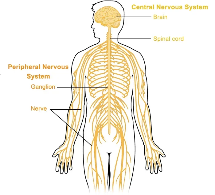
The peripheral nervous system is made up of nerves that branch from the brain and spinal cord. These nerves connect the central nervous system to the rest of the body. The peripheral neural system is further separated into the somatic and autonomic nervous systems.
The somatic nervous system is made up of nerves that connect to the skin and muscles and are engaged in conscious processes. The autonomic nervous system is made up of nerves that connect the CNS to visceral organs including the heart, stomach, and intestines. It facilitates unconscious actions.
Structure
Peripheral nerves
The peripheral nerves serve as the peripheral nervous system’s workhorses. Each neuron is made up of a bundle of nerve fibers (axons) and the connective tissue that covers them. Each nerve fiber is an extension of a neuron, whose cell body is located in either the grey matter of the CNS or the ganglia of the PNS. The analogous structure of the CNS is known as a ‘tract.’
Afferent or sensory neurons are peripheral nerves that bring information to the CNS, whereas efferent or motor neurons transfer impulses from the CNS.
Afferent neurons convey a wide range of impulses from sensory receptors/sense organs. They provide general feelings such as touch, pain, temperature, and spatial location (proprioception). Some also communicate more specific sensory information, such as the senses of smell, vision, hearing, and balance. In contrast, efferent neurons provide general neurological information to effector organs such as skeletal muscles, visceral organs, and glands. They initiate voluntary and involuntary motor processes such as muscle contraction and gland secretion.
Nerves are also classed as ‘cranial’ or’ spinal’ based on where they exit the CNS. Cranial nerves come from the skull (brain/brainstem), whereas spinal nerves exit the CNS through the spinal cord. There are 12 pairs of cranial nerves and 31 spinal nerve pairs, for a total of 43 paired nerves that comprise the PNS.
Cranial nerves
The first set of peripheral nerves is the twelve cranial nerves: olfactory (CN I), optic (CN II), oculomotor (CN III), trochlear (CN IV), trigeminal (CN V1, CN V2, CN V3), Abducens (CN VI), facial (CN VII), vestibular (CN VIII), glossopharyngeal (CN IX), vagus (CN X), spinal accessory (CN XI), and hypoglossal (CN XII).
Cranial nerves are peripheral nerves that primarily supply anatomical regions of the head and neck. The vagus nerve is an exception as it also innervates several thoracic and abdominal organs. Cranial nerves originate in particular nuclei of the brain. They exit the cerebral cavity via foramina and project to their designated target structure. Cranial nerves are classified into three kinds based on the type of information delivered by their fibers.
- Sensory
- Motor
- Mixed
Spinal nerves
The second group of peripheral nerves are spinal nerves, which come in 31 pairs: eight cervical, twelve thoracic, five lumbar, five sacral, and one coccygeal. Their numbering corresponds to the vertebral column exit level; cervical spinal nerves are numbered according to the vertebra positioned below, while the remainder are numbered according to the vertebra located above.
Each spinal neuron begins with numerous rootlets that connect to produce two major roots. The anterior root transports motor fibers from neurons found in the spinal cord’s anterior horn. The posterior root transmits sensory fibers from neurons with cell bodies in the dorsal root ganglion.
In the thoracic and upper lumbar regions, the anterior root also transports autonomic fibers from preganglionic sympathetic neurons, the cell bodies of which are situated in the lateral horn of the spinal cord. The anterior and posterior roots then combine to produce the spinal nerve proper, which contains mixed (sensory, motor, and autonomic) fibers.
Spinal nerves exit the vertebral column via intervertebral foramina positioned between two consecutive, neighboring vertebrae. Each spinal nerve is subsequently divided into two branches: posterior/dorsal and anterior/ventral rami. Both rami contain mixed fibers. The posterior rami move backward and branch to serve post-vertebral tissues. The anterior rami supply the skin and muscles of the limbs as well as the anterior trunk.
Immediately after the spinal nerve is divided into two rami, tiny communication fibers branch out. These white and grey rami communicantes connect the spinal nerves to the two sympathetic trunks of the autonomic nervous system that span the length of the vertebral column. Grey rami communicantes are present at all levels of the spinal cord, but white rami are exclusively seen at the T1-L2 levels.
Somatic nervous system
Skin, muscles, and sensory organs receive sensory and motor innervation from the somatic nervous system, which is voluntary. In other words, it transmits bodily sensations (pain, touch, temperature, and proprioception) and innervates skeletal muscles under conscious or voluntary control. In addition, the somatic nervous system is involved in spinal reflexes, such as the withdrawal reflex. This allows you to quickly move your palm away from a hot object.
Both the cranial and spinal nerves contribute to the somatic nervous system. Cranial nerves govern voluntary motor function and sensation in the head and face. Spinal nerves, as previously said, feed the trunk and limbs. The posterior rami moves rearward to feed the spinal column, muscles, and skin of the back, whereas the anterior rami nourishes the limbs and anterior trunk. The bulk of anterior rami join to create nerve plexuses, from which many main peripheral nerves originate. The exception is the anterior rami of the thoracic region, which moves relatively independently without creating plexuses, such as the trunk’s intercostal and subcostal nerves.
The nerve plexuses created by the anterior rami of the spinal nerves are as follows.
- C1- C4 form the cervical plexus
- C5-T1 combine into the brachial plexus
- T12-L4 form the lumbar plexus
- L4 – S4 join to form the sacral plexus.
The lumbar and sacral plexuses can be united into a single lumbosacral plexus. Each nerve plexus sends out many peripheral nerves that transport sensory and motor fibers to and from their respective target structures.
Autonomic nervous system
Finally, we’ve arrived at the autonomic division of the peripheral nervous system (ANS). It is involuntary, which means we have no cognitive control over it. The ANS provides sensory and motor innervation to smooth muscles, blood vessels, glands, and internal organs. As such, it regulates visceral and glandular activities in a coordinated manner, therefore contributing to homeostasis maintenance.
The autonomic nerves are likewise peripheral in nature, hence, the overall anatomy of a peripheral nerve outlined previously still applies. However, there is one caveat: all autonomic nerves connect to a sympathetic or parasympathetic ganglion. The section of the nerve that comes before the ganglion carries the impulse to the cluster of cell bodies. The section from the ganglion onwards is known as postganglionic and transports the impulse away from the cell bodies.
The ANS has three primary branches: sympathetic, parasympathetic, and enteric. The sympathetic division prepares the body to deal with times of heightened physical activity by controlling blood vessels (often, but not always, vasoconstriction), dilation of pupils, increased heart rate and blood pressure, and decreased peristalsis. The parasympathetic division helps the body conserve energy by performing’ rest and digest’, feeding, and breeding activities.
This is accomplished by acts that slow the cardiovascular system, stimulate gland secretion, and promote peristalsis. The PNS also regulates sexual desire and lacrimation (weeping). The myenteric (Auerbach) and submucosal (Meissner) plexuses make up the enteric nervous system (ENS), which is located inside the gastrointestinal tract’s walls. They collaborate to regulate peristalsis inside the digestive system. This system is frequently referred to as the second brain since it operates independently and is only impacted by ANS signals.
Sympathetic division
The preganglionic fibers of the sympathetic nerves exit the spinal cord via the T1 to L2 anterior roots and enter the respective spinal nerves. The fibers then pass through the white rami communicantes to the sympathetic trunk’s paravertebral ganglia, which are positioned on each side of the vertebral column.
Some fibers synapse here, while others pass through without synapsing and exit the sympathetic trunks as splanchnic nerves (greater, lesser, least, lumbar, sacral). These splanchnic nerves connect to their target organs in prevertebral ganglia named coeliac, aorticorenal, and mesenteric (superior and inferior). The postganglionic fibers then project onto their target sites, either directly or via the grey ramus communicantes and the course of spinal nerves throughout the body.
Blood arteries, sweat glands, arrector pili, the iris, and internal organs are also potential targets. The adrenal gland is one of the sympathetic nervous system’s targets. Sympathetic nervous system activity activates the sympathetic-adrenal medullary system, which then releases epinephrine/adrenaline.
Parasympathetic division
The parasympathetic nerve system is separated into cranial and sacral outputs. The preganglionic fibers of the cranial outflow leave the brainstem via the oculomotor, facial, glossopharyngeal, and vagus cranial nerves. They form synapses in the ciliary, pterygopalatine, otic, submandibular, and enteric ganglia. The postganglionic fibers eventually innervate the salivary glands of the head, the iris and ciliary muscles of the eyes, and, in the case of the vagus nerve, the thoracic and abdominal viscera.
The sacral outflow’s preganglionic fibers are significantly more confined, entering the spinal cord via the anterior roots of the S2- S4 spinal nerves. They follow the pelvic splanchnic nerves, eventually innervating the pelvic viscera (descending colon, sigmoid colon, rectum, bladder, and penis/clitoris).
Function
What exactly does the peripheral nervous system do?
Your peripheral nervous system is divided into two major subsystems: autonomic and somatic.
Autonomic: These are nervous system activities that your brain does without your conscious involvement.
Somatic: These are functions that you control by thinking about them.
These two subsystems are how your peripheral nervous system performs its three primary functions:
Senses: Your PNS is an important aspect of how your brain receives information from the environment around you. This job belongs to the somatic nervous system.
Movement: Your peripheral nerves provide command impulses to all of the muscles in your body that you can actively control. This job is also categorized as the somatic nervous system.
Unconscious processes: This is how your brain manages crucial functions that do not require your conscious thought. Examples of this include heart rate and blood pressure. This task relies on your autonomic nervous system.
Senses
Your brain functions like a sophisticated supercomputer. However, without external information, it has no knowledge of the world beyond its body. This is why the peripheral nervous system is so crucial. A computer requires peripheral devices such as a camera, microphone, or keyboard to receive information from outside origins, much like your brain does.
The peripheral nervous system is how your brain receives information from the outside world. The majority of your peripheral nervous system communicates with the rest of your body via the spinal cord. The cranial nerves are unique in that they attach directly to your brain. These nerves transmit impulses from your nose, ears, mouth, and numerous other organs. Your cranial nerves also control your sensation of touch in the skin of your face, head, and neck.
Other peripheral nerves connect to every region of your body. They spread out everywhere, even to the tips of your fingers and toes. The sensory nerves in your hands and feet also contribute to your brain’s ability to receive information from the outside environment. The motor nerves enable you to move different areas of your body.
Movement
Your peripheral nerves, which branch outward throughout your body, provide instructions and impulses from the brain to the muscles. That allows you to move about and do a variety of actions, from basic ones like scratching your nose to complex ones like juggling.
Unconscious processes
Your autonomic nervous system works without your conscious input. Part of your brain is always active, overseeing activities that keep you alive. Your brain relies on your peripheral nerve system to control those functions. Examples of these processes include your heart rate, respiration, blood pressure, and the digestion of food by your stomach.
Types of nerve signals
Your nerves are made up of bundles of nerve cells connected by long, arm-like projections known as axons. Nerve cells and axons twist and tangle to produce nerve fibers. This is analogous to how numerous strands of spun fabric fibers twist together to make sewing thread. Some of the nerves in that bundle send information to your brain, while others send it out.
Sensory: These nerves transmit information to the brain and spinal cord. They either link directly to your brain via your cranial nerves or provide information to your spinal nerves, which then flow into your spinal cord. Your spinal cord’s sensory nerve connections are located on its back.
Motor: These nerves transmit command messages from the brain to numerous regions of the body. They merely transport information away from the brain. The motor nerve connections are located on the front of your spinal cord, which means that they are exclusively used to convey muscle movement orders.
Autonomic: These nerves regulate the automatic operations of the organs and systems in your body. Your autonomic nerves frequently include mixed nerve fibers, some of which convey orders from your brain to their destination, while others send information about an organ’s function back to your brain.
How does the PNS interact with other organs?
Your autonomic nervous system, a component of the peripheral nervous system, assists your brain in controlling all of your body’s critical organs. This also allows your brain to care for itself. Your brain controls your heartbeat, ensuring that your heart pumps blood to your body and brain. Without that blood supply, your brain would perish in minutes.
Your peripheral nervous system also transmits nerve impulses from those organs to the brain. Examples include feeling warm in your stomach after drinking a hot beverage and feeling full after eating.
Conditions and Disorders
What are the most prevalent conditions and disorders affecting this bodily system or organ?.
Peripheral neuropathy, or sickness or injury to your peripheral nerve system, can be caused by a variety of disorders. Some of the more prevalent cases are:
Type 2 diabetes. Unmanaged or uncontrolled type 2 diabetes gradually harms your peripheral nerve system. This is why diabetics are at risk of losing sensation in their feet and lower legs. This kind of diabetes is the primary cause of peripheral neuropathy.
Autoimmune and inflammatory conditions. Lupus, Guillain-Barré syndrome, and rheumatoid arthritis are a few examples.
Hansen’s disease (better known as leprosy). While the symptoms of this condition, which is uncommon in industrialised nations, are most obvious on your skin, it also affects your peripheral nerves.
Congenital and genetic conditions. These are the difficulties you face when you are born. Genetic illnesses can be inherited from either one or both parents.
Infections. Viruses such as HIV and bacteria like Borrelia burgdorferi, which causes Lyme disease, can also cause nerve injury. Another prominent example is getting shingles, which can cause persistent nerve discomfort.
Medications and medical procedures. Certain antibiotics and chemotherapy treatments for cancer might harm peripheral nerves. This type of nerve injury can also occur as a consequence of surgery.
Poisons and toxins. Toxic heavy metals like mercury and lead can damage peripheral nerves. Many industrial chemicals can produce this type of harm.
Trauma. Injuries can result in nerve damage, which can be long-term or even permanent. Injuries can also cause swelling, which puts too much strain on peripheral nerves. Nerve compression illnesses include sciatica and carpal tunnel syndrome.
Tumors. Both malignant (cancerous) and benign (harmless) tumours can cause damage to your peripheral nervous system.
Your peripheral nerves may also exhibit symptoms of disorders affecting any component of your central nervous system. While they have no direct effect on your peripheral nervous system, they can nonetheless disturb its function.
Disease
Diseases of the PNS can affect a single nerve or the entire system.
Any peripheral nerve or nerve root might be injured, resulting in mononeuropathy. Such injuries can result from injury, trauma, or compression. Nerve compression can be caused by a tumor or an accident. Alternatively, if a nerve is in a region with a fixed size, it may get trapped if the other components grow in size, as in carpal tunnel syndrome and tarsal tunnel syndrome. Carpal tunnel syndrome frequently causes discomfort and numbness in the thumb, index, and middle fingers.
Peripheral neuropathy occurs when one or more nerves are injured through several causes. Toxic damage can be caused by diabetes (diabetic neuropathy), alcohol, heavy metals, or other poisons; some infections; and autoimmune and inflammatory disorders such as amyloidosis and sarcoidosis. periphery neuropathy is characterized by sensory loss in a “glove and stocking” distribution that begins at the periphery and gradually proceeds upwards. It may also be linked with acute and chronic pain. Peripheral neuropathy affects both the somatosensory and autonomic nervous systems.
Examination
Common tests to determine the health of a bodily organ?
Many tests can assist in identifying peripheral nervous system problems. The most typical beginning step is a neurological exam, in which your healthcare professional asks you to use different parts of your body, including your arms, hands, legs, and feet, in certain ways.
Some of the most frequent tests are:
- Blood tests (may reveal a wide range of abnormalities, from immune system issues to toxins and poisons, particularly metals such as mercury or lead).
- Nerve conduction tests.
- Electromyogram.
- Nerve ultrasound.
- Nerve biopsy.
- Genetic testing.
- Magnetic resonance imaging (MRI).
FAQs
What are the differences between CNS and PNS?
Our nervous system is divided into two parts: the central nervous system (CNS), which contains the brain and spinal cord, and the peripheral nervous system (PNS), which comprises nerves other than the brain and spinal cord.
What are the three 3 main organs of the nervous system?
The nervous system consists of the brain, spinal cord, and a complex network of nerves.This system transmits information back and forth between the brain and body. All of the functions of the body are controlled by the brain.
What are the two primary kinds of peripheral nerves?
The peripheral nerve system is split into two major components.
The autonomic nervous system (ANS) controls involuntary body activities and regulates glands.
The somatic nervous system (SNS) regulates muscle action and transmits information from the ears, eyes, and skin to the central nervous system.
What is PNS used for?
PNS is a regularly utilised treatment for chronic pain. PNS is appropriate for patients who have localised nerve pain caused by trauma, previous surgery, amputation, or even diabetes. This medication has also been utilised on individuals suffering from complex regional pain syndrome (CRPS).
What are the benefits of PNS?
Benefits include the following: Effective Pain Relief: PNS can help manage a variety of chronic pain conditions, including back pain, neck pain, and neuropathic pain. Non-invasive therapy: PNS is a therapy that does not involve surgery or the use of opioids.
References
- Professional, C. C. M. (2025e, March 19). Peripheral Nervous System (PNS). Cleveland Clinic. https://my.clevelandclinic.org/health/body/23123-peripheral-nervous-system-pns
- Peripheral nervous system. (2023, October 24). Kenhub. https://www.kenhub.com/en/library/anatomy/the-peripheral-nervous-system