Blog

  • Supraspinatus Muscle Pain

    Supraspinatus Muscle Pain

    Supraspinatus Muscle Pain occurs due to strain, overuse, or injury to the supraspinatus muscle, a key component of the rotator cuff in the shoulder. Pain is often felt on the top or side of the shoulder and may worsen with overhead movements.

    Common causes include repetitive stress, poor posture, or rotator cuff injuries. Early management with rest, stretching, strengthening, and proper biomechanics can help alleviate discomfort and prevent further complications.

    What is a Supraspinatus Muscle Pain?

    The supraspinatus muscle in the shoulder plays a crucial role in arm movement and stability. It is one of the four rotator cuff muscles responsible for lifting the arm and supporting the shoulder joint. Supraspinatus muscle pain is a common issue that can arise due to overuse, injury, or degenerative conditions. This type of pain can significantly affect daily activities, causing pain, weakness, and limited range of motion in the shoulder.

    Understanding the causes, symptoms, and management strategies for supraspinatus muscle pain is essential for effective treatment and prevention.

    One essential component of the shoulder’s rotator cuff is the supraspinatus muscle. It stabilizes the shoulder joint and aids in arm abduction, which is the raising of the arm away from the body. Pain, weakness, and limited shoulder movement are prominent symptoms of supraspinatus muscle pain, which can be caused by overuse, injury, or degenerative changes.

    Anatomy of the Supraspinatus Muscle

    Together with the teres minor, subscapularis, and infraspinatus, the supraspinatus muscle is one of the shoulder’s four rotator cuff muscles. It is essential for supporting shoulder movements and stabilizing the glenohumeral joint.

    Location and Structure

    • The supraspinous fossa of the scapula, or shoulder blade, is where the supraspinatus muscle begins.
    • It travels through a small opening known as the subacromial space beneath the scapula’s acromion.
    • It attaches to the humerus’s (upper arm bone) greater tubercle.
    • The supraspinatus tendon, which covers the muscle, is vulnerable to damage and deterioration.

    Function of the Supraspinatus Muscle

    • Arm Abduction: The deltoid muscle takes over after the supraspinatus performs the first 15 degrees of arm raising.
    • Shoulder Stability: It prevents dislocations by keeping the humeral head in the glenoid cavity.

    Causes of Supraspinatus Muscle Pain:

    Overuse, trauma, and degenerative changes are some of the causes of pain in the supraspinatus muscles. The most frequent causes are listed below:

    Injuries from Overuse and Repetitive Motion

    • Throwing, swimming, and lifting weights are examples of repetitive overhead exercises that can cause strain on the supraspinatus muscle.
    • prevalent in manual laborers (painters, construction workers) and athletes (tennis players, baseball pitchers).
    • causes weakening, irritation, and micro tears over time.

    Supraspinatus Tendinitis

    • inflammation of the supraspinatus tendon caused by incorrect mechanics or excessive use.
    • Tenderness, stiffness, and pain when moving the arms are some of the symptoms.

    Tears in the Rotator Cuff

    • rips in the supraspinatus tendon, either partial or total.
    • can be caused by gradual degeneration or abrupt trauma (such as falls or carrying large objects).
    • causes weakening, restricted motion, and excruciating shoulder pain.

    Shoulder Impingement Syndrome

    • Between the acromion and the humeral head, the supraspinatus tendon is squeezed.
    • causes pain, swelling, and limited mobility, particularly when moving upwards.
    • Muscle imbalances or bad posture may make it worse.

    Unbalanced muscles and bad posture

    • Extra strain is placed on the supraspinatus by forward head posture and round shoulders.
    • Improper mechanics may be caused by weakness in the scapular stabilizers, such as the trapezius and serratus anterior.

    Aging and Degenerative Changes

    • The tendon gets less elastic with time and is more vulnerable to damage.
    • common in those over 40, resulting in deterioration of the rotator cuff.

    Direct Injury and Trauma

    • Damage to the supraspinatus may result from falls onto an outstretched arm or dislocation of the shoulder.
    • Tendon tears or inflammation may be the outcome of sudden impact injuries.

    Nerve compression (entrapment of the suprascapular nerve)

    • The supraspinatus muscle may become weak and painful if the suprascapular nerve is compressed.
    • may be caused by cysts, bone spurs, or recurrent shoulder strain.

    Symptoms of Supraspinatus Muscle Pain:

    Depending on how severe the injury or ailment is, several symptoms can accompany supraspinatus muscle pain. Typical signs and symptoms include:

    • Pain in the Shoulders
    • Pain in the upper and outer regions of the shoulder that is dull or painful.
    • Although it typically doesn’t go past the elbow, pain can travel down the upper arm.
    • Activities that involve lifting, reaching, and throwing are worse.
    • Weakness in the Arm and Shoulder
    • Lifting or holding objects away from the body can be challenging.
    • diminished strength when moving above the ground.
    • may have trouble doing everyday tasks like getting dressed, combing their hair, or carrying a bag.
    • Restricted Motion
    • Raising the arm above shoulder level is difficult.
    • shoulder stiffness and soreness, particularly after rest.
    • Pain between 60° and 120° of arm abduction is known as the painful arc sign.
    • The sensation of clicking or popping
    • popping, clicking, or grinding noise as the shoulder is moved.
    • maybe a sign of bursitis, tendon injury, or impingement.
    • Pain at Night and Uncomfortable Sleep
    • increase pain while the affected shoulder is lying down.
    • gets worse at night, making it harder to fall asleep.
    • Tenderness and Swelling
    • soreness or swelling in the vicinity of the shoulder joint.
    • The supraspinatus tendon may be sensitive to pressure or contact.

    Treatment of Supraspinatus Muscle Pain

    The severity of the condition determines the course of treatment for supraspinatus muscle pain, which can range from conservative measures to surgery in extreme situations. Reducing pain, regaining shoulder function, and avoiding more injuries are the objectives.

    You might use home remedies to reduce pain and promote recovery in addition to shoulder workouts.

    Compress, ice, and rest your shoulder with the RICE technique. Raise your shoulder above your heart whenever you can. Additionally, you can take an Epsom salt bath or apply a heating pad.

    You can use over-the-counter painkillers like acetaminophen or ibuprofen to reduce pain. Or try using natural remedies like cloves, willow bark, or turmeric to ease pain. Several times a day, use an essential oil combination, menthol rub, or arnica cream on the affected area.

    Frequent acupuncture and massage therapies might help your body regain equilibrium and ease pain. Additionally, you can experiment with manipulative therapies like rolfing, osteopathic adjustments, and chiropractic adjustments.

    • Modification of Rest and Activity
      keep away of hard lifting and repetitive overhead motions.
      Adjust your activities to lessen shoulder strain.
    • Heat and Ice Therapy
      Ice packs (15–20 minutes, three times a day) help to relieve pain and inflammation.
      Heat therapy promotes blood flow and eases tense muscles.
    • Medicines Pain relievers (like acetaminophen) for mild to moderate pain and nonsteroidal anti-inflammatory drugs (NSAIDs) (like ibuprofen and naproxen) to reduce pain and inflammation.
    • Injections of Corticosteroids
      used to treat chronic pain and inflammation.
      injected to lessen edema in the subacromial space.
      should be applied with caution because the tendon may get weaker with repeated injections.

    Physical Therapy Treatment of Supraspinatus Muscle Pain:

    Across-the-chest stretch
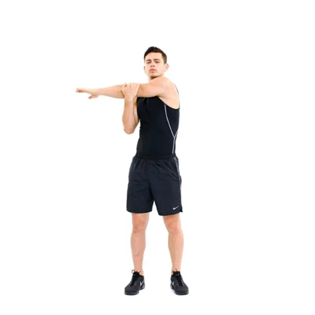
    across-the-chest-stretch
    Across-the-chest stretch
    • Cross your right arm over your chest.
    • Hold this position for no more than a minute.
    • On the other side, repeat.
    • Repeat 3–5 times on each side.

    Neck release

    • Your shoulders and neck might become less tense with this moderate exercise.
    • Put your chin down close to your chest. Your neck will be strained near the nape.
    • Keep your posture for no more than a minute.
    • On the other side, repeat.
    • Repeat 3–5 times on each side.

    To make this stretch deeper:

    • Keep your chin pressed on your chest.
    • Attempt to hold this position for no more than one minute.
    • On the other side, repeat.
    • Repeat 3–5 times on each side.

    Chest expansion
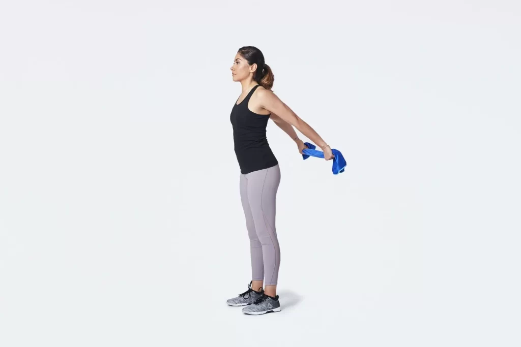
    Chest-expansion-workout
    Chest-expansion-workout
    • Spread your shoulder blades out over your chest and move them nearer each other.
    • Look up at the ceiling with your chin up.
    • Hold for a maximum of thirty seconds.
    • Do this three to five times.

    Eagle arms spinal rolls

    • If you have trouble with the arm position, you can do this exercise by holding the opposing shoulders.
    • Sitting, extend your arms to the sides.
    • For fifteen seconds, maintain this posture.
    • As you exhale, pull your elbows in toward your chest and roll your spine.
    • For one minute, keep doing this motion.
    • On the other side, repeat.

    Seated twist

    • Your hips should remain facing forward during this exercise.
    • Your right hand should be placed wherever it feels most comfortable.
    • On the left side, repeat.
    • Repeat 3–5 times on each side.

    Shoulder circles
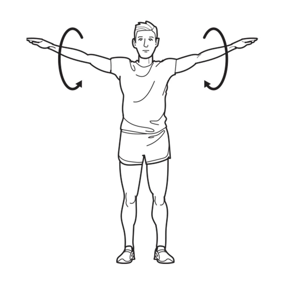
    Shoulder Circles
    Shoulder Circles
    • Let your right-hand dangle downward.
    • Do the same on the opposite side.

    Doorway shoulder stretch

    • Bend forward while using your core.
    • Stretch again with your left foot forward.
    • Do each side two or three times.

    Downward Dog Pose

    • Get on your hands and knees to begin.
    • Bring your head toward your feet so that your shoulders are flexed overhead while maintaining a straight spine.

    Child’s Pose
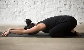
    Extended Child’s Pose on Fingertips stretch
    Child’s Pose
    • Your neck, shoulders, and back will all feel less tense after doing this calming pose.
    • Let your shoulders and spine relax as your chest drops heavily toward the floor.

    Thread the needle

    • Your shoulders, upper back, and chest will all feel less tight after doing this pose.
    • Lift your right hand toward the heavens with the palm pointing away from your body.
    • Lower your arm until it reaches to your left side and is beneath your chest, palm up.
    • To prevent collapsing into this space, engage your right arm and shoulder.
    • Before doing this stretch again on the left side, unwind in Child’s Pose.

    Prevention of Supraspinatus Muscle Pain:

    • To prevent shoulder impingement, keep your posture straight.
    • To increase stability, strengthen your rotator cuff muscles.
    • Stretching and good ergonomics can help prevent repetitive strain.
    • Before lifting weights or doing sports, warm up.

    FAQs

    What is the supraspinatus muscle orthopedic test?

    Test for Supraspinatus
    We can ask the patient to abduct both arms to 90° and then bring them anteriorly with a 30° forward flexion to check the integrity of the supraspinatus. The patient will be asked to push both arms upward against our resistance while in this position.

    When the supraspinatus is stretched, how long does healing take?

    If you work a desk job, it can take four weeks to get back to work. You will have to wait at least three months if you have to lift at work. Following rotator cuff surgery, you will require rehabilitation. This usually lasts 4 to 9 months and begins about 2 weeks following surgery.

    Can a massage help with a supraspinal tear?

    There are numerous muscle adhesions in your teres minor, subscapularis, infraspinatus, and supraspinatus. For the elimination of knots, particularly in the subscapularis, a combination of trigger point therapy and deep tissue massage can be highly beneficial.

    What makes the supraspinatus weak?

    People over 40 are more susceptible to degeneration and rotator cuff tears because of the body’s natural weakening of the soft tissue over time, which is commonly caused by misuse of the supraspinatus tendon.

    How can I determine whether my supraspinatus is tight?

    Localized pain and sensitivity on the front side of the shoulder are the primary symptoms of supraspinatus tendinosis. When you raise your arm, you may experience pain and stiffness. When lowering your arm from the elevated posture, you may also experience pain. Symptoms may be modest at first.

    Which medications are effective for supraspinatus?

    Summary of Medication
    A brief course of nonsteroidal anti-inflammatory medicines (NSAIDs) is appropriate as an adjuvant to the therapy program and other treatment modalities during the acute to subacute phases of shoulder impingement syndrome due to its analgesic and anti-inflammatory properties.

    Is the muscle of the supraspinatus deep?

    The rotator cuff muscles include the subscapularis, teres minor, infraspinatus, and supraspinatus. Stretching from the supraspinous fossa of the scapula to the proximal humerus, the supraspinatus is situated deep in the trapezius muscle in the posterior scapular area.

    For supraspinatus pain, which massage is most effective?

    One of the most effective treatments for shoulder and back aches and pains is sports massage, a firm, focused type of therapy that targets the soft tissues. Both professional and amateur athletes, as well as those who do not routinely exercise, can find it to be quite beneficial.

    What is supraspinatus physical therapy?

    Physical Therapy
    Supraspinatus, internal and external rotators, prone extension, horizontal abduction, forward flexion to 90°, upright abduction to 90°, shoulder shrugs, rows, push-ups, press-ups, and pull-downs are examples of isotonic resistance exercises that are used to strengthen the scapular stabilizers.

    How should I sleep if I have pain in my supraspinatus?

    Dozing Off on Your Back
    By raising the shoulder’s ball and socket joint, relieves pressure on the muscles, ligaments, bursae, and joint structures while also offering appropriate anatomical support. According to recent research, adopting this sleeping position can help reduce shoulder pain and encourage deeper, more restful sleep.

    What is the duration of supraspinatus pain?

    Over time, the majority of shoulder tendonitis instances resolve on their own. Depending on how severe it is, recovery could take weeks or months. Consult your physician if you: Feel pain that makes it difficult for you to carry out your daily tasks.

    How is supraspinatus released?

    Raise the arm to slightly above 90 degrees of shoulder abduction to start the active release. Pull the shoulder into adduction behind your back with your other hand after lowering the arm to a neutral posture. Repeat up to two or three different locations and up to six repetitions.

    Can the supraspinatus muscle be massaged?

    Self-massage is known as supraspinatus. I suggest using the Trigger Fairy to massage this muscle. This is the simplest and most secure method of applying pressure. Although it is a little more challenging and inconvenient, using a massage ball is still feasible and beneficial.

    How is a supraspinatus strain treated?

    Physical therapy, rest, cold packs, and nonsteroidal anti-inflammatory medications (NSAIDs) are all part of this treatment. Physical treatment can also be supplemented with corticoid injections. If, after three to six months of conservative treatment, there is no improvement, surgery may be the answer.

    What signs of pain in the supraspinatus are present?

    A feeling of burning in the shoulder. weakness when pushing a door open or lifting something heavy. Sleep was disturbed by the ache. difficulty carrying out daily tasks like putting on a jacket or brushing one’s hair.

    References

    • ProHealth Prolotherapy Clinic. (2025, January 25). Supraspinatus tendonitis – causes & best treatment options in 2025. https://prohealthclinic.co.uk/blog/supraspinatus-tendonitis/
    • Faoa, T. M. D. M. F. (n.d.). Supraspinatus tendonitis: Practice Essentials, etiology, Epidemiology. https://emedicine.medscape.com/article/93095-overview
  • Interphalangeal Joints

    Interphalangeal Joints

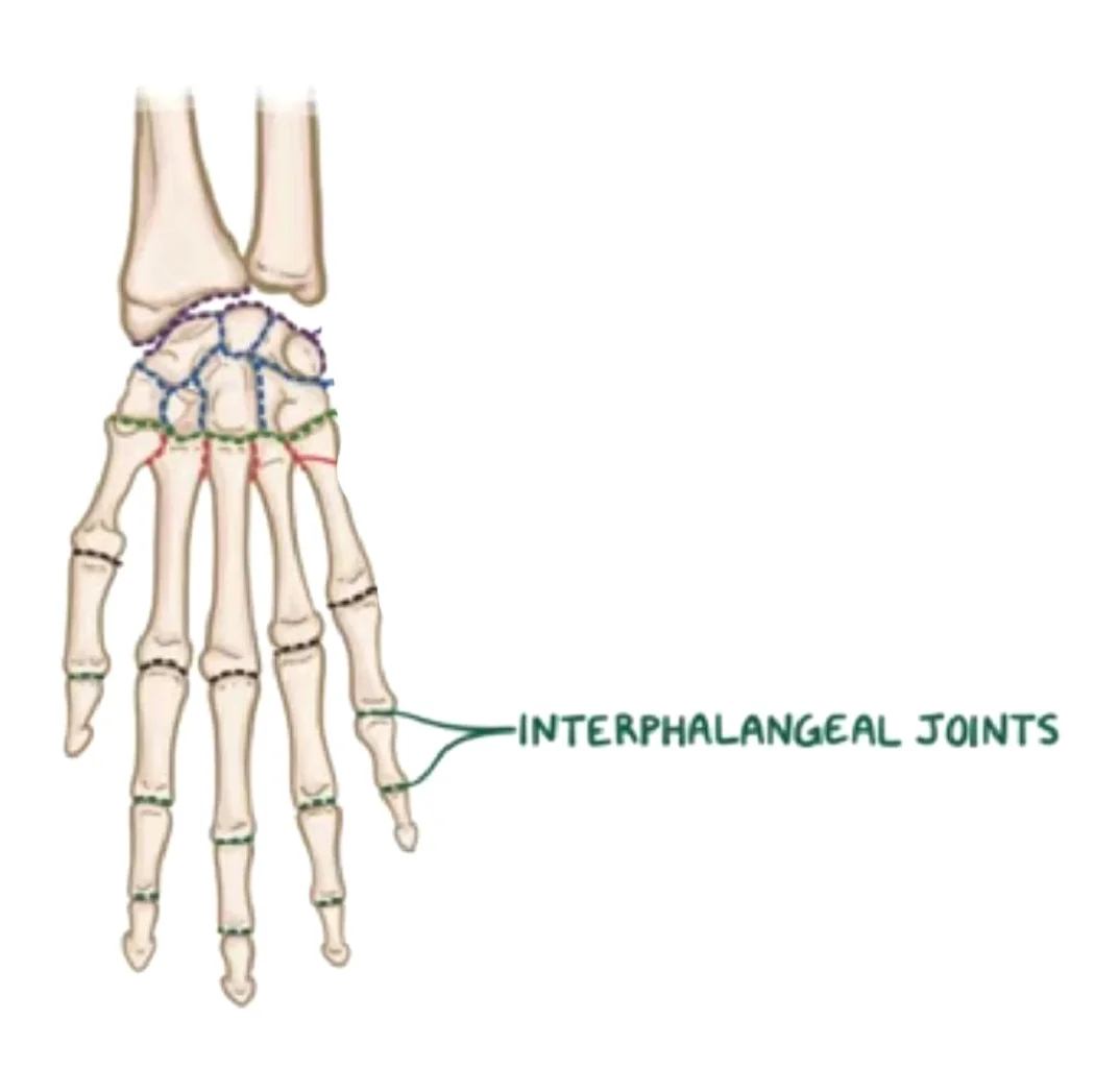
    The hand’s interphalangeal joints are the hinge joints that allow flexion in the direction of the palm between the finger phalanges.

    Introduction

    The hand’s interphalangeal joints are synovial hinge joints connecting the proximal, middle, and distal phalanges. These joints can be categorized into digits 2–5 according to the bones involved.

    The middle and distal phalanges are separated by the distal interphalangeal joint (DIPJ or DIJ), and the proximal interphalangeal joint (PIPJ or PIJ) is situated between the proximal and middle phalanges. The thumb’s interphalangeal joint is simply called that because the first digit only has a proximal and distal phalanx.

    The ability of the hand’s interphalangeal joints to support fine motor movements in the fingers. These joints enable movement in just one degree of freedom—flexion-extension—to achieve this.

    Anatomy

    Every finger (apart from the thumb, which has a single joint) has two sets.

    • “Proximal interphalangeal joints” (PIJ or PIPJ), which are located between the first
    • The distal interphalangeal joints (DIJ or DIPJ) are located between the third (distal) and second (intermediate) phalanges.

    The proximal and distal interphalangeal joints share many anatomical similarities. The distal joint’s smaller size and decreased mobility are the main differences, with a few minor variations in the segmentation of the flexor tendon sheath and the proximal attachment of the palmar plates.

    Joint structure

    There is a lot of lateral stability in the PIP joint. Its thick collateral ligaments are tight in all positions during flexion, unlike those in the metacarpophalangeal joint, and its transverse diameter is larger than its anteroposterior diameter.

    Dorsal structures

    Due to the extremely thin and flexible capsule, extensor tendon, and skin dorsally, both phalanx bones can flex more than 100° until the base of the middle phalanx touches the proximal phalanx’s condylar notch.

    Three bands separate the extensor mechanism at the PIP joint level. The dorsal tubercle of the middle phalanx, close to the PIP joint, is where the central slip is attached. After the PIP joint, the two lateral bands that support the extensor tendons continue dorsally to the joint axis. A transverse retinacular ligament, which connects the flexor sheath at joint level to the palmar border of the lateral band, holds these three bands together and stops the lateral band from dorsally shifting.

    The oblique retinacular ligament [of Landsmeer] is located on the palmar side of the joint axis of motion and extends from the terminal extensor tendon to the flexor sheath across the proximal phalanx. In extension, the oblique ligament pulls and tightens the terminal extensor tendon proximally, preventing passive DIP flexion and PIP hyperextension.

    Palmar structures

    A thick ligament, on the other hand, stops hyperextension on the palmar side. With a fibrocartilaginous structure, the palmar plate, the distal portion of the palmar ligament, is 2 to 3 millimeters (0.079 to 0.118 inches) thick. For the dorsal and palmar plates to withstand compression forces against the proximal phalanx’s condyles, chondroitin, and keratan sulfate are essential. The tendons that run in front of and behind the joint are shielded by these structures working together. Collagen fibers in these tendons allow them to maintain traction forces.

    Articulating Surfaces

    The head of the more proximal phalanx and the base of its distal counterpart make up each interphalangeal joint. This implies that, for instance, the union of the head of the proximal phalanx with the base of the middle phalanx forms the proximal interphalangeal joint. The articulation of the middle phalanx’s head with the distal phalanx’s base forms the distal interphalangeal joint, which follows the same pattern.

    Upon closer examination of the phalangeal head, two curved condylar processes with a shallow groove between them are visible. Two concavities of equal size and shape on the base of the distally lying phalanx receive these condyles.

    A raised ridge of bone sits between these two concavities and slides into the phalangeal head’s groove, enhancing intraarticular stability. The proximal articular surface is larger than the distal surface because of the layer of hyaline cartilage covering these joint surfaces, which extends farther palmary than dorsally. There is less articular surface on the dorsal aspect, which results in less hyperextension than in the metacarpophalangeal joints.

    The condylar processes’ inner sloping surfaces rather than their apex are where the point of articulation is located. The joint becomes noticeably out of alignment as a result of the radii of the phalangeal head’s condyles being larger than those of the phalangeal base’s convex surfaces. Most people have a small intercondylar joint space as a result of this incongruency.

    Joint capsule and ligaments

    Each interphalangeal joint has a fibrous joint capsule with a synovial membrane as its inner lining. Two collateral ligaments and a palmar ligament, sometimes referred to as a palmar/volar plate, strengthen each joint capsule. Dorsally, the extensor tendons broaden, strengthening the joint capsule. The phalangeal base’s articular surface is increased by this substantial ligamentous contribution to each joint capsule, enhancing joint congruence.

    Collateral ligaments

    The collateral ligaments originate from the head of the more proximal phalanx and extend to the palmar, or volar, aspect of its distal counterpart, running along either side of each interphalangeal joint. Each collateral ligament gives rise to an accessory ligament that attaches to the palmar ligament fibers by extending anteriorly. The excessive adduction-abduction movements of the interphalangeal joints are lessened by these ligaments.

    Palmar ligament

    Each interphalangeal joint’s palmar surface has a thick fibrocartilage plate called the palmar ligament, also known as the palmar or volar plate. The distal portion of this ligament, which blends with the accessory collateral ligaments, arches across the base of the distally lying phalanx, giving it the distinctive upside-down “U” shape. The periosteum of the body of the more proximally positioned phalanx incorporates the legs of the palmar ligament proximally. These ligaments, which are known as “check rein” ligaments, keep the joint from being overextended.

    When flexion occurs, the proximal margin of the palmar plate and the accessory ligament fold back upon themselves because they are both flexible. At the PIP joint, the proximal fibers of the C1 ligament and the A3 pulley connect the flexor tendon sheaths to the mobile volar ligament, while the annular pulleys A2 and A4 securely fasten the sheaths to the proximal and middle phalanges. This configuration creates a gap at the proximal phalanx’s neck during flexion, which the folding palmar plate fills.

    The collateral ligaments, which support the palmar plate, are located on either side of the joint and stop it from deviating from its normal position. The finger can be forced backward into hyperextension, causing the ligaments to tear partially or completely and avulse with a small fragment of fracture. A “palmar plate, or volar plate injury” is what this is known as.

    Together with the distal portion of the joint, the palmar plate creates a semi-rigid floor, and the collateral ligaments form the walls of a mobile box that stabilizes the joint throughout its full range of motion. The palmar plate also increases the moment of flexor action because it attaches to the flexor digitorum superficialis close to the muscle’s distal attachment. The two so-called check-rein ligaments that connect the palmar plate to the proximal phalanx in the PIP joint restrict its range of motion.

    Innervation

    The proper palmar digital nerves, which originate from the median and ulnar nerves, innervate the hand’s interphalangeal joints. The median nerve innervates the digits 1–3 and the lateral half of digit 4, whereas the ulnar nerve innervates the medial half of digit 4 and the entire fifth digit.

    Blood supply

    It is the distal extensions of the superficial palmar arch that supply oxygenated blood to each interphalangeal joint through the proper palmar digital arteries.

    Movements

    The hand’s interphalangeal joints’ morphology allows for only flexion and extension as active movements. Nonetheless, passive accessory movements to a limited extent are permitted, mostly at the distal interphalangeal joints of fingers 2–5.

    The thumb can flex and extend around a transverse axis that passes through the center of the proximal phalanx’s neck. The range of motion is up to roughly 90° flexion and 10° extension, with the possibility of passive hyperextension when the distal phalanx is subjected to significant force. The thumb’s interphalangeal joint is incapable of passive accessory rotation or lateral movement due to strong collateral ligaments.

    The entire flexion and extension of digit 2, also known as the index finger, takes place in the sagittal plane. However, the flexion and extension of more medially lying digits occur more obliquely to better oppose the thumb.

    The proximal interphalangeal joints’ degree of flexion increases slightly from digits 2 to 5, but it is generally considered to be between 100° and 110°. The digits 3 and 5 have the most and the least amount of flexion, respectively, in the distal interphalangeal joints (80° and 70°).

    The wrist joint’s location affects how strongly the hand’s interphalangeal joints can flex each other. The flexor muscles in the fingers lengthen when the wrist is extended, which increases the amount of tension that can be created in them and strengthens the grip. These finger flexors, on the other hand, become slackened and less able to produce tension when the wrist is flexed. This shows up as a weakened grip.

    The range of active extension for digits 2–5 is much smaller, reaching up to 2° in the proximal and 5° in the distal interphalangeal joints.

    The interphalangeal joints of every hand digit are in the closed-packed position of full extension and the open-packed (resting) position of slight flexion. These joints are more restricted in flexion than in extension due to their capsular pattern.

    Passive accessory movements, such as abduction-adduction, rotation, and anteroposterior gliding, are restricted to digits 2–5. The fingers can adjust to objects of different sizes and shapes while gripping thanks to these subtle movements.

    Muscles acting on the interphalangeal joints

    Many muscles can act on the fingers, which reflects their exceptional dexterity. The flexion-extension movements at the hand’s interphalangeal joints are produced by both intrinsic and extrinsic hand muscles.

    The flexor pollicis longus muscle is responsible for contracting the thumb’s interphalangeal joint. While the flexor digitorum profundus and flexor digitorum superficialis flex the proximal interphalangeal joints of digits 2–5, the latter also flexes the distal phalanx, making it the only muscle that can flex the distal interphalangeal joints.

    The extensor pollicis longus is responsible for extending the thumb’s interphalangeal joint. The extensor digitorum, lumbricals, and dorsal interossei all work together to extend the proximal and distal interphalangeal joints of digits 2–5. The palmar interossei aid in the extension of digits 2, 4, and 5, while the extensor indicis provides an extra extension for digit 2, the index finger.

    Through their tendinous aponeurotic insertion into the extensor expansion, the muscles that extend digits two through five do so. Anchored on both sides by the palmar ligament, this hood-like expansion stretches down the length of digits 2–5. In the middle of each finger, it serves to keep the extensor tendons pulling in the same direction.

    Clinical significance

    The distal interphalangeal joints are typically unaffected by rheumatoid arthritis. Therefore, osteoarthritis or psoriatic arthritis is strongly suggested when distal interphalangeal joint arthritis occurs.

    • Heberden’s nodes in the distal interphalangeal joint are one example of the degenerative joint disease known as osteoarthritis (OA), which affects IP joints and causes pain, stiffness, and deformity.
    • An autoimmune disease that causes inflammation and joint abnormalities, such as boutonnière and swan-neck deformities, is rheumatoid arthritis (RA).
    • Trauma-related fractures and dislocations are frequent (e.g., sports injuries, crush injuries).
    • The inability to extend the fingertip due to damage to the distal interphalangeal (DIP) joint is known as a mallet finger.
    • The condition known as trigger finger (stenosing tendinitis) is characterized by inflammation of the flexor tendons, which results in excruciating finger locking.
    • Nail changes and “sausage fingers” (dactylitis) are symptoms of psoriatic arthritis.

    FAQs

    Interphalangeal joints: what are they?

    The hand’s interphalangeal joints (green) The hinge joints between the finger phalanges that allow flexion in the direction of the palm are known as the hand’s interphalangeal joints.

    What is the finger’s IP joint?

    The interphalangeal joints are located between the middle and distal phalanges (distal interphalangeal joint, or DIP) and proximal and middle phalanges (proximal interphalangeal joint, or PIP) in each finger (except the thumb, which only has one interphalangeal joint and proximal and distal phalanges).

    What category do the interphalangeal IP joints fall under?

    The hand’s proximal, middle, and distal phalanges are separated by the interphalangeal joints, which are synovial hinge joints.

    What is the thumb’s interphalangeal IP?

    The tiny joints that connect the fingers and thumb are called interphalangeal (IP) joints. These are the joints that connect the fingers. The proximal interphalangeal joint (PIPjt) and the distal interphalangeal joint (DIPjt) are the two interphalangeal (IP) joints found in each finger.

    On the other hand, how many IP joints are there?

    The thumb only has two phalanges and one interphalangeal joint, whereas every other finger has three phalanges separated by two interphalangeal joints. The proximal interphalangeal joint (PIP joint) is the first joint near the knuckle joint.

    What is the thumb IP joint’s range?

    The average IPJ range of motion was 12 ± 9.2 ° (range 0–45 °) for extension and 88 ± 2.3 ° (range 80–90 °) for flexion.

    Which hand interphalangeal joint is the first one?

    The hand’s interphalangeal (IP) joints are hinge-style synovial joints that connect neighboring phalanges. The second to fifth fingers each have a proximal and distal interphalangeal joint, while the thumb has a single interphalangeal joint.

    What is IP arthritis?

    Arthritis of the fingers is known as interphalangeal joint arthritis. Every finger on your hand, except the thumb, is made up of three phalanges, or finger bones, which are divided by two joints called interphalangeal joints.

    References

    • Wikipedia contributors. (2025b, January 20). Interphalangeal joints of the hand. Wikipedia. https://en.wikipedia.org/wiki/Interphalangeal_joints_of_the_hand
    • Interphalangeal joints of the hand. (2023, October 30). Kenhub. https://www.kenhub.com/en/library/anatomy/interphalangeal-joints-of-the-hand
  • Intervertebral Joints

    Intervertebral Joints

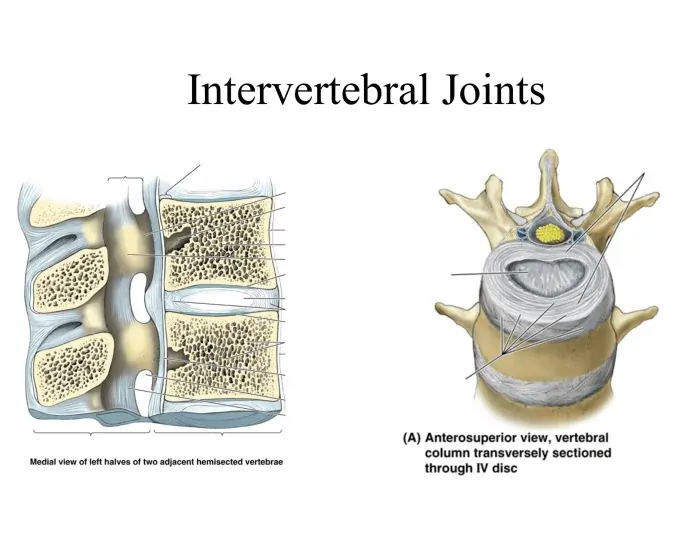
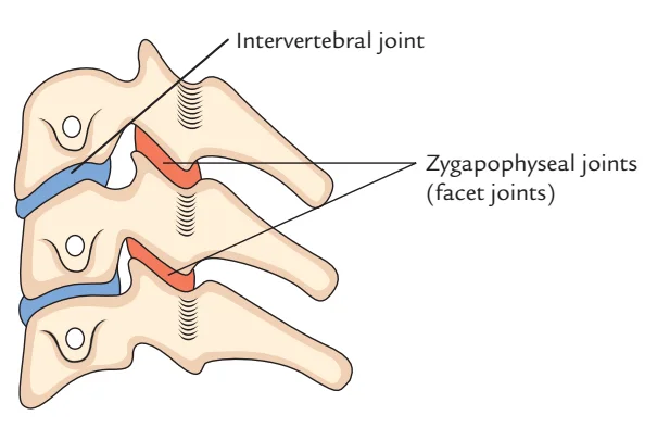
    Three intervertebral joints connect each neighboring vertebra from the axis to the sacrum: two between the facets of adjacent vertebral arches and one between the vertebral bodies (zygapophysial joints, also known as facet joints).

    Introduction

    The intervertebral joints join the vertebrae of the vertebral column which are closely nearby. Three distinct joints make up each intervertebral joint: two zygapophyseal (facet) joints and one intervertebral disc joint (intervertebral symphysis).

    Articular surfaces

    The intervertebral symphyses, or intervertebral disc joints, go from the C2 to the S1 vertebral levels. Adjacent vertebral bodies and the intervertebral disc that sits between them create them. At the C0-C1 and C1-C2 vertebral levels, there are no intervertebral discs.

    Both the superior and inferior surfaces of the vertebral bodies have a lot of texture. Their centers are very asymmetrical, with a modest smoothness at the edges. Additionally, the vascular foramina that penetrates the joint surfaces exacerbates their irregularity. The basivertebral veins and intraosseous nutritional arteries, which deliver and remove blood from the vertebral bodies, are transmitted by these foramina.

    In the vertical plane, the joint surfaces can be sellar (concave) with raised borders that resemble lips or relatively flat. In the cervical vertebrae, where they create the uncovertebral joints, these raised borders, also known as uncinate processes, are particularly noticeable. As the spinal cord passes through the vertebral foramen, the articular surfaces appear convex anteriorly and concave posteriorly in the horizontal plane. The cervical vertebrae are an exception, as their posterior joint surfaces may be nearly flat.

    The vertebral bodies’ discal surfaces are covered with vertebral end plates. They are irregular and less than 1 mm thick, with the center being the narrowest. Both bone and hyaline cartilage make up vertebral end plates in the early stages of life. In adulthood, the cartilage mineralizes and is replaced by bone. The vertebral end-plate’s roles include strengthening the intervertebral disc joint and absorbing shock.

    Fibrocartilaginous intervertebral discs separate the articular surfaces of successively neighboring vertebral bodies. In addition to the bony vertebral rim, they also stick to the vertebral end plates. Through ring apophyses, it is attached to the vertebral bodies. The anterior region of the wedge-shaped intervertebral discs is especially thick. A nucleus pulposus and an annulus fibrosus make up each intervertebral disc.

    The inner core of the intervertebral disc is formed by the nucleus pulposus. It is very pliable, gelatinous, and soft. These features make it easier for the intervertebral disc and joint to move, particularly in the cervical area. The nucleus pulposus is surrounded by the peripheral annulus fibrosus. Comprising fibrocartilage and collagen, it has a lamellar structure. It is therefore less flexible and more rigid than the nucleus pulposus. The annulus fibrosus reinforces the vertebral column by enclosing the nucleus pulposus and connecting the vertebrae.

    Ligaments and joint capsule

    There isn’t a joint capsule around intervertebral disc joints. The anterior and posterior longitudinal ligaments give them stability.

    There are two longitudinal ligaments, although the anterior longitudinal ligament is the stronger. It reaches the pelvic surface of the upper sacrum from the root of the occipital bone. It connects to the anterior parts of the vertebral bodies, the annulus fibrosis of the intervertebral discs, and the vertebral end plates along the way. The anterior longitudinal ligament’s function is to provide anterior support for the vertebral column.

    In the vertebral canal of the vertebral column, directly in front of the spinal cord, is the posterior longitudinal ligament. From the second cervical vertebra to the first sacral vertebra, it stretches. The posterior parts of the intervertebral discs, the vertebral end plates, and the edges of the vertebral bodies are where it attaches along the route. This ligament’s function is to support the back of the vertebral column.

    Innervation

    From the anterior ramus of the spinal nerve, the sinuvertebral nerve innervates the zygapophyseal joints and the intervertebral disc. The spinal nerve’s gray communicating branch provides sympathetic input to the sinuvertebral nerve as it travels. The medial branch of the posterior ramus of the spinal nerve also innervates the zygapophyseal joint, unlike the intervertebral disc joint.

    Blood supply

    The segmental arteries of the vertebral column, including the costocervical trunk and vertebral, ascending cervical, posterior intercostal, lumbar, iliolumbar, and lateral sacral arteries, supply arterial blood to the intervertebral joints. The posterior spinal branches of these segmental arteries deliver blood to the zygapophyseal joints and the most peripheral region of the nucleus fibrosis. Additionally, they supply the articular surfaces of the vertebral bodies with peripheral, metaphyseal, and nutritional arteries. The avascular intervertebral discs receive oxygen and nutrients by diffusion from this point.

    Blood flows into the spinal and basivertebral veins from the intervertebral joints. These then enter into the intervertebral veins via the vertebral venous plexuses.

    Movements

    The intervertebral disc joint is classified structurally as a fused, fibrocartilaginous symphysis. Functionally speaking, nonetheless, it is regarded as an amphiarthrosis that allows for a restricted range of motion. The intervertebral disc can translate, tilt, rock, and compress due to its soft and pliable nature, which expands the spinal column’s range of motion.

    According to structural classification, the zygapophyseal joint is a plane synovial joint. It operates similarly to a uniplanar diarthosis, permitting nonaxial motions in a single plane (translation). However, because of the various orientations of the articular processes in each region of the vertebral column—forward flexion and extension in the lumbar spine, lateral flexion and rotation in the cervical spine, and lateral flexion and rotation in the thoracic spine—zygapophyseal joints allow the spine to move in multiple planes and directions. Thus, the zygapophyseal joints guide movement, whereas the intervertebral disc joints constitute the main constraint on the spine’s range of motion.

    When the intervertebral joints are combined, the vertebral column can move multiaxially in three degrees of freedom (the average maximum glenohumeral active RoM is displayed in brackets):

    • Cervical spine
      • (Anterior) flexion (25°) – Extension (85°)
      • Bilateral flexion (40°)
      • Bilateral axial rotation (50°)
    • Thoracic spine
      • (Anterior) flexion (30-40°) – Extension (20-30°)
      • Bilateral flexion (20-25°)
      • Bilateral axial rotation (35°)
    • Lumbar spine
      • (Anterior) flexion (55°) – Extension (30°)
      • Bilateral flexion (20-30°)
      • Bilateral axial rotation (1-2°)

    The superior vertebral body moves anteriorly during spinal flexion, leaning forward and squeezing the anterior intervertebral disc segments. As a result, the superior vertebra’s inferior articular surface moves both superiorly and anteriorly to the inferior vertebra’s superior articular surface, resembling a seesaw. The superior zygapophyseal joint widens posteriorly as a result. The articular process’s 45° position within the sagittal plane allows the lumbar spine to have a higher range of motion in flexion. The zygapophyseal joints are less restricted and the vertebral bodies can slide more anteriorly since flexion and extension also occur in the sagittal plane.

    The thoracic cage (thoracic spine only), the posterior longitudinal ligament, the ligament flava, the nuchal ligament (cervical spine alone), the joint capsule of the zygapophyseal joint, and the tone of the deep, or intrinsic, muscles of the back, which are placed paravertebrally all limit flexion.

    The reverse happens with spinal extension. The posterior part of the intervertebral discs is compressed, tipped back, and slid posteriorly by the superior vertebral body. The anterior region of the superior zygapophyseal joint widens as the inferior articular surface shifts inferiorly and posteriorly. Since the articular process affects the broad spinous processes of the vertebrae across the thoracic and lumbar spines, extension is much reduced in these regions as opposed to the cervical area. The anterior longitudinal ligament’s tension, the posterior vertebral processes’ impaction, and the tone of the anterior neck (only the cervical spine) and anterior abdominal muscles (only the thoracic spine) all limit extension.

    The ipsilateral (right) articular processes extend when the vertebral column is bent laterally to the right, whereas the contralateral (left) articular processes flex at the zygapophyseal joints. The superior vertebra’s right inferior articular process glides inferiorly and posteriorly with the inferior vertebra’s superior articular process. Concurrently, the intervertebral disc’s left side is stretched while its right side is compressed.

    The opposite happens when the vertebral column flexes contralaterally to the left; the right zygapophyseal joints flex and the left ones lengthen. Since the vertebral column cannot do pure lateral flexion, it must couple this movement with a small amount of axial rotation. Due to its extremely limited axial rotational capacity, the lumbar spine is an exception. Since the articular processes are placed obliquely, 45° between the transverse and frontal planes, the cervical area experiences the highest lateral flexion. Their ability to easily connect axial rotation (transverse plane) with lateral flexion (frontal plane) is therefore enhanced.

    Compression of the intervertebral discs, impaction of the articular processes, tension of the contralateral trunk muscles, ligamenta flava, and intertransverse ligaments (thoracic, lumbar spines only) all limit lateral flexion. The thoracic cage’s form changes in tandem with lateral flexion in the thoracic area of the spinal column.

    The inferior process of the superior vertebra slips outwardly and laterally to the superior process of the inferior vertebra during axial rotation. The result is a shared axis around which the vertebral bodies spin concerning each other. A simultaneous twisting of the intervening intervertebral disc draws the vertebrae closer together. It has the same effect as squeezing a washcloth repeatedly. There is never complete rotation in the spinal column; instead, there is always some lateral flexion.

    The restricted articular space of the articular processes in the lumbar spine severely restricts rotation. The rotation of the articular processes immediately affects the tiny space between them. The orientation of the lumbar articular surface in the sagittal plane is another factor contributing to the restriction. Rotation is restricted by the impaction of opposing articular processes, the tension in the ligamentS flava, supraspinous and interspinous ligaments, and opposing trunk muscles, such as the abdominal obliques. The lumbar spine’s articulated ribs rotate as a result of the vertebrae’s axial rotation.

    The intervertebral joints are in the open-packed (resting) position halfway between flexion and extension, and in the closed-packed position, they are fully extended. Extension and equally limited lateral flexion and rotation are the capsular patterns of the joints. Through manual manipulation, additional accessory (arthrokinematic) motions can occur at the intervertebral joints. At the cervical intervertebral joints, traction and lateral sliding may take place. The thoracic intervertebral joints allow for rocking, apposition, separation, and distance. The last auxiliary movements that can occur in the lumbar spine are rotation and anterior gliding.

    Muscles acting on the intervertebral joint

    The intervertebral joints move because of the muscles in the neck and trunk.

    FlexionScalenus anterior, rectus abdominis, longus capitis, longus colli, sternocleidomastoid, and abdominal oblique muscles
    ExtensionThe quadratus lumborum, rotatores, interspinal, semispinalis multifidus, iliocostalis, spinalis, trapezius, splenius capitis, splenius cervicis, and longissimus
    Lateral flexionLongus colli, levator scapulae, splenius capitis, splenius cervicis, iliocostalis, longissimus, splenius thoracic, semispinalis cervicis, semispinalis thoracis, multifidus thoracic, multifidus lumborum, intertransversarii, quadratus lumborum, trapezius, scalenus medius, scalenus posterior, sternocleidomastoid, longus colli, and splenius cervicis
    Axial rotationMultifidus thoracic, multifidus lumborum, rotatores, lavatories costarum, abdominal oblique, transversus abdominis, splenius capitis, splenius cervicis, longissimus capitis, sternocleidomastoid, and scalenus anterior
    Muscles acting on the intervertebral joint

    Muscles acting on the intervertebral joint

    At the intervertebral joints, the cervical prevertebral muscles (longus colli, longus capitis) are the primary flexors that move the vertebral column. The sternocleidomastoid, scalenus anterior, and anterolateral abdominal muscles (rectus abdominis and abdominal obliques) assist them farther inferiorly in the thoracic and lumbar regions. The spinal column is flexed and the extensors’ activity is countered by the bilateral tension of these muscles. An upright posture is maintained when these opposing muscles contract at the same time.

    The suboccipital and trapezius muscles assist the posterior cervical muscles (splenius capitis, splenius cervicis), which are the main extensors of the intervertebral joints. The deep (intrinsic) back muscles that work on the spine in the lumbar and thoracic areas are the interspinal, transversospinales, and erector spinae. The spinal column stretches and hyperextends when both sides contract. They are regarded as the primary postural muscles and work in opposition to the flexors.

    A unilateral contraction of the corresponding muscles can also cause the aforementioned muscles to stretch laterally. At the same time, the spine will flex ipsilaterally as the contralateral muscles relax. Since these muscles extend superiorly and either anteriorly or posteriorly for the intervertebral joints’ rotation axis, they can also rotate axially. The vertebrae will therefore spin to one another when the muscle fibers contract.

    Clinical significances

    For spinal mobility, stability, and load bearing, the intervertebral joints are essential. Their malfunction can result in some clinical disorders that impair movement, produce discomfort, and create neurological symptoms. The primary clinical relevance of the intervertebral joints is listed below:

    Degenerative Disc Disease (DDD)

    • Degeneration of the intervertebral discs with age.
    • Causes discomfort, osteophyte development, and disc height reduction.
    • Can be a factor in persistent stiffness and lower back discomfort.

    Herniated Disc (Slipped Disc)

    • Through an annulus fibrosus rip, the nucleus pulposus emerges.
    • L4-L5 and L5-S1 are frequently affected, resulting in sciatica, or pain that travels down the leg.
    • Can result in numbness, weakness, and compression of the nerve.

    Spinal Stenosis

    • Spinal canal narrowing that compresses the spinal cord or nerve roots.
    • Causes weakness, numbness, pain, and trouble walking.
    • Can happen in the lumbar or cervical spine.

    Spondylolisthesis

    • One vertebra slides forward over another as a result of facet joint instability.
    • Commonly observed in L5-S1, it causes sciatica, back pain, and abnormalities in gait.
    • In severe cases, surgical fusion can be necessary.

    Spondylosis (Spinal Osteoarthritis)

    • Spinal joint wear and tear arthritis.
    • Causes disc degeneration and bone spur (osteophyte) development.
    • May cause discomfort, rigidity, and compression of nerves.

    Vertebral Compression Fractures

    • Frequently caused by trauma, tumors, or osteoporosis.
    • Leads to kyphosis (a malformation of the hunchback), extreme pain, and height loss.

    Discitis & Spondylodiscitis (Spinal Infections)

    • Infection of the nearby vertebrae and intervertebral disc.
    • Leads to strong back pain, fever, and possible neurological deficits.
    • Frequent pathogens: Staphylococcus aureus, Mycobacterium tuberculosis (Pott’s disease).

    FAQs

    Intervertebral joints: are they mobile?

    Complex structures that provide multidirectional motion, and spinal intervertebral joints have been the subject of numerous experimental investigations that have shown moment-rotation response.

    Which two intervertebral joints are they?

    Each segment has two joints. The superior and inferior joints are the upper and lower, respectively. Each inferior joint fits into the superior joint of the vertebra immediately below it due to the alignment of the joints. A little bridge called the interarticular connects the two joints.

    What kinds of vertebral joints are there?

    The vertebral column may move in a variety of ways because of its many joints. The vertebral bodies, vertebral arches, craniovertebral joints, costovertebral joints, and sacroiliac joints constitute the usual organization for these joints.

    The intervertebral joints are what?

    Intervertebral synovial facet joint: each vertebra has two superior and two inferior articular facets that are coated in articular cartilage and are located on the vertebral arch, between the pedicle and lamina. Above and below, these articulate with the corresponding facets of the vertebrae.

    Which names are used for the vertebral joints?

    Craniovertebral joints, joints between vertebral bodies, zygapophyseal joints, costovertebral joints, and sacroiliac joints are all considered to be part of the vertebral column.

    Do intervertebral joints consist of cartilage?

    The manubriosternal joint, which connects the manubrium to the sternum’s body, intervertebral discs, and the pubic symphysis are a few instances of secondary cartilaginous joints in human anatomy.

    In what way do the intervertebral joints move?

    Complex structures and spinal intervertebral joints provide flexion-extension (FE), lateral bending (LB), and axial rotation (AR) action.

    References

    • Knipe, H., & Gunasena, R. (2016). Intervertebral joint. Radiopaedia.org. https://doi.org/10.53347/rid-44861
    • Intervertebral joints. (2023, October 26). Kenhub. https://www.kenhub.com/en/library/anatomy/intervertebral-joints
  • Proximal Radioulnar Joint

    Proximal Radioulnar Joint

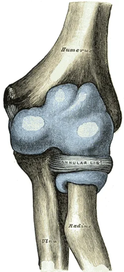
    The proximal radioulnar articulation, sometimes referred to as the proximal radioulnar joint (PRUJ), is a synovial pivot joint that occurs between the ring created by the annular ligament and the radial notch of the ulna and the circumference of the radius head.

    Introduction

    A synovial joint that joins the proximal ends of the radius and ulna is called the proximal radioulnar joint. The annular ligament and the radial notch of the ulna form a ring that contains the circumferent head of the radius in this joint. This joint is a pivot joint due to its configuration.

    Being a uniaxial joint, the proximal radioulnar joint can move with only one degree of freedom, from pronation to supination. This movement is specific to the human upper limb.

    Structure

    The proximal radioulnar joint is a pivot joint made of synovium. It happens between the radius’s head’s circumference and the ring that the annular ligament and the ulna’s radial notch make. The forearm’s interosseous membrane and the annular ligament stabilize the joint.

    The PRUJ is created by the ulna’s radial notch articulating with the medial side of the radial head. The PRUJ is stabilized by several ligaments during movement.

    Near the proximal radioulnar joint are several nerves, including:

    Embryology

    Shh secretion by the notochord controls the formation of the upper limb bud, which begins 26 days after fertilization.

    The bud’s lateral plate mesoderm creates the joint’s bone, cartilage, and tendon. These cells develop into chondrocytes and osteoblasts after the mesenchyme condenses into a blastema in the limb bud core. Proximally, the upper limb begins to chondrify at 36 days.

    At the locations of future joints, chondrification is suppressed. Studies have shown that these interzones express the proteins GDF5, WNT1, and WNT14. The annular ligament and proximal radioulnar joint interzone develop 51 days following fertilization. The formation of the articular cavity starts 56 days after fertilization. In the eleventh week, the elbow starts to mature. By week 12, the quadrate and annular ligaments are well-defined.

    Articular surfaces

    The radial fossa of the ulna and the head of the radius are the articular surfaces of the proximal radioulnar joint.

    Hyaline cartilage lines on both surfaces. While the radial fossa is reciprocally concave, the radial head is round and convex.

    The radial fossa, however, only touches one-fifth of the radial head. To ensure the completeness of this pivot joint, the radial head is tightly held against the ulna’s radial fossa by the annular ligament.

    Joint capsule

    The joint capsule, which surrounds the elbow joint, is continuous with the synovial membrane. The anterior portion of the capsule, which attaches the medial epicondyle and ulnar coranoid process anteriorly, makes it comparatively slender in comparison to other capsules. The capsule’s posterior part is thinner than its prior part.

    Ligaments

    Proximally, the fibrous capsule of the radioulnar joint is continuous with the capsule of the elbow joint, but distally, it links to the annular ligament. The articular surface margins and the annular ligament are where the synovial membrane is attached. Along the inside of the capsule, it forms a continuous line with the elbow joint’s synovial membrane. Therefore, there is a single, continuous synovial cavity shared by the elbow and proximal radioulnar joints.

    The quadrate and annular ligaments support the proximal radioulnar joint.

    • Annular ligament: robust ligament that is connected to the front and back of the ulna’s radial notch. The head of the radius is surrounded by it and the ulna’s radial notch.
    • Ulnar collateral ligament
    • Radial collateral ligament

    Annular ligament

    After encircling the radial head and attaching to the posterior margin of the radial fossa, the annular ligament extends from the anterior margin of the ulna’s radial fossa. The radial head can spin within this stable ring, which is made possible by the annular ligament.

    While the ligament’s distal boundary affixes to the radius’s neck, its proximal margin is fused with the joint capsule. The surface of the radius is directly in contact with the thin layer of cartilage covering the internal surface of the annular ligament. The supinator muscle attaches to the superficial surface, which is united with the radial collateral ligament.

    A brief fibrous band called the quadrate ligament extends from the neck of the radius, directly in front of the radial tuberosity, to the superior portion of the supinator fossa of the ulna.

    Interosseous Membrane

    A band of connective tissue called the interosseous membrane connects the ulna and radius between the radioulnar joints.

    The lateral ulnar border and the medial radial border are separated by it. The sheet has tiny pores that serve as a channel for the blood vessels in the forearm.

    Three main purposes are served by this connective tissue sheet:

    • Adds stability by holding the ulna and radius together when the forearm is pronated or supinated.
    • Serves as a point of attachment for the forearm’s anterior and posterior muscles.
    • Transmits forces to the ulna from the radius.

    Innervation

    The branches of the ulnar, radial, musculocutaneous, and median nerves supply the proximal radioulnar joint.

    Blood supply

    The deep brachial artery’s radial collateral branch, as well as the radial and recurrent branches of the radial and common interosseous arteries, create the periarticular network that supplies blood to the proximal radioulnar joint.

    Blood is supplied to the upper extremities by the brachial artery, which splits at the proximal cubital fossa into the radial and ulnar arteries. Medial, lateral, and posterior arcades are formed around the elbow by branches of the brachial, radial, and ulnar arteries. The proximal radioulnar joint is supplied by the lateral arcade of these arcades.

    The radial recurrent artery and the interosseous recurrent artery anastomose with the radial and middle collateral arteries from the profunda brachii artery form the lateral arcade. The radial head and neck are perfused by the radial and interosseous recurrent arteries, while the intraosseous branch of the ulnar artery supplies blood to the proximal ulna.

    Lymph is drained from deep lymphatic vessels in the palm by the radial and ulnar lymphatic vessels (RLV, ULV), which are deep lymphatic vessels of the forearm. This anterior interosseous lymphatic vessel, which originates in the pronator quadratus, drains into the cubital fossa as the ULV continues proximally towards it. The lymph of the RLV and ULV enters the cubital fossa, drains into the cubital lymph nodes, and then enters the brachial lymphatic vessel (BLV). Proximally, the BLV continues and enters the axillary lymph nodes.

    Nerves

    The PRUJ shares innervations with the humeroulnar and humeroradial joints because they share a joint capsule. The elbow capsule’s innervation is separated into anterior and posterior capsules.

    A tiny branch of the musculocutaneous nerve makes its way to the anterior capsule’s center via the brachialis muscle. The branch overlaps with the medial and radial nerves in certain places as it moves laterally and medially, respectively.

    The median nerve innervates the medial part of the anterior capsule through tiny branches it produces before passing the pronator teres. Additionally, a branch from the median nerve may emerge just proximal to the elbow and descend to the anterior capsule. The radial nerve branch that innervates the posterior capsule also innervates the lateral part of the anterior capsule.

    The radial nerve travels down the lateral head of the triceps and innervates the lateral part of the posterior capsule. After passing through the supinator muscle, it innervates the anterior capsule as well. The medial part of the posterior capsule, which surrounds the medial epicondyle and olecranon, is innervated by joint branches of the ulnar nerve that emerge just above the cubital tunnel. The central part of the posterior capsule receives innervation from the radial and ulnar nerves that overlap.

    Muscles

    Pronation and supination are the PRUJ’s main movements, and they happen in tandem with the distal radioulnar joint.

    The pronator teres and pronator quadratus are the forearm’s two main pronators. The humeral head of the pronator teres originates on the medial epicondyle, while the ulnar head is derived from the coronoid process. These heads unite before inserting onto the mid-radius lateral surface. The pronator quadratus is connected to the lateral radius and the distal quarter of the anterior ulna. The median nerve innervates both the quadratus and the pronator teres from levels C6-7 and C8-T1, respectively.

    The main forearm supinators are the supinator and biceps brachii muscles. The long and short heads of the biceps brachii originate from the coracoid process and supraglenoid tubercle, respectively, and cover two joints. The heads then attach to the medial part of the radial tuberosity and the anterior capsule via the distal biceps tendon and the bicipital aponeurosis, respectively.

    The biceps can flex the elbow and supinate the forearm thanks to these attachments. From nerve root levels C5–6, the musculocutaneous nerve innervates the biceps muscle. The anterior proximal ulna, lateral epicondyle, RCL, and annular ligament are the origins of the supinator. The dorsolateral proximal radius is where it inserts after encircling the radius. The radial nerve innervates it at nerve root level C6.

    Movements

    To allow for forearm rotation, including pronation and supination, the proximal and distal radioulnar joints function as a single unit. Pronation and supination ranges of motion are 61–66° and 70–77°, respectively, while the arm is lying next to the torso.

    The head of the radius rotates inside the ring that the radial fossa and annular ligament form in the proximal radioulnar joint. The location of the forearm determines the dynamic axis of rotation. The axis goes through the ulnar attachment of the articular disc in the distal radioulnar joint and the center of the head of the radius proximally when the forearm is in a supinated position. The distal end of the axis travels medially and passes through the ulna’s head when the forearm is pronated.

    Supination and pronation are followed by a series of additional motions in the proximal radioulnar joint in addition to the rotation of the radial head;

    • The superior surface of the radial head revolves in opposition to the humeral capitulum.
    • The ridge of the radial head slides against the humeral capitulum-trochlea groove.
    • Laterally and inferiorly, the head of radius tilts in the transverse plane.
    • The radial head is displaced laterally because the ellipsoid cross-section of the radius causes its broader axis to enter a transverse plane.

    When the supination angle is 5°, the proximal radioulnar joint assumes a closed-packed configuration. The forearm is in the open-packed (resting) posture when it is flexed at 70° and supinated at 35°. Pronation and supination define and restrict the joint’s capsular arrangement. The supplementary movements of the anteroposterior gliding of the radial head against the ulna and the humeral capitulum are made possible by the proximal radioulnar joint.

    Muscles acting on the proximal radioulnar joint

    The pronator quadratus and pronator teres are the muscles that work on the proximal radioulnar joint to cause pronation. The pronator teres is involved in rapid movements and movements against resistance, whereas the pronator quadratus is sufficient for little motions.

    The contraction of the supinator muscle during forearm extension results in supination. During resistance exercises and/or forearm flexion, the biceps brachii muscle serves as an accessory supinator.

    Clinical significances

    A Monteggia fracture occurs when the radial head dislocates and the ulna fractures at the same time. Although these fracture patterns can happen to adults, they are most frequently seen in children aged 4 to 10. Although olecranon fracture-dislocation and radial head fracture-dislocation variant patterns are also recognized, the ulnar fracture conventionally occurs distal to the olecranon, and the dislocation involves both the PRUJ and the radiocapitellar joint.

    There are four types of Monteggia fractures. Forced pronation of the forearm, such as falling on an extended and pronated upper limb, is the most common cause of the injury. The most prevalent is type I, in which the ulna’s diaphyseal fracture occurs along with an anterior dislocation of the radial head. Type II posterior dislocation of the radial head frequently exhibits a concomitant radial head fracture.

    Children who have an ulna metaphyseal fracture and either a lateral or anterolateral radial dislocation are most likely to experience type II. Finally, an anterior dislocation that includes a proximal third of the radius fracture and an ulna diaphyseal fracture is a type IV. Restoring motion can be accomplished through principal treatment using open reduction and internal fixation (ORIF).

    Children can also develop a nursemaid’s elbow, a dislocation of the radial head without a concomitant fracture. When the forearm is pulled axially, the radial head subluxes outside the annular ligament, which becomes positioned between the radial head and capitellum. This is a common consequence of this subluxation. Isolated radial head subluxation is significantly less frequent in children older than five as the annular ligament’s structural integrity increases and becomes thicker.

    Pediatric patients typically arrive with their arms held in a slightly flexed and slightly pronated position. Both the hyperpronation technique and the supination and hyperflexion maneuver, which apply the thumb reduction force over the radial head, are examples of reduction techniques. Although rare, recurrence is possible, particularly in patients under three. For initial events, immobilization is not required. Consider cast immobilization with the arm held in flexion and in a neutral to slight forearm supination position if recurrence is observed several times in a short time.

    FAQs

    The proximal radioulnar joint: what is it?

    The proximal ends of the radius and ulna are joined by a synovial joint called the proximal radioulnar joint. The annular ligament and the radial notch of the ulna form a ring that contains the circumferent head of the radius in this joint. This joint is a pivot joint due to its configuration.

    Which joint type is the proximal joint?

    The articulation between the hand’s proximal and middle phalanx is known as the proximal interphalangeal joint (PIPJ). All fingers except the thumb have it. Fine motor control is aided by this synovial hinge joint, which allows for flexion and extension in the middle of the fingers.

    Which ligament is the most crucial component of the proximal radioulnar joint?

    A sequential transection of the soft tissue constraints of the proximal radioulnar joint revealed that the central band of the interosseous membrane and the annular ligament serve as the primary stabilizers in pronation, while the central band is the major stabilizer in supination.

    What is the proximal radioulnar joint used for?

    The ulna on the medial side and the radius on the lateral side are the two forearm bones that connect to form the radioulnar joints. Pronation and supination are made possible by the combined action of the superior, or proximal, and inferior, or distal, radioulnar joints.

    Which two bones make up the joint between the proximal radioulnar bones?

    Immediately distal to the elbow joint, and contained within the same articular capsule, is the proximal radioulnar joint. The radial notch of the ulna and the head of the radius articulate to form it.

    Which rule of the proximal radioulnar joint is concave-convex?

    According to the convex-concave rule, rolling and gliding happen in opposite directions during forearm pronation and supination when the convex radial head articulates with the concave radial notch on the ulna. This is thought to be the case with the arthrokinematics of the proximal radioulnar joint (PRUJ).

    What is the proximal ligament of the radioulna?

    The proximal radioulnar joint is a pivot joint made of synovium. It happens between the radius’s head’s circumference and the ring that the annular ligament and the ulna’s radial notch form. The joint is stabilized by the annular ligament and the forearm’s interosseous membrane.

    At the proximal radioulnar joint, which muscles are present?

    Pronator teres muscle.
    Flexor carpi radialis muscle.
    Palmaris longus muscle. palmar aponeurosis.
    Flexor carpi ulnaris muscle.

    What distinguishes the distal radioulnar joint from the proximal one?

    Together with the humeroulnar and humeroradial joints, the proximal radio-ulnar joint (PRUJ) makes up the elbow’s articulating components. Pronation and supination of the forearm are made easier by the PRUJ, which is situated in the proximal forearm and works in tandem with the distal radio-ulnar joint (DRUJ).

    How does the proximal radioulnar joint look clinically?

    The proximal ends of the radius and ulna are joined by a synovial joint called the proximal radioulnar joint. In this joint, the annular ligament and the radial notch of the ulna form a ring that contains the circumferent head of the radius. This joint is a pivot joint because of its configuration.

    What are the proximal radioulnar joint landmarks?

    The radioulnar joint at the proximal end is a synovial pivot joint. It is situated between the ring created by the annular ligament the radial notch of the ulna and the circumference of the radius head. The joint is stabilized by the annular ligament and the forearm’s interosseous membrane.

    What is the proximal radioulnar joint also known as?

    The synovial pivot joint between the radius’s head circumference and the ring created by the ulna’s radial notch and the annular ligament is called the proximal radioulnar articulation, or proximal radioulnar joint (PRUJ).

    References

    • TeachMeAnatomy. (2020b, November 7). The Radioulnar Joints – TeachMeAnatomy. https://teachmeanatomy.info/upper-limb/joints/radioulnar-joints/
    • Proximal radioulnar joint. (2023, November 3). Kenhub. https://www.kenhub.com/en/library/anatomy/proximal-radioulnar-joint
    • Wikipedia contributors. (2025, January 11). Proximal radioulnar articulation. Wikipedia. https://en.wikipedia.org/wiki/Proximal_radioulnar_articulation
    • Williams, M. R., & Varacallo, M. A. (2023, July 24). Anatomy, shoulder and upper limb, proximal Radio-Ulnar joint. StatPearls – NCBI Bookshelf. https://www.ncbi.nlm.nih.gov/books/NBK551614/
    • Hacking, C., & Stewart, M. (2017). Proximal radioulnar joint. Radiopaedia.org. https://doi.org/10.53347/rid-53632


  • Facet Joints (Zygapophyseal Joints)

    Facet Joints (Zygapophyseal Joints)

    The facet joints, which are a group of synovial, plane joints between the articular processes of two neighboring vertebrae, are also known as zygapophysial joints, zygapophyseal, apophyseal, or Z-joints. Each spinal motion segment has two facet joints, and the recurrent meningeal nerves innervate each facet joint.

    Introduction

    This synovial connection, called the apophyseal or zygapophyseal joint, connects the superior articular process of one vertebra to the inferior articular process of the vertebra located directly above it. For every segment of spinal motion, there are two facets.

    The facet joints serve as the articular pillars that give the entire vertebral column structural stability. They are located between the pedicle and lamina of the same vertebra.

    The bilateral facet joints, in conjunction with the disc, transfer loads and, because of their mechanical function and geometry, guide and constrain motions in the spine.

    Articulating Surfaces

    The articular facet is on the superior process of the vertebra below, and the articular facet is on the inferior process of the vertebra above. The superior facet of the inferior vertebra is more convex in the lumbar area and relatively flat in the cervical and thoracic regions.

    The superior vertebra’s opposite inferior facet is concave, forming an arch with its peak pointing in the direction of the vertebral body. Depending on the location, facet joints can have varying orientations:

    The cervical region is 45 degrees in the frontal plane, and all motions, including rotation, flexion, extension, and lateral flexion, are available.

    The cervical vertebrae have articulating facets that face 45 degrees to the transverse plane and parallel to the frontal plane. The inferior articulating processes face down and anteriorly, while the superior articulating processes face up and posterior.

    No flexion or extension, lateral flexion and rotation, frontal plane, and thoracic region = 60 degrees

    While the inferior facets face medially, down, and anteriorly at the facet joints of adjacent thoracic vertebrae, which are inclined at 20° to the frontal plane and 60° to the transverse plane, the superior facets face posteriorly, as well as somewhat up and laterally.

    The lumbar region is 90 degrees in the sagittal plane, with just flexion and extension.

    The facet joints in the lumbar area are located in the sagittal plane; the articulating facets are 45° from the frontal plane and at right angles to the transverse plane. The superior and inferior faces are oriented laterally and medially, respectively.

    This is altered at the lumbosacral junction, where the inferior facet of L5 faces forward and the apophyseal joint shifts into the frontal plane. The spinal column cannot slide forward on the sacrum due to this change in orientation.

    Ligaments and Joint Capsule

    In addition to stabilizing the vertebral column, the posterior ligamentous complex maintains the facet joints of the adjacent vertebrae in a fixed relationship with one another. The following structures comprise it:

    • Posterior longitudinal ligament
      The facet joint capsule, which surrounds the joint like other synovial joints in the body, creates synovial fluid to lubricate and nourish the joint. The spinal nerves posterior ramus (dorsal primary rami) produce paired medial branches that innervate each facet joint. Both the spinal nerve below and the spinal nerve above the facet provide the facet with a medial branch. The medial branch nerve is triggered by pain in facet joints that are degenerated or inflamed. The limbs’ muscles and sensations are not under the control of these nerves.
    • ligamentum flavum
    • Interspinous ligament
    • Supraspinous ligament

    Motions Available

    Anatomical features including ligaments, facets, and intervertebral discs restrict the motion of each spinal segment. The coupling of the spine’s motions, or synchronous movements, is specifically caused by anatomical features.

    There is a physiological connection between flexion, extension, translation, axial rotation, and lateral bending. Anatomical structure differences by location determine the precise coupling pattern. Axial rotation and side bending occur in the same direction in the cervical and upper thoracic spines.

    Lateral bending and axial rotation in the opposite direction are combined in the lumbar spine. The pattern of connection in the middle and lower thoracic spine is irregular. However, according to which movement is started first, the coupling pattern will vary. If lateral bending occurs initially in the lumbar spine, it will be followed by axial rotation in the same direction. On the other hand, axial rotation will be accompanied by lateral bending in the opposite direction if it occurs first.

    Innervation

    Although innervation to the facet joints varies throughout spinal segments, medial branch nerves originating from the dorsal rami often innervate them. These nerves are believed to receive primarily sensory information, although there is some evidence that they also get motor input from the local musculature.

    The medial branch nerve, a branch of the dorsal rami, innervates the majority of the cervical spine’s joints from the same levels. That is, the C4 and C5 medial branch nerves innervate the facet joint between the C4 and C5 vertebral segments. There are two exceptions, though:

    • The C3 medial branch nerve and the third occipital nerve innervate the facet joint between C2 and C3.
    • The medial branch neurons of C7 and C8 innervate the facet joint between C7 and T1.

    The medial branch nerves from the vertebral segment above the upper segment and the upper segment innervate the facet joints in the lumbar and thoracic spines. For instance, the medial branch neurons of T1 and C8 innervate the facet joint between T1 and T2.

    Facet joint between L1 and L2; medial branch nerves of T12 and L1. However, the L5 dorsal ramus and the L4 medial branch nerve innervate the L5 and S1 facet joint. The facet joint is not innervated in this instance by the L5 medial branch.

    Function

    The spinal motion segment’s movement is guided and limited by the biomechanical function of each pair of facet joints. The facet joints, for instance, serve to shield the motion segment in the lumbar spine from excessive rotation, flexion, and anterior shear pressures. The range of side bending (lateral flexion) seems to be mostly unaffected by facet joints.

    Degeneration, dislocation, fracture, injury, trauma-induced instability, osteoarthritis, and surgery can all impair these capabilities. The purpose of the facet joints in the thoracic spine is to facilitate rotation and limit the degree of flexion and anterior translation of the corresponding vertebral segment.

    Cavitation of the synovial fluid in the facet joints causes the popping sound (crepitus) that is often associated with manual spinal manipulation, sometimes known as “cracking the back.”

    Both the superior and inferior facet joints are positioned to restrict rotation and permit flexion and extension. This is particularly valid for the lumbar region.

    Clinical significance

    The facet joints, which are the only genuine synovial joints connecting neighboring spinal levels, are affected by osteoarthritis (OA) of the spine. They are situated on the posterior side of the vertebral column.

    One of the most common causes of back and neck pain in older persons is facet joint osteoarthritis (FJ OA). As people age, facet-mediated pain becomes more common in clinical populations, indicating that FJ OA may play a significant role in treating spinal pain in elderly folks.

    A positive diagnostic facet joint block may suggest that chronic spinal pain is coming from the facet joints. For these people, certain treatments like neurolysis using radiofrequency or cryoablation may help remove facet joint pain. According to a 2013 study that examined the evidence supporting lumbar facet joint injections and physiotherapy therapies, the injections produce a brief duration of pain relief. For patients with chronic lower back pain, physiotherapy therapies such as soft tissue massage and land-based lower back mobility exercises may be helpful during this period to enhance long-term results.

    Facet joint arthritis

    All joints experience degenerative changes with age, largely because of the mechanical nature of their function. The facet joint in particular and other joints in the spine are especially affected by this.

    Facet joint arthritis or facet arthropathy are the frequent names for this condition. The degenerative process might cause the joint to expand, just like with any other type of arthritis. Small alterations to the facet joint may cause the intervertebral foramen to narrow, potentially putting pressure on the spinal nerve roots inside. Severe inflammatory reactions in the Z-joint, similar to a swollen arthritic knee, may occur in more severe cases.

    Summary

    The stability and movement of the spine depend heavily on the facet joints. The facet joints, like other synovial joints, experience a series of degenerative characteristics, such as osteoporosis, articular cartilage thinning, subchondral bone sclerosis, cartilage fibrillation, joint space narrowing, and the formation of juxtafacet cysts.

    Facet tropism, gender, rising BMI, and anomalies of the ligamentum flavum are additional potential contributors to facet joint degeneration, along with lower spinal level, age, sagittal orientation, and IVD degeneration.

    Facet joint degeneration can be detected and its severity evaluated using a variety of pathologic and imaging grading systems, each with unique advantages and disadvantages. The natural history of facet joint degeneration and its correlation with clinical symptoms should be further investigated to detect and quantify its severity.

    A deeper comprehension of the facet joints, how they interact with other spinal structures, and how pain and impairment develop can help to improve the precision of spine patient management and treatment.

    FAQs

    Which joints are zygapophyseal?

    The superior articular process of one vertebra and the inferior articular process of the vertebra directly above it form a synovial joint that is also referred to as the zygapophyseal or apophyseal joint. In every spinal motion segment, there are two facet joints.

    What kind of joint does the facet joint belong to?

    The articular facets of the vertebrae are joined by facet joints, which are symmetrical synovial-lined joints with a fibrous capsule. The inferior I facet of the vertebra above articulates with the superior facet of the lower vertebra.

    Are joints with facets gliding?

    You can bend and twist your body thanks to the facet joints of your spine, which slide and glide. As we age, our facet joints may develop adhesions that limit our range of motion. Adjustments aid in dissolving adhesions around the facets, restoring your range of motion and reducing pain.

    What is the facet joint’s true name?

    True synovial joints, facet joints—also referred to as zygapophyseal or apophyseal joints—can experience degenerative changes in a manner akin to that of other synovial joints.

    What is the total number of facet joints?

    The facet joints, which are a group of synovial, plane joints between the articular processes of two neighboring vertebrae, are also known as zygapophysial joints, zygapophyseal, apophyseal, or Z-joints. Each spinal motion segment has two facet joints, and the recurrent meningeal nerves innervate each facet joint.

    A zygapophyseal joint is what kind of synovial joint?

    Zygapophyseal joints, which make up the postero-lateral articulation between vertebral levels, are the only synovial joints in the spine. They are composed of a joint capsule, a synovial membrane, and hyaline cartilage covering the subchondral bone.

    What is the facet joint’s size?

    For the central zone, the facet joint space width was 1.93±0.51 (mean ± standard deviation) mm, the superior zone was 1.75±0.48 mm, the inferior zone was 1.63±0.49 mm, the medial zone was 1.48±0.44 mm, and the lateral zone was 1.65±0.48 mm. The right and left facet joints did not differ much.

    What is a facet joint’s typical angle?

    The articular surfaces of the facet joint, in the sagittal plane, often have an inclination angle between 20° and 78° in the cervical region, 55° and 80° in the thoracic region, and 82° and 86° in the lumbar region.

    What features distinguish facet joints?

    A capsule containing a trace amount of synovial fluid envelops the joint. To lessen friction between rubbing bones, the synovial fluid serves as an extra lubricant. In addition to providing support for the spine, healthy facet joints permit a great deal of bending and twisting.

    What form does a facet take?

    A facet is a characteristic of a polyhedron, polytope, or other analogous geometric structure in geometry that is typically one dimension smaller than the structure itself. More precisely, any polygon whose corners are the polyhedron’s vertices but are not a face is referred to as a facet of a polyhedron in three-dimensional geometry.

    Which ligaments make up the facet joint?

    Along the spine’s length, the facet joints are located on either side of the spinous processes on the back of each vertebra. The ligamentum flavum and the FCL surround the joint, which is made up of two facet surfaces from neighboring vertebrae. They help control joint motion by limiting synovial fluid flow.

    In what location are facet joints found?

    The facet joints serve as the articular pillars that give the entire vertebral column structural stability. They are located between the pedicle and lamina of the same vertebra.

    What is the significance of facet joints?

    These joints prevent the back from twisting excessively or slipping too far forward while allowing the spine to flex and twist. When these joints experience tension and damage from an injury, normal wear and tear, or disc degeneration, facet joint syndrome develops.

    Why is a facet joint used?

    The facet permits mobility while stabilizing the spine. Due to damage or “wear and tear” (degenerative change), these joints may become uncomfortable. The facet joints are the source of pain, which might spread.

    References

    • Wikipedia contributors. (2024c, June 2). Facet joint. Wikipedia. https://en.wikipedia.org/wiki/Facet_joint#:~:text=The%20facet%20joints%20(also%20zygapophysial,by%20the%20recurrent%20meningeal%2
    • Zygapophyseal joints. (2023, October 30). Kenhub. https://www.kenhub.com/en/library/anatomy/zygapophyseal-joints
  • Vastus Medialis Muscle Pain

    Vastus Medialis Muscle Pain

    Vastus Medialis Muscle Pain occurs due to overuse, muscle imbalances, or strain, often leading to discomfort around the inner thigh or knee. It can affect knee stability and movement, requiring proper stretching, strengthening, and recovery for relief and prevention.

    What is a Vastus Medialis Muscle Pain?

    On the inside of the thigh, the vastus medialis is a crucial muscle in the quadriceps group. It is essential for knee stability and extension, especially for preserving appropriate patellar tracking. Overuse, strain, muscle imbalances, or underlying knee issues can all cause vastus medialis muscle pain, which frequently manifests as pain in the inner thigh or around the knee. Daily activities like walking, crouching, and climbing stairs may be impacted by this pain, which calls for appropriate management to avoid more issues.

    The more common of the two, lower vastus medialis soreness with a trigger point is found just above and inside the knee joint. The front of the knee has an aching sensation that may radiate into the lower medial thigh region. The pain from this trigger point may go away after a few weeks, but a sudden weakness follows it in the muscle that causes the knee to buckle when walking suddenly.

    On the inside of the lower thigh, a few inches above the lower trigger point is the upper vastus medialis trigger point. Pain is directed directly above the knee and down the inside of the leg.

    Anatomy of Vastus Medialis Muscle pain?

    • Often referred to as the teardrop muscle, the Vastus medialis is a large quadriceps muscle located on the inside of the leg. It is the most prominent of the three quadriceps (thigh) muscles. It is positioned behind the vastus lateralis and in front of the rectus femoris.
    • The muscle is responsible for the knee joint’s ability to straighten. It is a component of the knee’s extensor mechanism.
    • When the knee is at a nearly 30-degree angle, especially when the leg is partially extended, the muscle is operating more fully. Consequently, the ideal workouts for strengthening the quadriceps are those that incorporate a final 30-degree angle motion exercise.
    • Because it works on the terminal range of knee extension (Last 30-degree knee Extension), physical therapists focus more on strengthening the Vastus medialis than other quadriceps muscle groups.

    Origin: Along with the spiral line to the medial lip of the linea Aspera, the medial intermuscular septum, and the aponeurosis of the adductor magnus, it originates from the lower portion of the intertrochanteric line.

    Insertion: After joining the rectus femoris, vastus lateralis, and vastus intermedius muscles, it penetrates the medial side of the quadriceps tendon, encircling the patella before entering the tibial tuberosity through the patellar ligament.

    Nerve supply: One element from the back is the femoral nerve. The L2, L3, and L4 are the nerve roots.

    Blood supply: Three muscular branches of the femoral artery supply the vastus medialis. Additionally, the deep femoral and down genicular arteries provide it with some little support.

    Structure: One of the four muscles that make up the quadriceps muscle is the vastus medialis, which is located in the thigh’s anterior compartment. The rectus femoris, vastus lateralis, and vastus intermedius are the remaining muscles. Of the muscles in the vastus group, it is the most medial. Along the whole length of the femur, the vastus medialis begins medially and joins the other quadriceps muscles in the quadriceps tendon.

    A continuous line of attachment on the femur, beginning on the front and middle side (anteromedially) on the intertrochanteric line, gives rise to the vastus medialis muscle. It then plunges down the inner (medial) lip of the linea Aspera and onto the medial supracondylar line of the femur after continuing down and back (posteroinferiorly) along the pectineal line. The fibers attach to the inner (medial) border and the inner (medial) portion of the quadriceps tendon on the patella.

    The many distal part of the vastus medialis muscle is known as the obliquus genus muscle. Its specific function is essential for maintaining the patella portion and preventing knee injury. It is merely the vastus medialis’s most distal fiber kind, and it lacks a good depiction.

    Function: There are four muscles in the thigh’s anterior compartment, including the vastus medialis. Together with the other quadriceps muscles, it is used in knee extension. Additionally, the vastus medialis muscle aids in the patella’s appropriate tracking.
    It has been suggested that the vastus medialis muscle conflicts into two groups of fibers: the vastus medialis longus, which is a long and relatively inline group of fibers with the quadriceps ligament, and the vastus medialis obliquus, which is a shorter and more obliquely oriented set of fibers. However, there is insufficient evidence to definitively support or contradict this theory.

    Causes of Vastus Medialis Muscle pain?

    Quadriceps muscle pain may be triggered or reactivated by the following situations or activities:

    • Usually, “catching oneself” after stumbling or unintentionally walking into a hole overloads the quadriceps muscle group as a whole. These circumstances cause the muscles to contract strongly in an eccentric (lengthening) manner in an attempt to slow down the body’s entire weight during the fall.
    • These muscles may also be overloaded by a new training regimen that includes deep knee flexion or squats.
    • Chronic tightness in the hamstrings might result from untreated trigger point activity in these muscles. The quadriceps, the hamstrings’ biomechanical twin, will continuously produce trigger point activity in response to the hamstrings’ persistent muscle stress.
    • When attempting to stoop down and lift anything, a patient with tension in their soleus muscle will typically compensate by overusing their quadricep muscles. When squatting, the tight soleus prevents individuals from properly dorsiflexing, or flexing their ankle.
    • In general, skiing and skiing-related incidents are more taxing on the quadriceps muscles.
    • People with Morton’s Foot Structure (see Connected Disorders) and those who pronate their feet (toes outward) exhibit more frequent trigger point activity in the vastus medialis.
    • Common sports activities like basketball, football, soccer, running, and jogging can also put too much strain on this muscle.
    • The vastus medialis trigger points may be triggered in auto accidents if a person’s knee strikes the steering wheel.

    Symptoms of Vastus medialis muscle pain

    The following are three typical signs of vastus medialis injury:

    • Pain at the front and inside of the knee, as well as in the inner thigh
    • persistent knee joint pain
    • Knee buckling, also known as when the knee gives out

    Other signs and symptoms include:

    • Thigh cramping or tightness
    • Tenderness when the area is touched
    • Bruising
    • Swelling
    • When the toes on the affected leg point outward, this is known as foot pronation on the affected side.
    • A common definition of pain is an ache that occurs both when moving and when at rest. A muscle that is hardly injured will hurt a lot. But after a few weeks, the pain can go away, only to be replaced by weakness in the knee when moving.

    Any of the following signs or symptoms will be present in this muscle:

    • Patients often report deep knee pain that seems like a toothache, especially while they’re trying to fall asleep.
    • Over time, people might also develop buckling knee disease, which is characterized by an abrupt weakening in the knee that causes patients to fall when walking.
    • Pronation is the constant pointing of the toes on the foot of the artificial leg outward.

    Diagnosis of Vastus medialis muscle pain?

    Examination

    Palpation

    • It is palpable all the way around. The quadriceps tendon, which connects to the patella’s proximal border (base), can be felt farther away.

    Related Knee Pain Disorders

    Knee Joint Injury: In both professional and recreational sports, ligament and meniscal strains and tears are rather common. The quadriceps, hamstrings, and gastrocnemius muscles are the muscles that drag the knee joint, and like any other joint, they are susceptible to aberrant stress from long-term tension. Regular (maintenance) trigger point therapy for the hamstring, vastus lateralis, and vastus medialis muscles could prevent many of these injuries. This is especially true for athletes at both extremes of the activity spectrum (those who are highly prepared and those who are badly conditioned). Many of these problems are inevitable in the conventional sense since they result from physical trauma to the leg and knee.

    • It is thought that the main cause of knee pain is weakening or exhaustion in some quadriceps muscles, especially the vastus medialis obliquus (VMO). It is known as tiredness and can be caused by a wide range of processes, from the buildup of metabolites in muscle fibers to the development of insufficient motor control in the motor cortex. The actuality of knee joint pain (patellofemoral pain syndrome) is correlated with the vastus medialis’s characteristics, especially its angle of insertion. This syndrome is challenging and conclusive, nonetheless.
    • Vascular medialis obliquus (VMO) foundering and fatiguing results in mal tracking of the patella and subsequent damage to surrounding structures, which puts advanced force on the knees and frequently causes injuries like tendinitis, anterior cruciate ligament rupture, chondromalacia, and patellofemoral pain syndrome. Researchers can assess and document the electrical movement generated by the skeletal muscle of the vastus medialis obliques to investigate the biomechanics and identify any potential anomalies, weakness, or exhaustion by using electromyography to analyze the muscle activity of the VMO and appropriate rehabilitation plans.
    • It is possible to provide objectives that would not only rectify the irregularity that has already been identified but also, if tested earlier, prevent such damages. Keeping injuries under control and teaching appropriate training methods are crucial to preventing valgus collapse forces from putting undue strain on other knee structures, creating asymmetry, and making a person more prone to injury.
    • Pain in the patellar region and the patellofemoral is linked to a deficiency of the vastus medials. Strengthening the medial oblique fibers and assessing the degree of foot supination and pronation are two aspects of a therapy plan that aims to restore the equilibrium between the vastus medialis and lateralis.
    • Due to the poor quality of the evidence supporting isolated activities, VMO strengthening has lost favor as a treatment for anterior knee pain. After that, students question whether VMO exists and discover that any quadriceps exercise will also stimulate the vast muscles. Khayambashi et al. confirmed that hip strengthening was more effective than knee strengthening in reducing patellofemoral pain, supporting the idea that strengthening further up the kinetic chain is a more realistic strategy.

    Patellofemoral Dysfunction: The patella, or knee cap, tracks abnormally when the knee joint moves, which is a sign of this condition. Patella tracking requires appropriate conformation of the contraction of the vastus lateralis and vastus medialis muscles. This coordination may be hampered by trigger points in either of these muscles, leading to a laterally positioned knee cap.

    Phantom Limb Pain: In persons who have had a limb amputated, trigger point pain is frequently the origin of phantom limb pain because it is typically felt in a different part of the body. These individuals may be informed that their pain is “not real” and is a mental fabrication, but in fact, it is just as genuine as anybody else’s and originates from trigger points in the remaining part of the limb or trunk. Being a neurological reflex designed to instruct the brain to alter one’s physical activity so the muscle containing the trigger point may rest and heal, directed pain does not require the existence of the body part it is targeting.

    Nerve Entrapment: The saphenous nerve becoming trapped can cause pain in the medial region of the knee.

    Morton’s Foot Structure: When the base of the big toe is closer to the bottom of the second toe, which is adjacent to it, the foot’s bony structure deviates. Usually, where the foot joins the second toe, a thick callus develops. The vastus medialis muscle may be overworked as a result of this illness, which produces side-to-side swaying or instability of the foot and knee.

    Growing Pains: While trigger point movement in the quadriceps (and other muscles) is the true reason, children’s unexplained knee and thigh pain is sometimes mistaken for “growing pains.” Children can easily overstretch their leg muscles when playing since they are always pushing their bodies to new boundaries.

    Treatment of Vastus Medialis Muscle Pain:

    The degree to which Rectus Femoris is severe will determine how it is treated. Acute rectus femoris should be treated using the RICE (rest, ice, compression, and elevation) principles.

    • Rest: refrain from doing painful activities (such as running, leaping, climbing or descending stairs, kneeling, and squatting).
    • Apply ice to the irritated tendon or region. This method is among the most rapid ways to lessen pain, inflammation, and edema. After applying it immediately, do so for 20 minutes at a time during breaks. Avoid putting ice on the skin right away.
    • Compression, like an ace bandage, can assist relieve the strain on the injured muscle. Use gentle compression if you’re using ice. This is especially useful if there is edema.
    • Elevation: assist reduce edema by promoting the area.

    If there are ruptures or significant damage to the rectus, hamstrings, or adductors, crutches may be necessary for walking or moving around.

    In moderate cases, medication, ice, and rest might be sufficient to ease the pain. Physical therapy is advised to create a regimen of strengthening and stretching exercises once the pain has subsided to stop the injury from happening again. To avoid a relapse of symptoms, return to the movement gradually.
    The next step should be to speak with your healthcare practitioner if the issue persists. To determine which tendon or tendon(s) are affected, the severity of the condition, and the best course of therapy, your physical therapist will do a thorough evaluation.

    Medical treatment:

    • The initial line of treatment is to avoid the actions that cause the engaged tendon to become painful or stressed.
    • RICE: Compression, Ice, and Rest To alleviate the strain on the bursa, elevation is recommended.
      Nonsteroidal anti-inflammatory medications, or NSAIDs, are used to reduce inflammation and pain.
    • Steroid injections could be necessary to reduce inflammation in the affected tendon.
      To relax the tendon and encourage healing, immobilization, strapping, or bracing may be beneficial.

    Physical therapy Treatment

    Physical therapists are experts who have received the necessary education and training to support interventions.

    To assess and choose the following, a physical therapist will do a comprehensive evaluation:

    • Tendon: To determine which tendon is impacted, a series of tests will be carried out.
    • Strength: To determine whether there are related weaknesses or strength discrepancies, opposing testing is carried out.
    • Flexibility: Tight muscles can contribute to weak muscles and poor mechanics, which can lead to inequality and make the hip more vulnerable to tendinitis.
    • Technique: Our running, jumping, cycling, or rowing motions can frequently cause issues. To improve technique, look at and notice the activities you engage in that might have caused the issue.
    • workout: review your workout regimen and any hasty adjustments that might have caused or contributed to the current state of affairs.
    • To determine whether there are any disparities, a physical therapist will assess your alignment, foot mechanics, and leg lengths. A vital component of balancing the strains placed on your body and legs is making sure you are wearing appropriate footwear.
    • To prevent making the situation worse, physical therapy for rectus femoris/quadriceps tendinitis must start traditionally. To promote healing, the focus will be on rest, reducing inflammation, and boosting blood flow. After the initial inflammation has subsided, a stretching and strengthening program will be started to improve the muscles’ flexibility and strength and lessen the strain on the hip and tendons. It could be necessary to use strapping or taping to rest, reduce strain on the tendon, and encourage recovery.

    The following are typical physical therapy techniques for rectus femoris pain:

    • The Manual Therapeutic Technique (MTT) is a hands-on approach used by physical therapists to improve knee and hip alignment, mobility, and range of motion. This includes soft tissue massage, stretching, and joint mobilization. Pain management is also made possible by the application of mobilization techniques.
    • Stretching and strengthening activities to restore range of motion and build stronger knee and lower extremity muscles to support, stabilize, and lessen the strain on the hip joint’s bursa and tendons are examples of therapeutic exercises (TE).
    • To alleviate the strain on the bursa and tendons during everyday motions, neuromuscular reeducation (NMR) is used to retrain the lower extremities and improve their movement mechanics and techniques (such as jogging, kneeling, squatting, and leaping). To relax the tendon and promote healing, bracing, strapping, or taping may be helpful.
    • To reduce pain and inflammation in the tough tendon and bursa, modalities include ultrasound, electrical stimulation, ice, cold laser, and others.
    • a home program that includes exercises for stabilization, stretching, and strengthening as well as guidance on how to advance to the next functional level and accomplish everyday tasks.

    Vastus Medialis muscle Stretching exercise:

    A physical therapist recommends stretching to relieve muscle tightness after electrotherapy has been used for two to three days to relieve muscle pain.
    When you feel comfortable and your pain has subsided, you apply this stretching technique.
    You can relieve tightness and soreness in your muscles with this stretching exercise.

    • The Lying Quad Stretch
    • The Simple Quad Stretch
    • The Kneeling Quad Stretch
    • Vastus Medialis Stretch

    The Lying Quad Stretch:

    half lying quad stretch
    The Lying Quad Stretch
    • With your left hand supporting your head, you are lying face down.
    • To finish this stretch, you can also lie on your side.
    • To stabilize yourself, flex your left knee joint and pull your right foot toward your butt after a few seconds.
    • After that, hold onto your ankle joint and maintain this stretch for 30 seconds.
    • Do it three times at a time, three times a day.

    The Simple Quad Stretch:

    • Standing on your left leg, you have one knee joint in contact with the other.
    • If necessary, you can keep a chair and the wall in place to support the ongoing.
    • With your right hand, grasp the right foot and draw it toward the direction of your butt.
    • Make sure your hips and chest are pressed forward.
    • Avoid worrying about putting your foot too near your butt.
    • To accomplish a decent hip flexor muscle stretch, however, you must concentrate on feeling the stretch in your quadriceps and moving your hip joint forward.
    • Hold this posture for thirty seconds.
    • Do it three times in a row and three times a day.

    The Kneeling Quad Stretch:

    Kneeling-quadriceps-stretch
    The Kneeling Quad Stretch
    • The starting position for this stretching exercise is a high lunge with the left foot forward.
    • Next, carefully lower your right knee joint to the floor and pause to regain your equilibrium.
    • When you’re ready, extend your right arm back and grasp your toes or ankle joint.
    • For 30 seconds, hold this stretching stance while maintaining bodily stability.
    • Next, Return to the lunge stance slowly, switching from the left foot to the right.
    • Do this stretching exercise three times at a time, three times a day.

    Vastus Medialis Stretch:

    • Quadriceps Stretching Exercise While Standing
    • For balance, you are standing with a chair and the wall.
    • Without bending forward, bring your heel up behind you, grab your ankle joint, and draw your heel toward your buttocks until you feel a stretch.
    • All four quadriceps muscles are stretched, but the vastus medialis muscle is also stretched by shifting the foot across the body to the opposite buttock.
    • Pushing your hip joint forward during this exercise will improve the stretch.
    • Do this stretching exercise three times at a time, three times a day.

    Vastus medialis muscle Strengthening Exercises:

    Following two to three days of electrotherapy and massage to relieve muscle pain, the physical therapist recommended strengthening activities to address muscle weakness.
    It is always advised to perform this strengthening exercise when you feel comfortable and pain-free.

    Muscle weakness and soreness can be alleviated with this all-strengthening exercise:

    • Lying Pigeon Progression
    • The Frog Pose
    • Floor extension
    • Lateral heel drop
    • Step downs
    • Leg extension
    • Single leg raises
    • Terminal knee extensions (TKEs)
    • Vastus Medialis activation Exercise
    • Ball Clench Extensions
    • Twisted Leg Raise
    • Ball Bridges
    • Ball Wall Squats
    • Isometric Contraction of the vastus medialis muscle
    • Seated Isometric vastus medialis muscle & Adduction
    • Externally Rotated ½ Squats
    • Wall/Ball Squat
    • Split Squats/Static Lunges
    • Step-Ups

    Lying Pigeon Progression:

    • The initial posture of the Lying Pigeon Progression involves lying face down on a mat on the floor.
    • An opposition band must then be securely placed around the injured foot, keeping the spare band within easy reach.
    • Keep your right leg outstretched or flex your left knee joint while you grasp the band with your left hand.
    • You have to point your toes toward the ceiling.
    • Then, until you feel the strain, pull forward with the opposition band.
    • Hold this workout posture for ten seconds.
    • Do this exercise three times a day and repeat the strengthening ten times in one sitting.

    The Frog Pose:

    • The first step in the Frog Pose exercise is to lie on your stomach, or in a prone position, with your elbow joints supporting your body.
    • Reach back to grasp your feet after flexing both of your knee joints.
    • This is where you feel the stretching.
    • Next, To point to the roof, carefully raise your elbow joint after switching your fingers to point in the same direction as your toes.
    • When you have any hip or knee pain, you should stop doing this exercise.
    • For ten seconds, hold this workout position.
    • Do this strengthening exercise three times a day and ten times in one sitting?

    Floor extension:

    • You have a lofty stance and are sitting on the ground.
    • With your chest proud, drag your shoulder joint down the back.
    • Next, with your left foot flat on the floor, flex your left knee joint toward your chest.
    • With your foot pointing slightly out to the side, raise your leg in front of you.
    • Keep both hands clasped beneath the left knee joint and keep your muscles bent throughout the workout.
    • Exhale while maintaining proper posture, leaning away from the wall, and raising your right leg as high as you can.
    • Hold this posture for ten seconds.
    • After that, take a breath and slowly descend to your starting posture.

    Heel drop:

    heel-drop
    Heel drop
    • Your left leg is straight but not locked, and your foot is resting on a little object while you stand erect.
    • Your left foot should lie flat on the floor, and your right knee should be slightly bent.
    • You have to be running your right knee over your toes.
    • Next, To maintain balance, squeeze your core muscle.
    • Push up off the right leg and exhale until both legs are fully extended.
    • As you step forward, try to maintain your hip joint state.
    • Take a breath, contract your left vastus medialis muscle, and then slowly go back to where you were before.
    • Hold this exercise position for ten seconds.
    • Do this strengthening exercise three times a day and ten times in one sitting?

    Step downs:

    • With your left foot over to the side and your right foot on the stage, you are standing.
    • Take a deep breath and flex your vastus medialis muscle.
    • Once your left foot is flat on the ground, flex your right knee joint.
    • Surely Additionally, make every effort to maintain a level hip joint.
    • Exhale and engage your core muscles.
    • After that, push off your foot and go back to where you were before.
    • For ten seconds, hold this workout position.
    • Do this strengthening exercise three times a day and ten times in one sitting?

    Leg extension:

    knee-extension
    knee-extension
    • moving to the front of a chair while seated in it.
    • After that, put an antagonistic band around your ankle joint and place it underneath the chair. Then, reach back and grasp the band with your hand.
    • Exhale, then slowly stretch your leg to its full length in front of you in a single motion.
    • After that, take a breath, tighten your muscles, and gradually return the leg to its 30-degree position.
    • For ten seconds, hold this workout position.
    • Do this strengthening exercise three times a day and ten times in one sitting?

    Single leg raises:

    • Your foot is flat on the mat and your knee joint is flexed as you lie on your back.
    • You must be placing an ankle joint weight on your thigh as you fully extend your right leg in front of you.
    • Raise the right leg roughly 2 inches off the mat by contracting the vastus medialis and squeezing your core muscles.
    • The leg duration of this exercise must be supported.
    • Be careful not to arch your back.
    • There should be no gap between the mat and the back.
    • For ten seconds, hold this workout position.
    • Do this strengthening exercise three times a day and ten times in one sitting?

    Terminal knee extensions (TKEs):

    • To meet the requirement, tie an antagonism band around a firm object and slide the other end up just over the rear of your right knee joint.
    • Remain back until the band is snug.
    • Next, straighten your left leg while maintaining a modest flexion in your right knee joint.
    • Maximize the contraction in your vastus medialis muscle by exhaling and pushing your right knee joint back to match your left knee joint.
    • For ten seconds, hold this workout position.
    • Do this strengthening exercise three times a day and ten times in one sitting?

    Vastus Medialis activation Exercise:

    • Your knee joint is flexed and you are sitting upright on a chair.
    • Lay the ball flat on the floor between your knees and your feet.
    • Then, firmly press down with your thumbs on the soft, squashy spot on the inside of the knee joint, just past the patella and kneecap.
    • Then Squeeze the ball lightly with your glutes contracted.
    • Instead of using your inner thigh, make sure the movement originates from your knee joint.
    • Try clenching your buttocks, clenching your knee joint, and pressing the backs of your thighs into the chair if you don’t feel stretched.
    • For ten seconds, hold this workout position.
    • Do this strengthening exercise three times a day and ten times in one sitting?

    Ball Clench Extensions:

    • Place the ball between your knee joints while you lay on your back with a rolled-up towel beneath your knees.
    • Next, lift one heel off the ground, grasp the ball slowly, and tighten your buttocks until your knee joint is straight.
    • Surely Continue to grasp the ball for ten seconds, then slowly go back to your starting posture.
    • Do this strengthening exercise three times a day and ten times in one sitting?

    Twisted Leg Raise:

    • One leg is straight out in front of you while the other knee joint is bent as you lie on your back.
    • As you work the straight leg, it relieves strain on the lower back.
    • Raise your foot until your thighs are parallel and turn it outward about 20 degrees into external rotation.
    • For ten seconds, hold this workout position.
    • Do this strengthening exercise three times a day and ten times in one sitting?
    • Surely This exercise helps you start the vastus medialis muscle by keeping the leg bent outward.

    Ball Bridges:

    Glute bridge and hamstring curl
    Ball Bridge
    • You are on your back with your feet hip-distance apart and your knee bent.
    • The ball should be placed between your knees.
    • After that, slowly squish the ball by clenching your glute muscles.
    • Without arching your back, raise your buttocks as high as you can.
    • For ten seconds, hold this workout position.
    • Do this strengthening exercise three times a day and ten times in one sitting?

    Ball Wall Squats:

    • Place a squashy ball between your knees and lean your back against a wall.
    • The toes point forward and away from the wall.
    • To activate the vastus medialis muscle, clench your glutes and gently squash the ball. Then, carefully slide down the wall while flexing your knee joint.
    • For ten seconds, hold this workout position.
    • Do this strengthening exercise three times a day and ten times in one sitting?

    Muscle contraction of the vastus medialis isometric:

    • You’re Place a towel under your knee joint and sit on your bed and ground with your legs straight out.
    • With the hip joint and leg slightly outwardly rotated, flex your quadriceps muscles.
    • To make sure your vastus medialis muscle is firing and triggering, hold this contraction for ten seconds while placing your fingertips on your VMO.
    • Do this strengthening exercise three times a day and ten times in one sitting?

    Seated Isometric Vastus Medialis Muscle & Adduction:

    • You are seated on a platform and chair with your feet hanging loosely.
    • To activate your vastus medialis muscle, place a ball between your thighs and squeeze it together.
    • Ten seconds should be spent maintaining this muscle contraction.
    • Do this strengthening exercise three times a day and ten times in one sitting?

    Externally Rotated ½ Squats:

    • You are standing with your feet turned outward and your knees shoulder-width apart.
    • To return to a standing position, squat halfway down and rise slowly, concentrating on engaging your vastus medialis muscle.
    • Do this strengthening exercise three times a day and ten times in one sitting?

    Wall/Ball Squats:

    Bosu ball squats
    Ball Squats
    • You are leaning against the wall with a Swiss ball on your back.
    • After that, crouch down gradually until your thighs are parallel to the floor, almost sitting.
    • Return to standing slowly and do not lock your knee joint.
    • Do this strengthening exercise three times a day and ten times in one sitting?

    Split Squats/Static Lunges:

    • Starting with your feet shoulder-width apart, take a big step forward to begin the Split Squats/Static Lunges workout.
    • You may touch your hip joint with your hands.
    • You must keep dumbbells at your side to make this exercise more difficult.
    • Leap down and up while maintaining an upright stance, making sure your front knee joint does not move in front of your great toe.
    • However, avoid letting your knee joint give way and concentrate on bearing the majority of your weight on your front heel.
    • Do this strengthening exercise three times a day and ten times in one sitting?

    Step-Ups:

    step-ups
    Step-Ups
    • staring at a chair and a bench.
    • Ascend onto a platform and use your gluteal muscles rather than your toes.
    • As long as the knee joint isn’t buckling inward.
    • It has been pushed or shifted.
    • After that, take a gentle step down, being careful not to strain your knee joint.
    • Do this exercise three times a day and repeat the strengthening ten times in one sitting?

    Prognosis

    Patients often respond favorably to conservative hip tendonitis treatment. It is crucial that the patient progressively resume all of their activities after the pain and inflammation have subsided and their strength and range of motion have returned.

    Training for everyday activities or athletic performance reduces the likelihood of tendinitis reoccurring. Depending on how severe the tendinitis is, it usually takes two to six weeks to fully recover from the activity.

    To avoid this, people should:

    • Warm Up: It’s important to warm up before engaging in physical exercise to get the muscles and tendons ready for the task at hand. A five to ten-minute warm-up increases heart rate improves circulation to muscles and tendons, and raises body temperature.
    • Stretching will be easier for your muscles and tendons if you raise your body temperature and improve circulation.
    • The risk of tendinitis can be reduced by stretching frequently in addition to before and after physical activity. Additionally, stretching will preserve and improve the elasticity and flexibility of tendons and muscles. Do not bounce while maintaining stretches for 20 seconds. Keep in mind that tendons become less flexible with age. It’s a natural aspect of growing older.
    • Strength: Maintaining muscles strong enough to withstand the strains placed on them requires a regular strength training regimen. Driving or participating in sports alone does not prepare your muscles for the stresses involved. Keep in mind that people naturally become weaker as they age.
    • Training: Don’t make quick progress in your regimen. To prevent injury, increase your training regimen gradually.
    • Footwear: To effectively minimize and distribute forces during weight-bearing or impact activities, appropriate footwear is essential.

    FAQs

    What is the vastus medialis used for?

    There are four muscles in the thigh’s anterior compartment, including the vastus medialis. Together with the other muscles that comprise the quadriceps muscle, it plays a role in knee extension. Additionally, the vastus medialis aids in proper patella tracking.

    Which exercise is effective for the vastus medialis?

    Training with the Vastus Medialis Teardrop Muscle for Large, Powerful Legs
    Your move: Include a variety of squats, lunges, and step-up exercises in your weekly regimen. These exercises will work the vastus medialis and all four quadriceps muscles. However, be sure to use your entire range of motion when executing the exercises.

    What causes the pain in my vastus medialis?

    Walking and stair climbing require the vastus medialis since it is vital for knee stability and extension. Overuse, direct injuries, and problems with foot anatomy are common causes of vastus medialis pain, which can cause severe knee instability and pain.

    Which exercises are recommended for pain in the vastus lateralis?

    Stretches for the Vastus Lateralis
    Stretch for the Quadriceps Kneel on one knee and place the other foot in front of you. Pull the rear leg’s ankle toward your glutes by pushing your hips forward. Hold for 20–30 seconds. Stretch for Side Lunge: Step sideways into a lunge position, bending one leg while extending the other.

    The vastus medialis is controlled by which nerve?

    The nerve in the femur
    A branch of the femoral nerve (FN) that runs along the vastus medialis (VM) muscle provides innervation to the muscle, which is part of the quadriceps femoris (QF).

    How is pain in the vastus lateralis treated?

    A compression bandage, ultrasound, massage, ice, elevation, rest, and stretching and strengthening exercises are some of the treatments.

    How is the vastus medialis repaired?

    In addition to over-the-counter painkillers, treatment for a vastus medialis injury usually consists of rest, ice application, compression, and leg elevation. Physical treatment can help with healing regardless of the severity of the injury, although surgery may be required if the tear is severe.

    How can vastus medialis pain be treated?

    VMO pain can be managed with rest, stretching, elevation, ice packs, and over-the-counter pain relievers for minor injuries. To find out if physical therapy or surgery is necessary for more serious injuries, speaking with a doctor is frequently necessary.

    Does backward walking make VMO stronger?

    Incorporating backward walking or running into your recovery program may be a helpful strategy to increase the strength and stability of the vastus medialis oblique (VMO), which is one of the main causes of a runner’s knee.

    When VMO is weak, what happens?

    The other quadriceps muscles drag the kneecap to the outside of the groove when the VMO is weak. The exterior or lateral patellofemoral joint region may experience friction and additional joint consistencies wear and tear as a result.

    Can knee pain result from tight VMO?

    A tight VMO (teardrop) muscle is frequently the cause of anterior knee pain, which affects a lot of people.

    How much time does it take to recover from a VMO strain?

    The phase of rehabilitation: 72 hours–6 weeks
    It’s time for your tissues to begin rebuilding after the initial inflammatory response. It may take six weeks or longer to fully heal.

    How long does it take to heal the vastus medialis?

    If a person with grade 1 damage rests their muscle as much as possible, they should recover in 1-2 weeks. Grade 2 or 3 strains can take longer to heal up to a month in some situations.

    Why can my vastus medialis be affected?

    A new (or impulsive) increase in a workout regimen involving recurrent squats, lunges, leg extensions, or wall sits, or an abrupt increase in the volume of jogging or cycling, are all common situations where the VMO is overloaded with repeated use.

    References

    • Ladva, V. (2024i, December 11). Vastus medialis muscle pain at the knee: Cause, Treatment, Exercise. Samarpan Physiotherapy Clinic. https://samarpanphysioclinic.com/vastus-medialis-muscle-pain-at-the-knee/
    • Valand, B. (2024, December 4). Vastus medialis muscle: Origin, Insertion, Function, Exercises. Samarpan Physiotherapy Clinic. https://samarpanphysioclinic.com/vastus-medialis-muscle-anatomy/
  • Tensor Fascia Latae Muscle Pain

    Tensor Fascia Latae Muscle Pain

    The Tensor Fascia Latae (TFL) is a small muscle located on the outer hip, playing a key role in hip stability, movement, and knee support. Pain in the TFL can result from overuse, poor biomechanics, or muscle imbalances, often leading to discomfort in the hip, thigh, or knee.

    Common causes include excessive running, prolonged sitting, or improper posture. Treatment typically involves stretching, strengthening, and myofascial release techniques to relieve tension and restore function.

    What is the Tensor Fascia Latae muscle pain?

    • A little muscle on the outside and top of the hip joint is called the Tensor Fascia Latae.
    • Tightness and soreness in the muscle cause Tensor Fascia Latae pain, primarily affecting runners.
    • Numerous conditions, including hip osteoarthritis and knock knee (valgus) posture, might induce this Tensor Fascia Latae pain.
    • The hip joint and the anterolateral part of the thigh, which reaches as far as the knee joint, are the locations of tensor fasciae latae pain trigger points.
    • Walking or lying down on the affected side exacerbates this pain, which manifests as deep hip pain.
    • Stretching, strengthening, and exercise, together with the RICE concept, help to relieve this pain.

    Anatomy of the Tensor Fascia Latae muscle?

    Situated in the lateral part of the thigh, the Tensor fasciae latae muscle is a fusiform muscle.
    A little muscle located on the exterior of the hip joint is called the Tensor Fascia Latae.
    Together with the gluteus medius, gluteus maximus, and gluteus minimus, this muscle is a member of the gluteal area.

    In the anterolateral side of the thigh, which extends from the anterior part of the iliac crest to the superior part of the tibia bone, where it inserts through the iliotibial tract, the tensor fasciae latae muscle is superficial.

    The Tensor Fascia Latae muscle’s primary job is to generate the leg’s external rotation, or extension and lateral rotation, at the knee joint. Furthermore, the thigh also influences the hip joint’s movements by functioning as a comparatively weak abductor and medial rotator-internal rotator.

    Cause of the Tensor Fascia Latae muscle?

    • Tensor fascia is the primary source of TFL pain. Overuse of the muscle causes latae muscular pain.
    • This pain is also a result of compensating for the leg’s weaker surrounding muscles.
    • Overuse of the TFL muscles is suggested and can lead to muscle pain when they are compensated with another muscle and are used excessively.
    • This muscle compensation happens when the surrounding muscles malfunction as a result of inhibition or muscle weakening.
    • When the hip joint is flexed to 90 degrees or more, the patient finds it difficult to sit for extended periods.
    • Tightness in the IT band causes the Tensor fasciae latae to tighten, which in turn causes pain in the patient.
    • When the Tensor Fascia Latae muscle becomes tight, it results in pelvic and iliac dysfunction.

    The following circumstances also cause the TFL Tensor Fascia Latae muscle to become overworked:

    • Running and walking exercises
    • Swimming and cycling
    • If the patient has a swayed back while standing
    • The patient crosses their legs while meditating.
    • If excessively wearing high heels
    • when the patient spends a lot of time sitting, driving, or kicking.
    • If the patient sleeps in the position of a fetus

    Symptoms of the Tensor Fascia Latae muscle Pain?

    • The patient feels Pain in the outside thigh, and outer hip joint, and when lying on the hip joint that is affected.
    • When the patient bears weight on one side, this pain is exacerbated.
    • Walking swiftly is difficult.
    • Lying on the affected side causes the patient difficulty.
    • With the hip joint bent to 90 degrees or more, the patient finds it difficult to sit for extended periods.
    • The Tensor Fascia Latae muscle also contains the place in the patient’s sore.
    • Due to misuse of the muscle, the patient also exhibits muscle spasms.
    • The patient experiences weakness in the affected leg.

    Which conditions frequently relate to Tensor Fascia Latae (TFL) pain?

    Hip Osteoarthritis:

    • when the individual has TFL Tensor Fascia Latae, which can occasionally develop on the side of the hip that has osteoarthritis.
    • This happens because of the Tensor Fascia Latae muscle’s altered biomechanics, which put undue pressure and stress on the hip joint’s structural components.
    • Osteoarthritis develops as a result of the early onset of hip joint degradation and inflammation caused by this muscle stress and strain.

    Tension & tightness:

    • The tense and tight compensatory muscles are frequently linked to this muscle soreness.
    • The brain’s elevated alert is the cause of these muscles’ tension and rigidity.
    • Muscle tone is increased and it feels as though the muscle cannot fully stretch when it is always on high alert and ready to go into action.
    • The muscle will become chronically tight and shortened if it stays in this posture for an extended period because it cannot relax and recover from the tension.
    • TFL tightness can result from a variety of positions and movements if the patient has poor posture.

    Knock knee (valgus) posture:

    • A condition known as the knock knees posture occurs when one or both knees become excessively internally rotated due to the TFL muscle soreness and tightness, which is occasionally caused by the internal rotator of the hip joint turning the leg inward from the hip joint.

    Anterior pelvic tilt:

    • Muscle soreness with an anterior pelvic tilt is caused by tightness in the Tensor Fascia Latae, which results in another posture.
    • Because the TFL muscle performs the hip flexion movement, Tensor Fascia Latae muscle soreness may be the reason for this aberrant posture.
    • The anterior pelvic tilt is caused by the TFL muscle’s stiffness dragging the front of the pelvis, where it originates, downward, anchoring the legs when standing.

    Lateral pelvic tilt:

    • However, when unilateral TFL muscle stiffness occurs on one side, it pulls that side of the pelvis downward, causing the pelvis to drop to one side.

    Treatment of the Tensor Fascia Latae muscle pain?

    Trigger Point Release:

    • Applying forceful pressure to a trigger point a hyperirritable area or taut band is a key component of trigger point therapy. By reducing the blood supply to this location and the pressure felt, this pressure helps to relieve the tension in the muscle.
    • Blood returns to the area and removes any toxins the muscle has released when the pressure is relieved.
    • In a matter of seconds, these sites are frequently released, causing agony to radiate to other areas of the body.

    Dry Needling:

    • Using individually packaged, sterile acupuncture needles, a soft tissue occupational therapist applies the needle directly to the trigger point, causing a transient reaction in the muscle that swiftly subsides and permits the muscle to relax.

    Myofascial Release:

    • The manipulative therapy known as myofascial release aims to relieve fascial tension caused by inflammation, trauma, and bad posture.
    • The body’s muscles, bones, nerves, and organs are encased in these connective tissues known as fascia.
    • points where the fascia is restricted, putting a lot of strain on the muscles and nerves and resulting in persistent pain.
    • Long stretching strokes are used by a soft tissue occupational therapist to improve joint range of motion (ROM) and reduce pain by balancing tissue and muscle mechanics.

    Heat:

    • Using a heat pack to apply heat to the sore Tensor Fascia Latae muscle helps reduce the pain. Heat is a cheap and efficient way to relieve pain because it relaxes muscles, increases blood flow to the area, and improves range of motion and flexibility.
    • By improving blood flow and circulation in the region, tensor fascia lata muscle pain symptoms can be lessened and healing properties can be supplied to the muscles.

    Physical Therapy Treatment for the tensor fascia lata muscle?

    Stretching and exercise are part of the physical therapy regimen to lessen tensor fascia lata muscle pain.

    Stretching for the tensor fascia lata muscle:

    • The easiest way to relieve tensor fascia muscle pain is to avoid activities that aggravate it.
    • Stretching, strengthening, and gentle exercise are crucial for reducing pain and hastening the healing process.
    • Stretching gently eases the pain of the tensor fascia muscles by promoting blood flow and circulation to the tissues, which lessens muscle stiffness and spasms.
    • To lessen the strain and demands on the tensor fascia lata muscle and, consequently, the pain associated with it, it is crucial to address the hip joint’s muscular imbalances and increase its strength.
    • TFL stork standing stretch
    • Outer hip stretch
    • foam roller to stretch
    • Static standing TFL stretch
    • Quadruped active TFL stretch

    TFL stork standing stretch:

    • To support their body, the patient stands next to the wall and rests their hand against it.
    • Next, To be in a stork standing position, lift the leg nearest the wall, bend the knee joint, and put it on the right side of the other leg, above the knee joint.
    • The tensor fascia lata muscle on the other hand should be placed over the stretching area.
    • Then slowly bend them toward the wall while standing on one leg.
    • Bend the torso while keeping the leg motionless.
    • Over the tensor fascia lata muscle, the patient experiences the tension force.
    • For 30 seconds, maintain this posture.

    Outer hip stretch:

    Outer-hip-piriformis-stretching
    Outer hip stretch
    • The patient is lying on his back on the floor.
    • Over the right knee joint, cross the left foot.
    • The left knee must remain bent.
    • Next, push and pull the left knee across the body using the right hand.
    • Maintaining the left shoulder on the floor is crucial.
    • For ten to twenty seconds, maintain this stretching position.

    Foam roller to stretch:

    • The foam roller is positioned on the upper lateral aspect of the thigh at the level of the side pocket, one inch below the anterior iliac crest, while the patient lies down on the right or left side.
    • Next, position the other leg in front of the right.
    • Use the elbow on the floor to support the body.
    • After that, begin carefully moving up and down on the foam roller for 5 to 10 seconds from the level of the anterior iliac crest to the upper third of the thigh.
    • For ten to twenty seconds, maintain this stretching position.

    Static standing TFL stretch:

    • To stretch, the patient is standing.
    • The patient is in a staggered stance when standing, with the foot behind them pointing outward and the hip joint rotated at a 45-degree angle.
    • Next, push the patient’s weight forward by contracting their glutes until they experience a stretch.
    • Using the arm on the affected side, reach up and back.
    • Maintain this posture for 30 seconds.
    • Do this three times in a row, three times a day.

    Quadruped active TFL stretch:

    • The patient is lowering himself to the ground, knees beneath the hip joint, and hands stacked beneath the shoulders.
    • Next, extend one leg behind the gluteal muscle contraction.
    • Toes are out at a 45-degree angle and the hip joint is externally rotated.
    • Surely Maintain a neutral spine and adduct the hip joint.
    • Maintain this posture for a maximum of 30 seconds.
    • Do this three times in a row, three times a day.

    Exercise for the TFL muscle pain:

    • Clamshell exercise with bands
    • Sidesteps with bands
    • Quadruped hip extension
    • Quadruped hip extension with bent knee
    • Single glute bridge

    Clamshell exercise with bands:

    Clamshell exercise with a resistance band
    Clamshell exercise with bands
    • The patient is lying sideways with knees bent at a 45-degree angle and both legs together.
    • Place your head on your lower arm.
    • Raise the patient’s upper leg as high as they can while keeping their feet together.
    • The patient is then given a brief respite at the peak.
    • Return to the starting position gradually after that.
    • Perform three sets a day with ten repetitions on each side.

    Sidesteps with bands:

    • The patient is standing in an athletic position with their feet hip-width apart, leaning forward, and their knees slightly bent.
    • Make sure your feet are a little wider than shoulder-width apart by taking a sidestep.
    • Approach the first foot with the other foot.
    • On the opposite side, repeat this practice.
    • Perform three sets a day with ten repetitions on each side.

    Quadruped hip extension with bands:

    hip extension in a quadruped
    Quadruped hip extension
    • The patient is placing the knees beneath the hip joint and the hands beneath the shoulders when you get on the floor.
    • maintaining a straight spine and using the core muscles.
    • To fully stretch one leg, press it up and back behind.
    • Next, Return to the starting position gradually.
    • On the opposite side, repeat this practice.
    • Perform three sets a day with ten repetitions on each side.

    Quadruped hip extension with bent knee:

    • The patient is placing the knees beneath the hip joint and the hands beneath the shoulders when you get on the floor.
    • maintaining a straight spine and using the core muscles.
    • Maintaining a 90-degree bend in the knee joint, press one leg back behind you.
    • Then slowly go back to where you were before.
    • On the opposite side, repeat this practice.
    • Perform three sets a day with ten repetitions on each side.

    Single glute bridge:

    Single-leg-glute-bridge-with-a-squeeze-
    Single glute bridge
    • The patient is lying on his back with his arms by his sides for support, his knees bent, and his feet flat on the ground.
    • Next, extend one leg in front of the patient by lifting it off the ground.
    • It is necessary to engage the glute muscles and maintain the upper back on the floor.
    • Push through the foot’s heel on the ground.
    • Hips and shoulders are formed in a straight line by lifting the hips off the ground until they reach the knee.
    • Maintain an active core throughout, pause for one to two seconds at the top, and then go back to the beginning position.
    • On the opposite side, repeat this practice.
    • Perform three sets a day with ten repetitions on each side.

    FAQs

    What is the TFL pain test?

    Tensor Fasciae Latae (TFL) and iliotibial band (ITB) tightness, contraction, or inflammation are assessed by Ober’s test. Other tests that are frequently used to identify iliotibial band syndrome include the Renne test and Noble’s test.

    How can someone with TFL pain sleep?

    In a similar vein, sleeping on the other side without a pillow beneath the knee will cause static compression by pulling the TFL/ITB against the greater trochanter. Try sleeping on your back as a solution. Place a pillow between your knees to lessen the strain on your upper hip if you can’t avoid side laying.

    Why are TFL strikes occurring?

    Travel on the London Underground has been disrupted by industrial action organized by the National Union of Rail, Maritime and Transport Workers (RMT), ASLEF, and other unions in response to disputes over job reductions, pensions, pay, safety, and other issues. The London Underground has been described as “one of Britain’s most strike-prone industries.”

    Under the TFL, which nerve is it?

    The superior gluteal nerve, L5, and S1 innervate the tensor fasciae latae. The greater sciatic foramen, which is located above the piriformis, is where the superior gluteal nerve leaves the pelvis at the sources of the anterior rami of the L4, L5, and S1 nerves. Additionally, the nerve runs between the gluteus medius and minimus.

    Which exercise is effective for the TFL?

    Exercise for Internal Hip Rotation
    Focus on engaging the TFL muscle on the outside of your hip as you slowly raise your foot behind you while twisting your knee inward. To activate the muscle, hold the position for one to two seconds. Return your knee to its starting position slowly.

    What is the process for TfL?

    Numerous laryngeal procedures, including the injection of botulinum toxin to treat spasmodic dysphonia, vocal fold augmentation, laser manipulations to treat laryngeal dysplasia and papillomatosis, and the removal of benign vocal cords, have been guided by transnasal fiberoptic laryngoscopy (TFL).

    Can TFL pain be caused by weak glutes?

    Improve Weak Glutes and Get Rid of Hip Pain and Stiffness
    TFL soreness, trochanteric bursitis, lateral meniscus problems, patellar tracking issues, and other ailments might result from a weak gluteus medius. Let’s first review some important information before moving on to workouts that can strengthen the gluteus medius.

    How can my TFL be loosened?

    Place yourself on your back. Cross the opposing foot over the leg at tibia level while maintaining one leg straight. You should feel a stretch on the outside of the straight leg after gradually pulling it inward with the foot. For the suggested amount of time, hold the stretch.

    How much time does TFL take to recover?

    The TFL can breathe again and the pain will go away as soon as you identify the underlying reason for the TFL overload and begin activating the appropriate muscles. To prevent the pain from returning, it will take four to six weeks to finish the four R’s.

    Which nerve regulates the TFL?

    superior gluteal nerve
    The superior gluteal nerve, L4, L5, and S1 innervate the TFL. The anterior rami of L4-S1 produces the superior gluteal nerve, which travels along with the superior gluteal artery and vein, passes over the piriformis, and leaves the pelvis by the larger sciatic foramen.

    I have TFL pain; can I still run?

    Since the TFL is used extensively to provide pelvic stability with the predominant one-sided bearing of weight, many runners have reported TFL tears or strains. Running and other activities that exacerbate the pain must be stopped to relieve the pain.

    How can someone with TFL pain sleep?

    In a similar vein, sleeping on the other side without a pillow beneath the knee will cause static compression by pulling the TFL/ITB against the greater trochanter. Try sleeping on your back as a solution. Place a pillow between your knees to lessen the strain on your upper hip if you can’t avoid side laying.

    What should I avoid if I have TFL pain?

    Ways to Treat Tensor Fasciae Latae (TFL) Pain
    You may believe that stretching or massaging the tensor fasciae lata is all that is necessary when you experience pain in this area. However, avoid falling into that trap. Occasionally, massage and stretching can exacerbate your TFL pain.

    Which workout is most effective for TFL pain?

    The Tensor Fascia Latae were most efficiently loaded by side leg lifts with external rotation, they discovered (research). This implies that we can strengthen the TFL by using this workout. Note: Pointing your toes toward the ceiling is known as the “external rotation.”

    How much time does it take for a strain of the tensor fasciae latae to heal?

    The extent of the injury determines this. Depending on the type and severity of the problem, it could take one to six weeks.

    How may pain in the tensor fasciae latae be relieved?

    Lying on your side, place your feet together and bend your knees to a 90-degree angle. While keeping your feet in contact, raise your upper leg as high as you can without moving your pelvis. Return your knee to the floor after a brief period of holding. On each side, repeat ten to fifteen times.

    References

    • Ladva, V. (2024h, December 11). Tensor Fascia Latae muscle pain: Cause, symptoms, Treatment, Exercise. Samarpan Physiotherapy Clinic. https://samarpanphysioclinic.com/tensor-fascia-latae-muscle-pain-cause-symptoms-treatment-and-exercise/#google_vignette
    • Valand, B. (2022b, December 14). Tensor fascia lata muscle Origin, Insertion, Function, Exercise. Samarpan Physiotherapy Clinic. https://samarpanphysioclinic.com/tensor-fascia-lata-muscle-anatomy/
  • List of the Nerves of the Human Body

    List of the Nerves of the Human Body

    The human nervous system consists of a vast network of nerves that transmit signals between the brain, spinal cord, and the rest of the body. These nerves are classified into:

    • Cranial Nerves (12 pairs): Control sensory and motor functions of the head and neck.
    • Spinal Nerves (31 pairs): Connect the spinal cord to the body, enabling movement and sensation.
    • Autonomic Nerves: Regulate involuntary functions like heartbeat, digestion, and breathing.

    This system ensures communication and coordination throughout the body.

    What are Nerves?

    Nerves function like cables, carrying electrical impulses between your brain and the rest of your body. You may move your muscles and feel sensations thanks to these impulses. They also maintain autonomic functions such as breathing, sweating, and digestion.

    Nerve cells are also known as neurons. Neurons are found all over your body, particularly in the brain and spinal cord. Nerves and your brain and spinal cord form the foundation of your nervous system. When doctors say “nerve,” they usually mean the part of your nervous system that isn’t connected to your brain or spinal cord. This is your peripheral nervous system.

    The different types of Nerves

    You have two major types of nerves:

    Sensory nerves send signals to the brain that enable you to touch, taste, smell, and see.
    Motor nerves send signals to muscles and glands, allowing you to move and function.
    There are two major groups of nerves that branch from your brain and spinal cord:

    Cranial nerves: These 12 nerve pairs originate in the brain and travel throughout your face, head, and neck. Cranial nerves can perform sensory functions, motor functions, or both. For example, cranial nerves assist you in making facial expressions, moving your eyes, and processing smells.

    Spinal nerves: 31 pairs of spinal nerves branch from your spinal cord. These nerves can perform either sensory or motor functions. For example, spinal nerves can transmit sensations from your joints and muscles to your spinal cord. Spinal nerves also control certain reflexes or involuntary responses, such as pulling your hand away from a hot stove.

    How many nerves are there altogether?

    There are hundreds of peripheral nerves throughout your body. The sensory branches of the cranial and spinal nerves are formed when the numerous sensory nerves that provide sensation from the skin and internal organs combine.

    There are smaller nerves within the motor portions of the cranial and spinal nerves, which are further subdivided into smaller nerves. One cranial or spinal nerve can therefore split into anywhere between two and thirty peripheral nerves.

    Function of Nerves

    Electrical signals are transferred between body parts by nerves. These signals control your

    • A voluntary movement.
    • Senses (touch, pain, feeling hot or cold, vibration, hearing, balance, taste, smell, and vision).
    • Blood pressure.
    • Breathing.
    • Digestion.
    • Heart rate.
    • Stress response.

    Structure of the Nerves

    Your nerves are composed of:

    • Axons are cord-like groups of fibres in the centre of your nerve.
    • Dendrites are branches that convey electrical impulses.
    • A layer of connective tissue called the endoneurium envelops axons.
    • The layer of connective tissue known as the perineurium envelops fascicles, which are collections of axons.
    • Your nerve’s outer layer is covered in a layer of connective tissue called the epineurium.
    • In your brain, oligodendrocytes surround axons. Outside of the central nervous system (brain and spinal cord), Schwann cells surround the axons.
    • Oligodendrocytes and Schwann cells both contain myelin, a type of fatty tissue. The axons are covered in a layered sheath (coating) of myelin. The myelin sheath functions similarly to electrical wire insulation. If it is damaged, your nerves are unable to send electrical signals as quickly. They occasionally stop sending electrical signals altogether.

    Location

    classification of sensory root areas on the body’s surface, along with nervous system anatomy.

    • Development of the Nervous System
    • The spinal cord and medulla spinalis.
    • The brain and encephalon
    • The hindbrain, or rhombencephalon
    • The midbrain, or mesencephalon
    • The forebrain, or prosencephalon
    • The spinal nerves connect the brain to the spinal cord through their composition and central connections.
    • The meninges, or outer covering of the brain, and the medulla spinalis
    • The cerebrospinal fluid

    List of Cranial Nerves

    There are 12 pairs of cranial nerves.

    • The Olfactory nerve
    • The optical nerve
    • The ocular motor nerve
    • The Trochlear Nerve
    • The trigeminal nerve
    • The abducens nerve
    • The facial nerve
    • The vestibulo-cochlear nerve
    • The glossopharyngeal nerve
    • The Vagus Nerve
    • The accessory nerve
    • The Hypoglossal Nerve

    A List of Spinal Nerves

    Spinal nerves are mixed nerves that communicate directly with the spinal cord, modulating motor and sensory input from the body’s periphery.

    • The posterior divisions
    • The anterior divisions
    • The Thoracic Nerves
    • The Lumbosacral Plexus
    • The Sacral and Coccygeal Nerves
    • The sympathetic nerves
    • The cephalic segment of the sympathetic system
    • The cervical segment of the sympathetic system
    • The thoracic segment of the sympathetic system
    • The abdominal part of the sympathetic system
    • The pelvic part of the sympathetic system
    • The sympathetic nervous system’s great plexuses

    Nerves of the Upper Limb

    • The brachial plexus
    • Axillary nerve
    • Dorsal Scapular Nerve
    • The dorsal branch of the ulnar nerve
    • Musculocutaneous Nerve
    • The nerves include the radial, dorsal scapular, thoracic, suprascapular, nerve to subclavius, and lateral pectoral nerve.
    • The medial pectoral nerve
    • The medial cutaneous nerve of the arm
    • The medial cutaneous nerve of the forearm

    Nerves in the lower limb

    • the lumbar plexus (L1-S4) and iliohypogastric branches.
    • The genitofemoral, obturator, lateral femoral cutaneous, and ilioinguinal nerves are the femoral nerves.
    • Sacral plexus branches (L5-S2) include superior and inferior gluteal nerves and posterior femoral cutaneous and pudendal nerves that connect to the piriformis, obturator internus, and quadratus femoris muscles.
    • Saphenous and femoral cutaneous nerves.
    • Obturator, gluteal, and cluneal nerves.
    • Superior genicular (medial and lateral).
    • inferior genicular (medial and lateral).
    • The middle genicular tibial nerve.
    • Deep fibular and dorsal digital nerves.
    • Proper plantar digital nerves.
    • Lateral dorsal cutaneous nerve and plantar nerves.

    The parasympathetic nerve is a network of nerves that relaxes the body after stress or danger.
    The sympathetic nerve system regulates “fight-or-flight” responses.

    Nerve supply to the heart

    • The cardiac plexus and vagus nerves.
    • Vagal cardiac nerves

    Nerve supply to the thoracic region

    • intercostal nerve.

    Nerve supply to the lungs

    • pulmonary plexus.
    • Vagus nerve [parasympathetic innervation].

    The nerve supply to the liver

    • the hepatic plexus.
    • coeliac plexus (sympathetic supply)
    • Vagus nerve [parasympathetic innervation].

    The nerve supply to the small intestine

    • the splanchnic vagus nerve, which has parasympathetic innervation.

    The nerve supply to the large intestine

    • the vagus nerve (parasympathetic innervation) and pelvic splanchnic nerves.
    • The mesenteric and hypogastric plexuses are superior and inferior, respectively.

    FAQs

    Which nerve in the human body is the most vital?

    The human body’s most important nerves are the cranial and spinal nerves.

    What are the twelve nerves in the human body?

    The olfactory nerve (CN I), optic nerve (CN II), oculomotor nerve (CN III), trochlear nerve (CN IV), trigeminal nerve (CN V), abducens nerve (CN VI), facial nerve (CN VII), the vagus nerve (CN X), vestibulocochlear nerve (CN VIII), glossopharyngeal nerve (CN IX), accessory nerve (CN XI), and hypoglossal nerve (CN XII).

    Where are the nerves located?

    The nervous system is divided into two parts: the central nervous system, which includes the brain and the spinal cord. The peripheral nervous system is made up of nerves that branch off the spinal cord and extend throughout the body.

    What is a nerve composed of?

    Your nerves are composed of axons, which are cord-like groups of fibres in the centre of the nerve. Dendrites are branches that convey electrical impulses. A layer of connective tissue called the endoneurium envelops axons.

    References:

    • Professional, C. C. M. (n.d.-t). Nerves. Cleveland Clinic. https://my.clevelandclinic.org/health/body/22584-nerves
    • Seladi-Schulman, J. (2019, August 7). How Many Nerves Are in The Human Body? Healthline. https://www.healthline.com/health/how-many-nerves-are-in-the-human-body#in-the-body
    • Admin. (2021, February 22). What Is A Nerve? – Structure, Function, Types of Nerves, Nerve Disorders. BYJUS. https://byjus.com/biology/nerves/
    • Poojasingh. (2023, December 13). List of the nerves in the human body – Mobile Physiotherapy Clinic. Mobile Physiotherapy Clinic. https://mobilephysiotherapyclinic.in/list-of-the-nerves-in-the-human-body/
  • Atlantoaxial Joint

    Atlantoaxial Joint

    The atlantoaxial joint is in the upper neck region between the first and second cervical vertebrae or the atlas and axis bones. The joint is essential.

    The synovial joint known as the atlantoaxial joint is categorized as a uniaxial pivot joint. It is classified as a pivot joint since it comprises a centrally located bony component that pivots around the craniovertebral ligaments and only permits rotation.

    Introduction

    The atlantoaxial joint (AAJ), which connects the first cervical vertebra (C1) to the second cervical vertebra (C2), is the most flexible part of the spine. Two lateral plane joints and one median pivot joint make up this joint’s three synovial joints. Approximately 50% of cervical rotation is produced by this intricate structure, which provides stability while permitting significant flexibility.

    It is susceptible to some diseases, such as instability, degenerative changes, and dislocation, due to its distinct structure.

    Rotation is the main motion of the atlantoaxial joint complex. The head and the atlas spin around the axis of rotation. With this movement, we can turn our head to see either left or right. We can also shake our heads in the well-known “no” pattern with this motion.

    Structure and Function

    With several different structural features that contribute to its vital role in cervical stability and mobilization, the atlantoaxial joint is a special kind of joint. Two lateral atlantoaxial joints and one medial joint make up its three separate synovial joints. The anterior and posterior ligamentous structures, as well as the dens of the C1 vertebrae, create the structure of the median. These are, respectively, the transverse ligament and the atlas’ osteoligamentous rings. Classified as gliding joints are the two bilateral atlantoaxial joints seen laterally.

    The atlantoaxial joint is composed of the axis (C2) and the atlas (C1), the top portions of the cervical spine. These two vertebrae are distinct from the other vertebrae in the cervical spine because of their particular anatomical structure, which is found near the craniovertebral junction, where the skull and cervical spine converge. This joint differs from the others in the cervical spine because it does not have an intervertebral disk.

    The atlantoaxial joint’s multifunctionality, which enables the head to rotate and turn to the left or right, is essential in real-world applications. The kinematic capabilities of the head and neck would not be able to support the over 600 movements per hour that humans perform without the atlantoaxial joint. This cervical spine joint has a variety of functions. The head is generally stabilized and the weight is supported in a neutral position. Additionally, this cervical unit helped shield the spinal cord from outside pressure.

    Additionally, it permits the vertebral artery to carry blood to the brain. Along with protecting and stabilizing the head, neck, and internal tissues, the atlantoaxial joint also helps the neck muscles and head-neck movements. A capsule of articular cartilage surrounds the atlantoaxial joint as well, serving to connect the posterior surface of the axis to the atlas margins at the lateral masses.

    Atlas (C1)

    The atlas is a round structure that articulates with the cranium above it through lateral masses or zygapophyseal joints. Through its bilateral condyles, the atlas articulates with the axis below. Another feature that sets the atlas apart from the other cervical vertebrae is the absence of a spinous process or vertebral body. The transverse ligament, which serves as a groove for the vertebral artery, is also found in the atlas between the anterior and posterior arches, two superior articular surfaces.

    Axis (C2)

    This vertebra is known as the C2 vertebra and is distinguished by the dens or odontoid process. This structure extends superiorly from the front section of the vertebrae and articulates with the axis above. The bilateral transverse foramina, bifid spinous process, and superior articular facets are further axis traits.

    Embryology

    Blastocyst development is the result of several cellular divisions that take place throughout pregnancy during the first two weeks after conception. The process of gastrulation, which involves the blastocyte forming into a stratified structure made up of the ectoderm, mesoderm, and endoderm, occurs near the end of the second week. Mesodermal structures termed somites start to migrate into their mature, functional form as vertebrae during the establishment of the cervical spine and atlantoaxial joint.

    The notochord involutes and somites develop into their mature forms at primary ossification centers, which are vertebrae. The following processes occur during normal cervical spine development in embryogenesis: gastrulation and somatic mesodermal creation, mesoderm condensation into somites, dermomyotome and sclerotome reorganization, somite segmentation, chondrification, and ossification of the vertebrae. One ossification center for the vertebral body, two for each of the neural arches, and one for the dens are all located along the axis.

    Since the junction between the dens and the body of the axis does not fully fuse until around age six, these dens ossification foci are especially significant because they can be mistaken for fractures in juvenile patients. The odontoid process and the axis body are known to unite by the ages of 4 and 6 and to fully develop by the age of 25.

    Lateral atlantoaxial joints

    Articular surfaces

    The inferior articular surface of the lateral mass of the atlas (C1) and the superior articular surface of the lateral mass of axis (C2) articulate to form the left and right lateral atlantoaxial joints. Hyaline cartilage covers the articular surfaces of these joints because they are synovial.

    Even though they are categorized as planar-type joints, the lateral atlantoaxial joints have somewhat more complicated articular surfaces. Their long axis runs from anteromedial to posterolateral at an oblique angle, and both the axis and atlas have oval articular surfaces.

    Additionally, the surfaces have a slight inferior tilt, with the anterior aspect being superior to the posterior. Finally, flexible meniscoids cover the anterior and posterior joint edges that are not congruent, and the articular surfaces are convex in the sagittal plane.

    Joint capsule

    The loose fibrous capsules that enclose the lateral atlantoaxial joints are coated with a synovial membrane. The lateral masses of the axis and atlas are enclosed by the joint capsule, which is attached to the lateral articular facets of the joints. An auxiliary atlantoaxial ligament reinforces each side of the joint capsule.

    Ligaments

    The posterior aspect of the lateral atlantoaxial joints is supported by the supplementary atlantoaxial ligament. It originates from the tectorial membrane’s deep laminae and extends laterally. On the posterior face of the atlas’ lateral mass, the ligament blends with the fibers of the posterior capsules and attaches superiorly close to its transverse ligament. From there, it travels medially and obliquely downward to join the rear of the axis body close to the dens’ base.

    Although its precise involvement in rotational stability at the craniocervical junction is unknown, the auxiliary atlantoaxial ligament is believed to be involved. It should be mentioned that others consider these fibers to be a part of the tectorial membrane rather than a distinct ligament.

    Median atlantoaxial joint

    Articular surfaces

    A pivot-type synovial joint is the median atlantoaxial joint. In general, the odontoid process, or dens of the axis, forms the joint. It is encircled by an osteoligamentous ring made up of the transverse ligament of the atlas posteriorly and the anterior arch of the atlas anteriorly.

    There are two sets of articulations within this osteoligamentous ring, each with its synovial cavity. The inner surface of the anterior arch of the atlas (cervical vertebra 1) and the anterior articular facet of the dens of the axis (cervical vertebra 2) articulate with one another. The transverse ligament of Atlas’s front surface and the posterior articular facet of the dens of axis form the second articulation.

    The dens’s anterior facet is rectangular and convex in both the transverse and vertical planes. It fits against the atlas’s front arch’s appropriately concave surface. Concave vertically and convex transversely are the dens’ rear articular faces. The transverse ligament’s fibrocartilaginous surface is where it rests.

    Hyaline cartilage covers every articulating surface of the median atlantoaxial joint.

    Joint capsule

    There are two synovial cavities in the median atlantoaxial joint, one on either side of the dens of the axis. Between the dens and the transverse ligament, the larger of the two is the posterior synovial cavity. The synovial membrane lines a thin joint capsule that encloses each synovial cavity. A significant range of motion is possible within the joint due to the relative looseness of this joint capsule, particularly in the superior section.

    Ligaments

    Many ligaments hold the median atlantoaxial joint in place. The atlas and axes are connected by the primary ligaments of the joint, which are referred to as the cruciform ligament complex.

    Three ligaments—two longitudinal and one horizontal—combine to form the cruciform ligament, which gets its name from the way they resemble a cross. The following are the three bands that make up the cruciform ligament:

    • Atlas’s transverse ligament is a robust, wide ligament that connects to tubercles on their medial faces and runs transversely between the atlas’ lateral masses. Also known as the transverse atlantal ligament, it broadens in its central region, where a layer of articular cartilage covers it anteriorly, and arches behind the dens. Through its posterior articulation, this ligament contributes to the development of the osteoligamentous ring surrounding the dens of the axis. The transverse ligament, which acts to prevent the atlas from translating forward to the axis, is the primary stabilizing factor of the dens of the axis.
    • The superior longitudinal band of the cruciform ligament extends upward to insert at the basilar portion of the occipital bone from the superior margin of the median portion of the transverse ligament of the atlas. The tectorial membrane and the dens’ apical ligament are where the attachment is located.
    • The cruciform ligament’s inferior longitudinal band descends to attach on the posterior aspect of the body of the axis after emerging from the inferior edge of the transverse ligament of the atlas’ median portion.

    The median atlantoaxial joint has multiple accessory ligaments in addition to its primary ligaments that unite the occipital bone and the axis (C2).

    • The cervical vertebral column’s tectorial membrane is the superior extension of the posterior longitudinal ligament. This robust, wide band begins on the back side of the axis’s body and rises to insert on the foramen magnum’s anterior border. Attached close to the hypoglossal canals, it blends in with the meninges or spinal dura mater. The posterior surface of the dens is covered by the tectorial membrane. After the cruciform and alar ligaments, it is located.
    • Alar ligaments: these two ligaments join to the medial portions of the occipital condyles after an oblique, superolateral path from the posterolateral borders of the apex of the dens of the axis (C2). The short but robust alar ligaments restrain excessive motion in the atlantoaxial joint.
    • Apical ligament of dens: attaches to the anterior edge of the foramen magnum by branching out superiorly from the apex of dens. The cruciform ligament’s superior longitudinal band is in front of the apical ligament, which inserts between the two alar ligaments. Also named the apical oral ligament, this reflects the vestigial cranial extension of the notochord. Its biomechanical importance is debated.
    • The anterior atlantoaxial membrane, which extends from the inferior border of the atlas to the inferior portion of the axis, is a continuation of the anterior longitudinal ligament. Continues as the anterior atlanto-occipital membrane to the occiput.
    • A superior continuation of the ligamentum flavum, the posterior atlantoaxial membrane spans the space between the atlas and axis, running from the upper margins of the axis’s laminae to the inferior border of the atlas’s posterior arch. The posterior atlanto-occipital membrane extends to the occiput.

    Innervation

    The ventral major ramus of the second cervical spinal nerve innervates the atlantoaxial joint by branches.

    Blood supply

    Branches of the deep cervical, occipital, and vertebral arteries anastomose deliver arterial blood to the atlantoaxial joint.

    Movements

    One of the most flexible joints in the spine is the atlantoaxial joint. It enables us to shake our heads in the well-known “no” pattern or swivel our heads to look left and right. All three of the atlantoaxial joints—the medial and two lateral—act together to produce axial rotation, which is the movement’s result.

    The right lateral mass of the axis translates anteriorly on the articular facet of the axis in the case of the left rotation. The left lateral mass of the axis exhibits reciprocal posterior displacement at the same moment. These motions take place at the lateral atlantoaxial joint of the plane type. Concurrently, the dens, which are contained inside its osteoligamentous ring, are centered by the atlas. The head turns leftward as a result of the head and atlas (C1) moving as a single unit over the axis (C2).

    Rotation to the right does the opposite. At the atlantoaxial joint, the rotational range of motion is 40° (range 39-49°). A modest “screwing down” of the atlas on the axis occurs during rotation because of the convex, obliquely oriented articular surfaces, where the atlas drops vertically by about 1 millimeter. The alar ligaments primarily restrict rotational mobility.

    There is also limited flexion, extension, and lateral flexion possible at the atlantoaxial joint. The front arch of Atlas rolls and slides somewhat on the dens of the axis to induce flexion and extension. The atlas glides anteroinferiorly on the axis during flexion, and there is also a small anterior translation. A slit backward tilting is made possible by the atlas sliding superoposteriorly in extension. The weak and loose joint capsule and the moderately flexible transverse ligament, which bends upward during extension and downward during flexion, allow for these movements.

    According to reports, the range of flexion-extension motion is 11–21°. With a reciprocal displacement on the other side, lateral flexion is created when the inferior articular facet slides along the convex oval facet on one side. As a result of the inferior inclination of the joint surfaces, the atlas experiences a slight lateral tilt, which causes lateral bending on the axis. It has been demonstrated that rotation and contralateral lateral flexion are related. Atlantoaxial joint lateral flexion has a reported range of motion of only 5 to 10°.

    The entire atlantoaxial joint is in an open-pack position when the head and neck are semiflexed, and it is in a closed-packed position when the head is fully extended. Equally limited lateral flexion, extension, and rotation are described by the atlantoaxial joint’s capsular pattern. Even though we can take into account the segmental motion at the C1-C2 joint, craniovertebral movements are not isolated. Instead, the upper cervical spine as a whole works as a single joint complex.

    Muscles acting on the atlantoaxial joint

    The rectus capitis posterior major and ipsilateral obliquus capitis inferior are the two main suboccipital muscles that generate rotation in the atlantoaxial joint. In addition, the contralateral sternocleidomastoid muscle and the ipsilateral splenius capitis muscle are engaged.

    The suboccipital muscles aid in posture maintenance and rotation, as well as proprioception. The obliquus capitis inferior, which attaches to the atlas’ transverse process, also stabilizes the atlantoaxial joint.

    Clinical significance

    A fracture or injury to the atlantoaxial joint can result in major issues due to its proximity to the brain stem and its significant instability. Typical trauma-related disorders consist of (but are not restricted to):

    The dens can push into the brainstem and cause death if there is a large depression in the skull. The dens itself can break as a result of ossification or trauma.

    Transverse ligament: If the atlas’s transverse ligament is damaged or diseased, the dens become unanchored and may glide up the cervical spine, paralyzing the affected area. Death may ensue if it gets to the medulla. Alar ligaments: The weaker alar ligaments can be stretched by stress or trauma, increasing the range of motion by about 30%.

    Genetic characteristics may occasionally cause ossification of the posterior atlantooccipital membrane, transforming the groove into a foramen.

    Arthritis

    The atlantoaxial joint is susceptible to osteoarthritis. This includes the traditional pathophysiology, which includes bone thickening with limited joint space, radiographic osteophytes, and loss of articular cartilage. Conservative treatment, which includes analgesics, is typically successful. In severe cases, surgery may be used, and the results may be favorable.

    Abnormal widening

    The transverse atlantal ligament is injured when the atlantoaxial joint widens, as assessed by the distance between the front of the odontoid process and the posterior surface of the anterior arch of the atlas. Although a maximum of 3 mm for men and 2.5 mm for women is occasionally acceptable, the atlanto-dental distance is typically less than 2 mm.

    FAQs

    How does one define the atlantoaxial joint?

    The synovial joint known as the atlantoaxial joint is categorized as a uniaxial pivot joint. This joint is situated between the first and second cervical vertebrae, also referred to as the axis and atlas, respectively, in the upper portion of the neck.

    What joint does C1 have with C2?

    The atlantoaxial joint (AAJ), which connects the first cervical vertebra (C1) to the second cervical vertebra (C2), is the most flexible part of the spine. This joint is made up of two lateral plane joints, one median pivot joint, and three synovial joints.

    Why is C1 referred to as Atlas?

    The most widely accepted reason for the name Atlas for the first cervical vertebra is because the vertebra supports the globe of the skull in the same manner that Atlas supports the globe of the sky. But the Titan was being punished, which is a significant aspect of the Atlas tale.

    What does an atlas serve as?

    While allowing for significant motion in flexion, extension, rotation, and lateral bending, the atlas and axis support the head on the lower cervical spine. Additionally, the cervical cord and vertebral arteries pass through the first two vertebrae.

    What are the “yes” and “no” joints?

    They are made up of two sets of joints: the atlantooccipital joints, which are referred to as “yes” or nodding movement joints, and the atlantoaxial joints, which are referred to as “no” or rotating movement joints, which are positioned between the atlas (C1 vertebra) and the axis (C2 vertebra).

    Which neck joint is the highest?

    These are, respectively, the transverse ligament and the atlas’ osteoligamentous rings. Classified as gliding joints are the two bilateral atlantoaxial joints seen laterally. In the cervical spine, the atlas (C1) and the axis (C2) are the highest segments that comprise the atlantoaxial joint.

    For what reason is Atlas unique?

    According to legend, Atlas was an expert in astronomy, mathematics, and philosophy. He was credited with creating the first celestial sphere in antiquity. According to some accounts, he even created astronomy itself.

    What is an atlantoaxial joint?

    single-axis pivot joint
    The synovial joint known as the atlantoaxial joint is categorized as a uniaxial pivot joint. This joint is situated between the first and second cervical vertebrae, also referred to as the axis and atlas, respectively, in the upper portion of the neck.

    What is the connection between AA and OA?

    The topmost joints in the spine, located directly below the head, are called occipito-atlantus joints (OA) and atlanto-axial joints (AA). Damage to the cartilage, trauma, osteoarthritis, rheumatoid arthritis, and other conditions can cause arthritis discomfort in either joint.

    What is the atlantoaxial joint’s issue?

    Since the atlantoaxial joint is the spine’s most movable articulation, instability and dislocation are common. The possibility of compression of vascular and neurological systems makes atlantoaxial rotatory dislocation, which can happen in the context of both traumatic and nontraumatic occurrences, extremely dangerous.

    References

    • Forbes, J., & Das, J. M. (2023b, August 28). Anatomy, head and neck: Atlantoaxial joint. StatPearls – NCBI Bookshelf. https://www.ncbi.nlm.nih.gov/books/NBK563271/
    • Atlantoaxial joint. (2023, October 30). Kenhub. https://www.kenhub.com/en/library/anatomy/atlantoaxial-joint
    • Wikipedia contributors. (2024c, August 23). Atlanto-axial joint. Wikipedia. https://en.wikipedia.org/wiki/Atlanto-axial_joint

  • Temporomandibular joint

    Temporomandibular joint

    The temporomandibular joint (TMJ) connects the jawbone (mandible) to the skull (temporal bone) on both sides of the face. It allows movements like chewing, speaking, and yawning.

    The joint consists of bones, cartilage, muscles, and a disc that cushions movements. Dysfunction in the TMJ can lead to pain, clicking sounds, or restricted jaw movement.

    Introduction

    A synovial joint that enables the intricate movements required for life is the temporomandibular joint (TMJ), also known as the jaw joint. This joint is between the temporal bone’s mandibular fossa and the mandibular condylar head. This system, which includes the TMJ, teeth, and soft tissue, is involved in speech, eating, and breathing.

    The TJM is classified as a ginglymoarthrodial joint due to its ability to rotate in the sagittal plane and translate on its axis, which causes additional movement. Numerous passive forces, including passive tension of the muscles and ligaments, limit these motions.

    TMJ dysfunction can lead to excruciating discomfort and restrictions in one’s lifestyle.

    Accurate diagnosis and suitable therapy depend on a thorough understanding of the anatomy of the TMJ and associated tissues.

    Structure

    These include the articular disc, mandibular condyles, temporomandibular ligament, sphenomandibular ligament, temporomandibular ligament, articular surface of the temporal bone, lateral pterygoid muscle, and joint capsule.

    Anatomy

    The squamous portion of the temporal bone, known as the glenoid fossa, hosts the jaw’s condyle and makes up the cranial side of the TMJ. We locate a bone piece termed the postglenoid process laterally to the posterior articular ridge, the fossa’s posterior section.

    A medial bone prominence at the posterior border of the zygomatic bone is formed by the articular eminence, which is the anterior limit of the temporal bone’s glenoid fossa. We locate a bone piece termed the postglenoid process laterally to the posterior articular ridge, the fossa’s posterior section. The upper wall of the external acoustic meatus is formed in part by the postglenoid process region.

    The medial bone prominence at the posterior border of the zygomatic bone is formed by the articular eminence, which is the anterior limit of the temporal bone’s glenoid fossa. Anterior to the fossa and the base of the skull is the articular eminence, which is reached by the slightly slanted preglenoid plane. The articular eminence is located anterior to the pit, as is the base of the skull, and is reached by the slightly slanted preglenoid plane. The articular disk and the condyle can move freely and easily in this region. A bone ridge called the articular tubercle is located close to the zygomatic process’s root on the lateral surface of the articular eminence.

    Comparing the mediolateral and anteroposterior regions of the glenoid fossa, the latter is wider. The upper region of the mandible is represented by the glenoid fossa’s inferior articular surface. It is composed of the mandibular condyle, which has a transverse dimension of roughly 15 to 20 mm and an anteroposterior measurement of roughly 8 to 10 mm.

    Both the front (about 2 mm) and posterior (approximately 3 mm) portions of the cartilaginous disc, with a narrower diameter in the middle, are part of the biconcave or oval articular disc that covers the condyle and interposes beneath the glenoid fossa. The disk’s anterior region is made up of a fibrous layer on the bottom and a layer of fibroelastic fascia on top. To keep the disc from slipping as the mouth opens, the upper part makes touch with the postglenoid process. The bottom part of the disk is responsible for preventing the disk from rotating too much about the mandibular condyle.

    The joint capsule, articular eminence, condyle, and upper region of the lateral pterygoid muscle are all in touch with the anterior part of the articular disk.

    Attached to the mandibular condylar structure are the medial and lateral aspects of the cartilaginous disc. The disc’s edges partially merge with the fibrous capsule that envelops the joint.

    Joint

    The TMJ is a condylar, hinge-type, synovial joint. Fibrocartilaginous surfaces and an articular disc, which separates the joint into two chambers, are components of the joint. These superior and inferior articular cavities are bordered by separate superior and inferior synovial membranes.

    Capsule

    Attached to the neck of the mandibular condyle below, and the perimeter of the mandibular fossa and the articular tubercle directly in front is the articular capsule, also known as the capsule ligament. Its flexible attachment to the mandibular neck permits unrestricted mobility.

    Articular disc

    It is the articular disc that makes the temporomandibular joint distinctive. The disc, which is situated between the mandibular fossa of the temporal bone and the head of the mandibular condyle, is made of dense fibrocartilagenous tissue. The temporomandibular and sternoclavicular joints are two of the few synovial joints in the human body that have an articular disc. Each joint has two compartments, the lower and upper chambers, separated by the disc. These two spaces are called synovial cavities, and they are made up of an upper and a lower synovial cavity.

    The fluid that fills these spaces is made by the synovial membrane that lines the joint capsule. The form of the disk is biconcave. Furthermore, the superior head of the lateral pterygoid inserts into the anterior part of the disc. It is attached to the temporal bone in the back. Unless the disk is damaged, the upper and lower compartments cannot communicate with one another.

    Since the central region of the disc is avascular and innervated, it receives its nourishment from the synovial fluid around it. In contrast, there are both blood vessels and nerves in the posterior ligament and the surrounding capsules. Fibroblasts and white blood cells are among the few cells that are present. In comparison to the outer section, which is thicker but more cushioned, the core portion is likewise thinner but denser in consistency.

    The avascular core region of the disc is nourished by the synovial fluid in the synovial cavities. As people age, the disc as a whole thins and may develop new cartilage in the middle, which can cause the joint to move less freely. Except for the surfaces of the articular disc and condylar cartilage, the inner surface of the articular capsule in the TMJ is covered by the synovial membrane.

    The first movement of the jaw when the mouth opens is called rotational movement, and it is facilitated by the lower joint compartment made up of the mandible and the articular disc. Translational movement is the secondary gliding motion of the jaw when it is widely expanded, and it is facilitated by the upper joint compartment made up of the temporal bone and the articular disc.

    In certain instances of anterior disc displacement, the condyle compresses this region on the articular surface of the temporal bone, causing pain as the jaw moves.

    Retrodiscal tissue

    The retrodiscal tissue is strongly innervated and vascular, in contrast to the disc itself. Therefore, when there is inflammation or compression in the joint, the methodical tissue frequently plays a significant role in the pain associated with Temporomandibular Disorder (TMD).

    Articulating Surfaces

    The articular tubercle (from the squamous portion of the temporal bone), the mandibular fossa, and the mandibular head are the three surfaces that make up the temporomandibular joint.

    An articular disk keeps the bones’ articular surfaces apart, preventing them from ever coming into contact with one another. This joint has a special mechanism. When such a disk is present, the joint is divided into two synovial joint chambers, each of which is bordered by a synovial membrane. Fibrocartilage, rather than hyaline cartilage, covers the articular surface of the bones.

    Ligaments

    Three ligaments—two minor and one major—are connected to the temporomandibular joints. These ligaments are crucial because they define the mandible’s boundary movements or the furthest ranges of motion. Normal function rarely permits mandibular movements beyond the functional limits permitted by the muscle attachments since doing so will cause unpleasant stimuli.

    Numerous proprioceptive afferents are sent by several ligaments that control the TMJ forces. Several components, including the capsule, masticatory muscles, skin receptors, and receptors within the periodontal ligaments, contribute to the joint’s proprioception. The way the TMJ functions is significantly influenced by the tension that the articular ligaments feel.

    TMJ ligament
    TMJ ligament

    Sphenomandibular ligament

    The Meckel cartilage remnant is the sphenomandibular ligament (SML). On its way to the jaw, it inserts itself into the medial wall of the TMJ joint capsule after emerging from the sphenoid spine, which is also the source of the pterygospinous ligament. It affects the malleus and forms some fibers of the malleus’ anterior ligament through the petrotympanic fissure.

    It descends farther to affix itself to the mandibular lingula (jaw, middle ear, and sphenoid). The pterygomandibular fascia is in contact with the mylohyoid nerve and some vessels that traverse the ligament. The inferior alveolar nerve, the medial meningeal artery, the internal maxillary artery, the auriculotemporal nerve, and the lateral pterygoid muscle are all in superior and lateral relationships with it. After 10 degrees of mouth opening, its primary function is to shield the TMJ from an excessive translation of the condyle.

    Stylomandibular ligament

    From the styloid process of the temporal bone to the posterior edge of the jaw or the jaw angle, the stylomandibular ligament (STML) develops. The deep cervical fascia, specifically the parotid fascia, is thought to have thickened. It helps to prevent the jaw from protruding too much. The middle ear stapes will develop from the first and second branchial arches, which are their embryological ancestors (via the Reichert cartilage). It crosses the inner part of the medial pterygoid muscle on its way.

    Pterygomandibular ligament

    The thickening of the buccopharyngeal fascia is known as the pterygomandibular ligament or raphe (PTML). It extends to the rear region of the mandibular bone’s retromolar trigone from the apex of the hamulus of the internal pterygoid plane of the skull. The buccinator muscle (anterior) and the pharyngeal constrictor muscle (posterior) are two muscles that come into contact with PTML. The ligament is derived from the mesenchymal attachment of the first and second branchial arches in embryology. PTML restricts overly forceful jaw motions.

    Pinto or malleolomandibular or discomalleolar ligament

    From the perspective of embryology, it originates in the tympanic region. There are two parts to the ligament. The extra-tympanic area, or the posterosuperior portion of the TMJ joint capsule in contact with the retro-discal tissues (passing through the petro-tympanic fissure), is the subject of the second. The first one discussed the middle ear and the malleus concerning the anterior ligament of the malleus. There are two purposes for it. The synovial membrane is shielded from the stresses of the surrounding structures by the TMJ. It would appear that it controls or influences the proper pressure for the middle ear.

    The collateral ligament is made up of two symmetrical fiber bundles that insert at the medial and lateral poles of the mandibular condyle after starting at the level of the articular disk’s intermediate fascia. The disk is anchored to the condyle by it.

    Nerve supply

    The mandibular (CN V), face (CN VII), and C1, C2, and C3 muscles innervate the muscles that act on the TMJ.

    As branches of the mandibular nerve (CN V3), which is itself a branch of the trigeminal nerve (CN V), the auriculotemporal and masseteric nerves supply sensory innervation to the temporomandibular joint. Free nerve terminals innervate the TMJ’s muscles, ligaments, and bones; many of these endings function as nociceptors. It is necessary to clarify that the fibrocartilage covering the TMJ condyle is not innervated.

    Blood supply

    Branches of the external carotid artery, primarily the superficial temporal branch, furnish its arterial blood flow. The arterial blood flow of the joint may also be influenced by other branches of the external carotid artery, including the maxillary, ascending pharyngeal, anterior tympanic, and deep auricular arteries.

    In healthy individuals, there is no blood flow to the fibrocartilage covering the TMJ condyle.

    Neurovascular supply

    The external carotid branches—primarily the superficial temporal branch—provide the arterial feed to the TMJ. The maxillary, ascending pharyngeal, and deep auricular arteries are additional branches that contribute.

    The auriculotemporal and masseteric branches of the mandibular nerve (CN V3) innervate the TMJ.

    Development

    The development of the articular disc and joint spaces at roughly 12 weeks in utero results in the formation of the temporomandibular joints. At around 10 weeks, the mesenchyme between the growing temporal bone and the mandibular condylar cartilage shows the component of the fetus’s future joint. In this area, two slits resembling joint cavities and an intermediate disk appear by 12 weeks. The fibrous capsule of the joint is formed by the mesenchyme surrounding it. The role of newly developing muscles in joint development is mostly unknown. The front part of the fetal disk is where the lateral pterygoid muscle’s growing superior head attaches. The disk also connects to the middle ear’s malleus after continuing posteriorly through the petrotympanic fissure.

    The head of each mandibular condyle contains a growth center before the individual reaches adulthood. This growth center is made up of hyaline cartilage on the condyle’s articulating surface, beneath the periosteum. As the body’s final bone growth center, it can grow in multiple directions, unlike a normal long bone. As the individual reaches adulthood, appositional growth causes this portion of the bone’s cartilage to lengthen.

    Through endochondral ossification, the bone gradually replaces the cartilage. This mandibular growth center in the condyle enables the mandible to grow longer, which is necessary for the larger permanent teeth and the adult’s larger brain. This mandibular growth is tracked and referred to throughout orthodontic therapy because it also affects the face’s overall form. The condyle’s growth center for bone has vanished by the time an individual achieves full adulthood.

    Function

    The right and left joints work in tandem and are dependent upon each other since the TMJ is attached to the mandible.

    The discomandibular space rotates and the translational discotemporal space acts simultaneously when the mouth opens; the rotation happens before the translation. Lateral movement of the condyle can occur through rotation, anterior sliding of the same condylar structure, and anterior translation/rotation of the opposing condyle in the medial direction. While the opposing condyle slides forward, the condyle can travel backward. The anterior sliding causes the bilateral or ipsilateral TMJ protrusion.

    The TMJ’s intricate motions enable several purposes:

    • Chewing
    • Sucking
    • Swallowing
    • Phonation
    • Facial expressions
    • Breathing
    • Protrusion, retrusion, lateralization of the jaw
    • Opening the mouth
    • Preserve the proper middle ear pressure

    Movements

    The hyoid muscles and masticatory muscles are responsible for movements at this joint. Different roles are played by the temporomandibular joint’s two divisions.

    Protrusion and Retraction

    The mandible can protrude and retract, or move anteriorly and posteriorly, thanks to the upper portion of the joint.

    The protrusion is accomplished by the lateral pterygoid muscle (with assistance from the medial pterygoid), while retraction is accomplished by the posterior fibers of the temporalis. The mandible alternately extends and retracts on each side to provide a lateral movement (i.e., for chewing and grinding).

    Elevation and Depression

    The mandible can be raised and lowered, opening and closing the mouth, thanks to the lower portion of the joint. Gravity is the main cause of depression. The mylohyoid, geniohyoid, and digastric muscles help, though if there is resistance. The contraction of the temporalis, masseter, and medial pterygoid muscles results in elevation, which is a very powerful movement.

    Jaw movement

    From the edge of the lower front teeth to the edge of the upper front teeth, the normal whole jaw openness is 40–50 millimeters. The vertical range of motion must be evaluated with consideration for the overbite. If the overbite is 3 millimeters and the distance between the edges of the upper and lower front teeth is 40 millimeters, for instance, the jaw openness is 43 millimeters.

    Only the mandible moves when the jaw moves.

    Excursions are normal mandibular movements that occur during activity, such as chewing or mastication. The forward excursion, called protrusion, is one of two lateral excursions (left and right). Retrusion is the opposite of protrusion.

    A transient underbite is created when the jaw is shifted into protrusion, causing the mandibular incisors, or front teeth, to transcend the maxillary (upper) incisors after first coming edge to edge with them. To do this, the condyle in the upper part of the joint is translated down the articular eminence, whereas, in the lower part of the joint, just a small amount of rotation occurs. In addition to what is required to prevent the mandibular incisors from colliding with the maxillary incisors.

    The two temporomandibular joints define the precise movement of the mandible during chewing. While the opposing side of the mandible is known as the balancing or circling side, the side that travels laterally is called the working or rotating side. The latter terminology is more accurate because it defines the sides based on the movements of the corresponding condyles while being a little out of date.

    When the jaw is moved into a lateral excursion, the balancing side condyle does the translation, and the working side condyle, which is the condyle on the side of the mandible that extends outwardly, only does rotation (in the horizontal plane). In real functional chewing, rotation (in a vertical plane) also plays a role in both condyles, as the teeth are moved not just side to side but also up and down when biting is included.

    The four masticatory muscles—the masseter, medial pterygoid, lateral pterygoid, and temporalis—are largely responsible for moving the jaw. These four muscles, which are all innervated by V3, the trigeminal nerve’s mandibular division, cooperate in various groups to move the mandible in various directions. With the help of gravity, the lateral pterygoid muscle contracts to move the disc and condyle forward within the glenoid fossa and down the articular eminence. This motion causes the jaw to protrude, and the digastricus muscle also opens the jaw. Three other muscles shut the mouth: the temporalis pulls up on the mandibular coronoid process, and the masseter and medial pterygoid pull up the mandibular angle.

    Clinical significance

    Pain

    There are four main causes of temporomandibular joint pain.

    • Myofascial pain dysfunction syndrome, which mostly affects the masticatory muscles. This is the most frequent reason.
    • An irregular connection between the disc and any other joint component is known as an internal derangement. Internal derangement includes, for instance, disc displacement.
    • A degenerative joint condition affecting the articular surfaces is osteoarthritis of the temporomandibular joint.
    • Temporal arteritis, for which it is regarded as an accurate diagnostic standard

    Temporomandibular joint dysfunction, often known as temporomandibular joint disorder (TMD), is the term used to describe pain or joint dysfunction. This phrase describes a collection of concerns about the temporomandibular joints as well as the blood vessels, ligaments, muscles, tendons, and other tissues that are connected to them.

    Other pathologic disorders, though uncommon, can also impair temporomandibular joint function, resulting in pain and swelling. Chondrosarcoma, osteosarcoma, giant cell tumors, and aneurysmal bone cysts are among these disorders.

    Disc displacement

    Disc displacement is the most prevalent condition affecting temporomandibular joints. This occurs when the articular disc, which is connected to the superior head of the lateral pterygoid muscle anteriorly and the methodical tissue posteriorly, extends from the space between the condyle and the fossa, causing the mandible and temporal bone to make contact with something other than the articular disc. This is typically extremely painful, as previously mentioned, because the center part of the disc lacks sensory innervation, in contrast to these surrounding tissues.

    The disc is typically displaced anteriorly following translation, or the condyle slides anteriorly and inferiorly forward within the fossa and along the articular eminence, in the majority of disorders. The condyle returns to the disk when the joint opens, causing a “pop” or “click” that is typically felt as well as occasionally heard. “Reducing the joint” refers to this procedure (disc displacement with reduction). Another “click” or “pop” will occur when the condyle slides off the rear of the disc upon closing, indicating that it is posterior to the disc. The bilaminar region, as well as the nerves, arteries, and veins, are compressed against the temporal fossa by the condyle as it clenches, resulting in discomfort and irritation.

    When opening in disc displacement without reduction, the disc remains in front of the condylar head. There is a restricted amount of mouth opening and no “pop” or “click” sound when opening.

    Congenital disorders

    • Aplasia of the cranium or mandible
    • Hypoplasia of the cranium or mandible
    • Hyperplasia of the cranium or mandible
    • Dysplasia and aberrant tissue growth

    Traumatic disorders

    • Mandibular dislocation
    • Fracture
    • Subluxation

    Inflammatory disorders

    • Synovitis
    • Capsulitis
    • Myositis

    Degenerative disorders

    Idiopathic disorders

    • Temporomandibular disorder (TMD, sometimes known as “temporomandibular joint pain-dysfunction syndrome”) is characterized by pain and dysfunction in the masticatory muscles (the muscles that move the jaw) and the TMJ. Due to its poorly known pathophysiology and heterogeneous origin, TMD does not easily fall into any one etiologic group. After dental pain (i.e., toothache), TMD is the second most common source of orofacial discomfort and is responsible for most TMJ pathology.
    • Fibromyalgia

    FAQs

    Which joints are temporomandibular?

    Your lower jaw is joined to your skull by two joints called the temporomandibular joints (TMJ). They are, more specifically, the joints that slide and rotate in front of each ear and are composed of the mandible (the lower jaw) and temporal bone (the side and base of the skull).

    How terrible is the temporomandibular joint?

    Individual differences exist in the consequences of TMJ dysfunction. For some, it’s a short-term problem that disappears within a week or two. Others have a chronic illness that lowers their quality of life. Inform your healthcare physician of any regular headaches, facial pain, jaw pain, or other TMJ symptoms.

    Do I need to be concerned about TMD?

    Failure of jaw bone mass and cartilage: Temporomandibular disorder, or TMD, can cause irreversible damage to the jaw if treatment is not received. Both the jawbone and the cartilage supporting the joint or junction may degenerate. In addition to increasing the risk of jaw dislocation, this produces painful friction in the jaw.

    Is TMJ’s condition a concern?

    After being diagnosed with temporomandibular joint dysfunction (TMD), many people in Nashville, Tennessee, wonder if the condition is serious. The reason for this is that, although the condition is not life-threatening, it can significantly harm your dental health and general fitness.

    What does TMJ mean?

    When you chew, yawn, or talk, your jaw discomfort may get worse. It might be dull, acute, or persistent.TMJ disorders can sometimes make it difficult to open the mouth completely, which can make speaking or eating challenging. It can also cause excruciating earaches and tension headaches.

    Can someone cure TMJ?

    The difficulty of treating joint issues is one factor. Additionally, we don’t fully comprehend the condition, which is part of the problem. However, there is also some positive news. Although there isn’t a cure for TMJ, many people can live pain-free lives after finding relief from their symptoms.

    References

    • TeachMeAnatomy. (2023b, January 19). The temporomandibular joint – Structure – Function – TeachMeAnatomy. https://teachmeanatomy.info/head/joints/temporomandibular/
    • Wikipedia contributors. (2024b, August 5). Temporomandibular joint. Wikipedia. https://en.wikipedia.org/wiki/Temporomandibular_joint
    • Bordoni, B., & Varacallo, M. A. (2023, July 17). Anatomy, head and neck, temporomandibular joint. StatPearls – NCBI Bookshelf. https://www.ncbi.nlm.nih.gov/books/NBK538486/