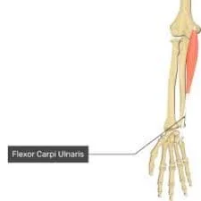
Flexor Carpi Ulnaris Muscle
The flexor carpi ulnaris muscle is a key anatomical structure located in the forearm. As its name suggests, it is primarily responsible for flexing the wrist joint, specifically in an ulnar direction (towards the little finger side).
What Is The Flexor Carpi Ulnaris Muscle ?
The superficial flexor muscle of the forearm that flexes and adducts the hand is called the flexor carpi ulnaris (FCU) muscle in the anterior forearm compartment. The only anterior forearm compartment muscle fully innervated by the ulnar nerve, this muscle is the strongest wrist flexor.
Due to their near lateral location to the muscle, the ulnar nerve and artery can be found during surgery using the FCU tendon as a mark.
Trauma and ongoing wear and tear are the most common reasons for FCU injuries. This muscle is essential for wrist surgeries such as cubital tunnel release and wrist and hand tendon transplants. When treating problems affecting the forearm, wrist, and hand, healthcare providers must comprehend the surgical significance of the FCU.
Structure Of Flexor Carpi Ulnaris Muscle
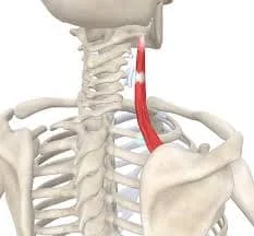
Situated on the ulnar side of the forearm, the Flexor Carpi Ulnaris (FCU) muscle is a long, thin muscle. The ulnar and humeral heads are its two points of origin.
The Humeral Head
The bony protuberance on the inside of the elbow joint, known as the medial epicondyle of the humerus bone, is the origin of this head.
The Ulnar Head
On the other hand, the olecranon and the proximal three-fourths of the ulna’s subcutaneous margin give origin to the ulnar head by aponeurosis.
- The FCU muscle’s two heads combine to form a lengthy tendon that travels through the ulnar groove in the wrist and down the forearm.
- The tendon then attaches to the wrist’s hamate and pisiform bones.
- One of the main nerves of the forearm, the ulnar nerve, innervates the FCU muscle.
- The ulnar artery, a branch of the brachial artery, supplies blood to it. When it comes to wrist and hand mobility, the FCU muscle is critical.
- Its main functions include flexing the wrist and adducting the hand to the forearm’s ulnar side.
- Additionally, it aids with wrist stabilization during lifting and gripping.
Function Of Flexor Carpi Ulnaris Muscle
In comparison to the other forearm flexors, this muscle can be strengthened by activities that prevent it from flexing. You can practice wrist curls with dumbbells and utilize a wrist roller. These workouts are meant to guard against damage to the elbow joint’s ulnar collateral ligament.
Associated conditions
Numerous conditions have been linked to the Flexor Carpi Ulnaris (FCU) muscle, including:
Tendinitis
- Overuse or repetitive strain injuries might result in FCU muscle tendinitis.
- This may result in discomfort and swelling in the surrounding tissues, muscles, or tendons.
Cubital Tunnel Syndrome
- The cubital tunnel is a small tissue tunnel in the elbow that the ulnar nerve—which supplies the FCU muscle—passes through.
- Pain, numbness, and weakness in the hand and forearm can be signs of compression of this nerve inside the cubital tunnel.
Ulnar collateral ligament injury
- The FCU muscle helps to stabilize the elbow joint, especially when pitching or doing other overhead motions.
- FCU muscle pain and weakness can result from injuries to the ulnar collateral ligament, which stabilizes the elbow joint by joining the humerus and ulna bones.
Fractures and dislocations
Pain, weakness, and a reduction in range of motion can result from fractures or dislocations of the elbow, wrist, or hand that impact the FCU muscle and its functionality.
Repetitive strain injuries
- Typing is one of the repeated motion-intensive activities that can strain and injure the FCU muscle, resulting in pain and dysfunction.
- Depending on the severity of the illness and its underlying cause, treatment options for FCU-related conditions may include rest, physical therapy, medication, or surgery.
Origin
There are two heads on the flexor carpi ulnaris: the ulnar and humeral heads. The common flexor tendon connects the humeral head to the medial epicondyle of the humerus. An aponeurosis connects the ulnar head to the upper two-thirds of the ulna’s dorsal border and the medial margin of the olecranon. The nerve and artery that runs between the two heads is the ulnar nerve.
Insertion
The pisiform, the attachment point of the hamate, and the anterior side of the base of the fifth metacarpal are where the flexor carpi ulnaris inserts.
Hamate bone:
It is a wedge-shaped bone that is situated close to the hand’s base on the medial side of the wrist.
The tendon of the FCU muscle inserts into these two bones, facilitating wrist flexion and hand adduction towards the ulnar side of the forearm.
The motions of the FCU muscle are crucial for performing tasks requiring wrist flexion, like typing on a keyboard, tossing a ball, and clutching things.
Nerves
The muscular branch of the ulnar nerve, which emerges from the C7 and C8 nerve roots, controls the facia centralis. Between the two heads of the FCU, the ulnar nerve enters the forearm. The ulnar nerve travels in three directions: within the Guyon canal, superficially to the flexor retinaculum, and lateral to the FCU tendon at the wrist.
Action
At the wrist joint, the flexor carpi ulnaris flexes and adducts.
Blood Supply
The FCU is supplied by the minor ulnar artery branches and the posterior ulnar recurrent collateral arteries. The ulnar nerve and FCU tendon are located laterally to the ulnar artery. It is possible to feel the pulse of this artery here.
Lymphatic Drainage
The superficial and deep lymphatic veins in the upper limb provide the lymphatic drainage for the FCU. The cubital lymph nodes, which are located close to the medial humeral epicondyle, receive drainage from the superficial arteries surrounding the basilic vein.
The axillary lymph nodes receive the lymph veins that surround the cephalic vein. Along with draining fluid from the FCU, the deep lymphatic arteries also follow the main deep veins until coming to an end at the humeral axillary lymph node.
Related Muscles
Together with the pronator teres, flexor carpi radialis, palmaris longus, and flexor digitorum superficialis (FDS), the FCU is one of the forearm’s superficial flexor muscles. Using a shared flexor tendon, the superficial forearm flexor muscles are attached to the medial epicondyle of the humerus. The most medial of these muscles is the FCU.
Tendon
On the anterior surface of the distal forearm, one can observe the flexor carpi ulnaris tendon. There are two or three tendons on a person’s distal forearm, right in front of the wrist. The tendon that is closest to the little finger, or the most medial among them, is the flexor carpi ulnaris tendon. The tendon of the flexor carpi radialis muscle is the most lateral, while the tendon of the palmaris longus, which is the middle tendon, is not always present.
Embryology
Around the fourth or fifth week, dorsolateral somite cells enter the limb bud and begin to build the muscles of the upper limbs. The lateral plate mesoderm’s freshly created connective tissue splits the muscle masses into extensor and flexor components as the limb buds expand.
Fibroblast growth factor promotes Sonic hedgehog expression in the zone of polarizing activity, synchronizing the developmental schema between the anteroposterior and proximodistal axes. The major ventral rami travels through the tissue once the limb buds develop. After this, the ulnar nerve develops to supply the FCU.
Anatomical Variants
In anatomical variety, the auxiliary FCU can originate in a plane between the FDS and the FCU or anteromedial to it, extending to the radial aspect of the FCU. This variant forms a distinct tendon 5 cm above the proximal border of the flexor retinaculum, originating from the medial epicondyle 1 cm posterolateral to the FCU origin, per one case report. The tendon inserts into the wrist bones, especially the triquetral and hamate, and the flexor retinaculum distally.
The abductor digiti minimi, pisiform, hook of the hamate, or fifth metacarpal are possible locations for the brevis variation to insert. Some may attach to the triquetral bone, the index finger tendon of the flexor digitorum profundus (FDP), or the flexor pollicis longus tendon. The ulnar nerve may be compressed by abnormal anatomical features in the Guyon canal. Particularly, aberrant FCU variants that extend distally to the Guyon canal have been linked to ulnar artery thrombosis. Understanding these anatomic variations greatly facilitates preoperative surgery planning.
Surgical Considerations
When doing procedures such as the following, the ulnar shaft can be exposed using the FCU, a surgical mark.
- Internal fixation and open reduction of ulnar fractures
- osteotomy of the ulna
- lengthening or shortening of the ulna
- ulnar clubhand treatment of the ulnar fibrous anlage
The internervous plane between the extensor carpi ulnaris and FCU is accessible by ulnar shaft exposure. Between the two heads of the FCU, the ulnar nerve enters the forearm and travels deep to the FCU tendon at the wrist. When removing the FCU from the ulna epiperiosteally, care must be used to avoid ulnar nerve injury.
When following a volar approach to Guyon canal compression surgery, the FCU is also critical. Through this process, the ulnar nerve is freed from its constriction. The deep palmar fascia and FCU fibers continue into the volar carpal ligament. To enable muscle retraction and expose the ulnar nerve and artery, the fascia of the FCU is cut. The volar carpal ligament and FCU are situated somewhat close to the ulnar nerve and artery. The ulnar nerve and arteries should not be injured during the superficial and deep surgical dissection of this region.
The proximal forearm region, where the ulnar nerve travels between the two heads of the FCU, is where cubital tunnel syndrome is caused by compression of the nerve. For subcutaneous ulnar nerve transposition, the two FCU heads’ deep and superficial aponeuroses must be released. To prevent accidental compression, a transverse fascial arc is also released distally to the FCU hiatus. When cutting into the FCU and related soft-tissue structures, there is a chance that the ulnar motor branches will unintentionally become attached.
Clinical Significance
The ulnar artery and vein, as well as the venae comitantes, comprise the neurovascular bundle of the forearm, which is covered by the FCU, FDS, and FDP. When flexing and ulnarly deviating the wrist, the wrist control unit is a critical anatomical guidance.
The space between the two FCU heads is known as the cubital tunnel. The fibro aponeurotic covering of the epicondylar groove is continued by the Osborn ligament, which joins the two FCU heads. Compression of the ulnar nerve between the two FCU heads and their aponeurosis results in cubital tunnel syndrome.
The second most prevalent form of compression neuropathy in the upper extremities is this one. These is the little finger, ulnar portion of the ring finger, and ulnar dorsal hand are among the symptoms. Sleeping with the arm flexed makes the issue worse at night because the flexion stretches the ligament, flattening the cubital tunnel and applying more pressure on the nerve.
Sensation in the ulnar 1-1/2 fingers may be reduced by the FCU’s continued compression of the ulnar nerve. Patients may also report thumb pinching and weak grasp. Patients may also exhibit the Froment’s paper sign, which is characterized by weak thumb adduction and metacarpophalangeal joint bending, making it difficult to hold thin objects between the thumb and index finger. Claws on the ring and little fingers and muscle atrophy are signs of chronic compressive syndrome.
Other Issues
Overuse injuries such as FCU tendinopathy cause focal volar and ulnar-sided wrist pain. Roughly speaking, ulnar deformity and wrist flexion might cause the same pain. A sesamoid found in the FCU tendon called the pisiform is responsible for tendonitis-related wrist pain.
Assessment Flexor Carpi Ulnaris Muscle
Palpation
Finding the bone landmarks connected to the flexor carpi ulnaris will allow you to palpate the muscle. These are the pisiform bone (insertion), the olecranon process (used to identify the ulna), and the medial epicondyle (origin).
Manual Muscle Testing
The patient is seated with their hand flat on a table and their posterior forearm exposed.
At the side of the upper limb being tested sits the therapist. The therapist feels the muscles and tendons while stabilizing the patient’s forearm. The patient’s fifth metacarpal and metacarpophalangeal joints are where the other hand is situated on the radial side of the hand.
The patient flexes the wrist against the therapist’s resistance while doing ulnar deviation.
- Activities that involve repetitive motions of the wrist and hand, such as typing, can cause strain and overuse injuries to the FCU muscle, leading to pain and dysfunction.
Treatment for FCU-related conditions may include rest, physical therapy, medications, or surgery, depending on the severity of the condition and the underlying cause.
Diagnosis
A medical practitioner, such as a doctor or physical therapist, can diagnose the flexor carpi ulnaris muscle by doing a physical examination and potentially imaging testing. The diagnosis of a flexor carpi ulnaris muscle injury or disease may require the following steps:
Medical history
The medical practitioner will inquire about any symptoms you may be feeling, such as wrist or forearm pain, weakness, or tingling.
Physical examination
- The medical expert will assess the injured region, searching for indications of bruising, pain, or swelling.
- They might also assess your wrist and hand strength and range of motion.
Imaging tests
Imaging tests like an MRI, ultrasound, or X-ray may be requested if necessary to help diagnose the issue and rule out other illnesses.
The flexor carpi ulnaris muscle is frequently affected by tendinitis, ulnar wrist pain, and strains or tears. Treatment options may include rest, ice, compression, and physical therapy exercises to stretch and strengthen the injured muscle, depending on the nature and degree of the injury. Surgery might be necessary in more serious situations.
Exercise
Stretching Exercise Of Flexor Carpi Ulnaris Muscle
Wrist Flexor Stretch
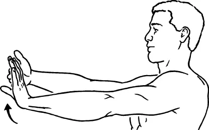
- While maintaining a straight elbow and your palm toward the floor, raise one arm in front of you while standing.
- With your other hand, gently bring the palm of your outstretched hand as close to you as feels comfortable so that you can point your fingers toward the ceiling.
- Stretch your fingers, bending and relaxing as you go.
- For ten to thirty seconds, hold the stretch.
- Release the tension and go back to where you were.
- Use the other arm to perform the same movement.
- Perform two to six repetitions on each side.
- You may experience a stretch in your forearm, wrist, and hand as you complete each repetition.
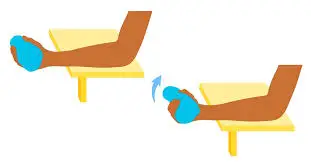
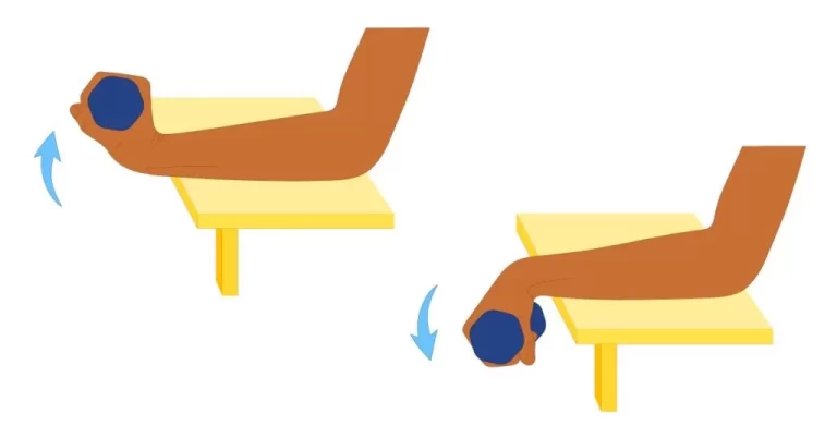
Wrist curl
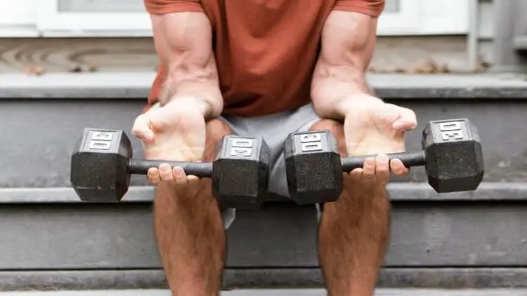
- Hold a small dumbbell in the palm of your hands and place your palms up or your forearm on a table. Another option is to use a weight bench if you’re at the gym. Then, let your wrist and hand dangle off the edge of the surfaces.
- Now, bending your wrist, gradually descend the dumbbell towards the floor.
- Curling your hand toward your forearm will help you bring the weight back up.
- As you lift the weight, contract your forearm flexors.
- Repeat for two to four sets of twelve to twenty repetitions (because the muscle fibers in this exercise are slow-twitch, higher reps are ideal).
Strengthening Exercise Of Flexor Carpi Ulnaris Muscle
Towel Crush
- To begin, place your forearm on the table. You can stand or sit.
- Holding the towel in your hand, roll it up, or gather it into a ball.
- Squeeze the towel and hold it for ten seconds when you’re ready.
- 3 sets of 10 repetitions; release and repeat.
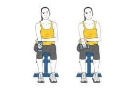
Wrist Curves
- Position yourself next to a desktop or chair and hold a dumbbell in your right hand. You should have a shoulder-width gap between your feet.
- Your hand and dumbbell should be facing up as you rest your right arm on the tabletop.
- When you’re ready, release the dumbbell while maintaining a firm grip.
- After you’ve descended to the lowest point, extend your arm muscles to the top of the action once more. Since your wrist is the only portion of your arm that should be moving, keep your forearm still.
- After ten sets of exercises, switch arms. finished for three sets.
Hammer curls

- Each hand should hold a dumbbell with the palms facing inward, or toward your body.
- Keeping your elbows close to your sides, curl the weight toward your shoulders.
- Do 12–15 reps in 3 sets.
Pronation/supination with dumbbell
- With your palm facing down and your elbow bent at a 90-degree angle, hold a dumbbell.
- Gently turn your forearm down so that your palm is facing the ground after you have turned it to face upward.
- Repeat for 3 sets of 12–15 repetitions should be done.
Conclusion
One of the forearm muscles in charge of wrist and hand movement is the Flexor Carpi Ulnaris (FCU) muscle. Its main actions are the adduction of the hand toward the body’s midline and flexion of the wrist joint. Additionally, the FCU helps to stabilize the wrist when grasping.
The Flexor Carpi Ulnaris muscle facilitates the wrist and hand’s flexion and adduction movements. It needs to function correctly for much of the daily work that involves the wrist and hand.
FAQ
What is the action of the carpi ulnaris?
Wrist extension and adduction are the main functions of the extensor carpi ulnaris, an extended fusiform muscle found in the posterior compartment of the forearm.
Why does my flexor carpi ulnaris hurt?
Your muscles might become irritated by tasks like hair cutting and using tools like hammers and screwdrivers, which can lead to flexor carpi ulnaris tendonitis. Because they employ the FCU, sportsmen who participate in sports like rugby, swimming, tennis, rock climbing, and climbing may be more susceptible to flexor carpi ulnaris tendonitis
Where is the flexor carpi ulnaris located?
The fusiform flexor carpi ulnaris muscle is situated in the forearm’s anterior compartment. Along with the pronator teres, palmaris longus, flexor digitorum superficialis, and flexor carpi radialis, it is a member of the superficial flexors of the forearm. The most medial of the superficial flexors is the flexor carpi ulnaris.
What is the origin of flexor carpi ulnaris?
A muscle in the superficial compartment of the anterior forearm is called the flexor carpi ulnaris. joins the pisiform bone, the hamate hook, and the base of the fifth metacarpal.
What exercise works the flexor carpi ulnaris?
Using a dumbbell or barbell in your hand, curl your wrists. Weight machine: To strengthen your flexor carpi ulnaris muscles (which operate both arms), grab the handles of the weight machines at the gym and pull them down toward your body.
How do you strengthen Carpi ulnaris?
Hold a 1-2 kg dumbbell while placing your arm resting palm down on a table. Extend your hand with your other hand, and then let the weight gently bring it back down. Repeat at the bottom by raising your hand above and allowing gravity to draw it back down.
How do you test flexor carpi ulnaris?
Then stand or sit with your elbows bent 90 degrees and your hands facing down.
As much as possible, bend your wrists downward, then return them to their natural posture.
Now, extend your wrist to the furthest extent possible so that your fingers point outward.
What nerve controls the flexor carpi ulnaris muscle?
The flexor carpi ulnaris and flexor digitorum profundus are two of the forearm flexor muscles innervated by the ulnar nerve. Additionally, it innervates the hand’s intrinsic muscles, such as the interossei, lumbricals, palmaris brevis, and hypothenar muscles.
What is the function of flexor carpi ulnaris?
The flexor carpi ulnaris muscle works with the flexor carpi radialis to flex the wrist and the extensor carpi ulnaris to abduct the wrist, allowing it to do both actions simultaneously.
Where is the flexor carpi ulnaris?
One of the muscles of the superficial anterior compartment of the forearm is the flexor carpi ulnaris. The muscular belly in the distal part of the forearm gives birth to a lengthy tendon that runs anteromedially.
Reference
- Acosta, J. R., Graefe, S. B., & Varacallo, M. (2023, August 8). Anatomy, Shoulder and Upper Limb, Hand Adductor Pollicis. StatPearls – NCBI Bookshelf. https://www.ncbi.nlm.nih.gov/books/NBK526059/
- Palmaris Longus. (n.d.). Physiopedia. https://www.physio-pedia.com/Palmaris_Longus#:~:text=The%20Palmaris%20longus%20(PL)%20muscle,the%20Flexor%20Carpi%20Radialis%20muscles.
- Flexor carpi ulnaris muscle. (2024, May 4). In Wikipedia. https://en.wikipedia.org/wiki/Flexor_carpi_ulnaris_muscle

5 Comments