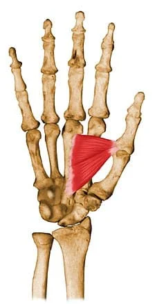
Adductor Pollicis Muscle
The adductor pollicis is a hand muscle located in the palm, specifically close to the base of the thumb. Its two main components are the transverse head and the oblique head. The distal portion of the ulna and the interosseous membrane give rise to the transverse head, whereas the capitate bone and the bases of the second and third metacarpal bones give origin to the oblique head.
What is The Adductor Pollicis Muscle?
The muscles in the human hand are made up of layers of muscles, tendons, and fascial compartments, making them incredibly complicated.
The tendon of the adductor pollicis is formed by combining these two heads when the fibers travel laterally; this tendon frequently has a sesamoid bone. Next, the tendon is attached to the thumb’s medial aspect and proximal phalanx via the upper extensor cover.
The triangle muscle of the hand is called the intrinsic adductor pollicis. It belongs to the same muscle group as the opponens pollicis, flexor pollicis brevis, and abductor pollicis. The thenar muscles on the radial (lateral) side of the hand create an elevation known as the thenar eminence.
There are two heads to the adductor pollicis: tangential and transverse. This muscle extends from the base of the first proximal phalanx to the capitate and third metacarpal bones. The main purpose of this muscle is the adduction of the thumb at the carpometacarpal joint.
Structure Of Adductor Pollicis Muscles
Oblique Head
The oblique head’s origins are the capitate bone, the bases of the second and third metacarpals, the intercarpal ligaments, and the sheath of the flexor carpi radialis tendon.
Most of the fibers from this origin descend obliquely and join to form a tendon inserted into the proximal phalanx base of the thumb on the ulnar side. It then connects the tendons of the flexor pollicis brevis medial section and the transverse head of the adductor pollicis.
Transverse Head
The transverse head sits on a deep floor.
It has a broad base and protrudes in a triangle form from the palmar surface of the lower two-thirds of the third metacarpal bone. Together with the medial portion of the flexor pollicis brevis and the oblique head, the fibers converge and are inserted into the ulnar side of the base of the proximal phalanx of the thumb.
Function Of Adductor Pollicis Muscles
Because of the thumb’s changed plane for the palm and other digits, adduction in this setting refers to putting the thumb into an opposite position in the middle of the palm. Using the adductor pollicis, the thumb can also be brought alongside the palm and index finger.
The thumb’s adduction ability is essential for hand mobility. The two primary activities for which the thumb’s adduction mechanisms must stay intact are pinching and gripping.
Relations
The adductor pollicis muscle is the biggest and deepest of the thenar muscles. The adductor pollicis is crossed on its ventral aspect by the flexor tendons of the index finger, first lumbrical, and flexor pollicis brevis muscles. On its dorsal side, the muscle is attached to, or occasionally united to, the first dorsal interosseous muscle. The channel formed by the two heads of the adductor pollicis is also used by the radial artery, the deep palmar arch, and the deep branch of the ulnar nerve.
Origin
The triangular-shaped adductor pollicis muscle has two heads.
- The bases of the second and third metacarpals are where the oblique head starts.
- The transverse head originates from the third metacarpal shaft.
Insertion
Laterally, the two heads join to create a tendon. The tendon is attached to the medial sides of the proximal phalanx of the thumb.
Nerve Supply
The ulnar nerve’s deep branch (C8-T1) innervates the adductor pollicis. Comprehending the ulnar nerve’s course is crucial to evaluating the clinical symptomatology of damage to the neuromuscular upper extremities.
The ulnar nerve runs distally, posterior to the elbow’s medial epicondyle at the cubital tunnel. It is the final branch of the brachial plexus’s medial cord. After penetrating the two heads of the flexor carpi ulnaris in the forearm, it produces several branches that innervate two forearm muscles.
After passing superficially through the flexor retinaculum on the wrist, it enters the hand by the Guyon canal. It splits its deep and superficial branches once it is in the hand.
Blood Supply
The adductor pollicis receives its primary blood supply from the deep palmar arterial arch. One important anatomical location for the radial artery is the adductor pollicis.
The radial artery enters the hand between the two heads of the adductor pollicis and proceeds anteromedially between them as it passes through the first dorsal interosseous muscle. The deep palmar arch is because of the artery passing between the two heads and entering the hand’s deep compartment.
Lymphatic Drainage
The cubital, epitrochlear, and supratrochlear lymph nodes on the elbow receive the hand’s superficial lymphatic channels’ discharge, which runs parallel to the basilic and cephalic veins. Before ending at the infraclavicular and axillary lymph nodes, these nodes drain into the deep brachial and deltopectoral lymph nodes.
Related Muscles
The thumb uses nine different muscles to perform its many functions. These muscles can be classified into two groups according to the location of the muscle fibers in the thumb: intrinsic muscles (those in the hand) and extrinsic muscles (those in the forearm).
The four extrinsic muscles that influence thumb movement are the abductor pollicis longus, which abducts/extends the MCP joint, the extensor pollicis brevis, which extends the MCP joint, the extensor pollicis longus, which extends the MCP/IP joints, and the flexor pollicis longus, which flexes the MCP/IP joints.
Embryology
From day 26 of week 4 of the early development period, the upper limb develops as a limb of dermal tissue from somites and the lateral plate. From proximally to distally, the limb develops and is controlled by several sensory centers that provide unique factors that control differentiation.
The creation of radioulnar limbs is regulated by the zone of polarizing activity (ZPA), which produces sonic hedgehog protein (SHH), and the apical ectodermal ridge (AER), which induces differentiation of the underlying pathway of Wnt regulates the dermis, the ventral/dorsal limb axis, and the interdigital tissue’s programmed cell death. Any one of these three stages can become disrupted, leading to anomalies in upper limb development.
When chondrogenic forms of the digits develop on day 36, the full building of the hand structures begins. In the sixth and seventh weeks, early muscle lumps begin to grow across the hand and upper extremities. At this point, the adductor pollicis and other intrinsic hand muscles start to develop. Week eight is when the upper limb bones start to o. Weeks 3 through 8 are the most likely times for upper limb developmental abnormalities to develop because most upper limb development is completed by then.
Anatomical Variation
In each of the adductor policy’s two heads, they can be then divided into nine different fascicles. If they are related to specific origins and insertions, there is a large lot of variation between the nine particular fasciae. Every muscle head has two layers of tendons. Differences in the biomechanical properties of different thumbs can be related to multiple small variations between the nine different fascicles.
Surgical Considerations
Surrogate Marker for Malnutrition
According to recent research, adductor pollicis muscle thickness (TAPM) is a rather excellent indicator of nutrition in postoperative patients.. Using this measurement could help in the diagnosis process and result in better outcomes, as nutrition is a key cause of disease and death in surgical patients. Though not yet supported by enough data to be used regularly, this assessment is still being researched as a potential future deficiency measurement tool.
Thumb-in-Palm Deformity
Thumb-in-palm deformity is one of the most common upper extremity indicators of cerebral palsy. What sets this apart is the thumb’s fixed contact with the palm at the MCP joint. Because so many muscles interact dynamically in the thumb, correcting significant abnormalities in this region is a unique surgical difficulty. Two primary muscle problems cause the thumb-in-palm deformity in cerebral palsy.
- Contraction and spasticity of the thenar adduction muscles
- Voluntary control of the thenar abduction muscles is weak.
Initially, modest thumb in-palm problems may be treated with a mix of occupational therapy, splinting, and botulinum toxin injections. Surgery may be necessary if the disease compromises the functional ability to grip and pinch.
Children between the ages of 7 and 10 generally become the ones who have it done because they typically recover from it. Deformity correction may not be appropriate in a patient who is not cognitively capable of recovering sufficiently from surgery.
The most common type of thumb-in-palm formation is type 1, when the first digit is adducted over the palm. The main surgical approach to treat this abnormality is three levels up:
- The adductor pollicis muscle in the palm can be relaxed to help stretch or calm spastic muscles.
- Extensor pollicis longus changing to the first dorsal compartment strengthens the muscle, which helps to strengthen weak or stiff muscles.
- Stabilize the joint if it is unstable.
The thumb-in-palm deformity comes in a variety of forms, and many different surgical techniques can be utilized to correct it. The prognosis for recovery ability following this procedure is typically quite good provided the patient adheres to post-operative therapy.
Clinical Significance
Some clinical illnesses can affect the adductor pollicis muscle, resulting in weakening or loss of function in thumb adduction. These conditions include nerve injuries and muscular abnormalities. The general dexterity and fine motor skills of the hand may be affected by this.
Depending on the patient’s health, physical therapists and hand experts may recommend particular exercises to strengthen and rehabilitate the adductor pollicis muscle.
Ulnar Nerve Entrapment
Examining the adductor pollicis function is a standard diagnostic technique for potential ulnar nerve entrapment in the elbow. The most typical manifestation for these patients is intrinsic muscle atrophy, which causes decreased grasp and pinch strength, difficulty turning keys, and difficulty with performing manual activities.
In clinical situations, individuals with decreased thumb adduction strength may show compensatory thumb IP joint flexion through the anterior interosseous nerve- innervated flexor pollicis longus.
Thumb Collateral Ligament Injury
Thumb ulnar collateral ligament (UCL) injury is caused by mechanisms imposed at the MCP joint that result in hyperabduction or hyperextension. Fractures, both nondisplaced or displaced, near the base of the proximal phalanx are among the injuries based on the thumb UCL.
Surgery is necessary when there is a displaced injury over the adductor aponeurosis and there is bony or ligamentous damage known as a Stener lesion. Surgery is required for the recovery from these injuries, which are also known as “Skier’s” thumb (acute) or “Gamekeeper’s” thumb (chronic).
Neuromuscular Blockade Monitoring
In the area of anesthesia, ulnar nerve stimulation is frequently used to measure the adductor pollicis motions to evaluate neuromuscular blocking. To avoid issues with ventilatory support, neuromuscular blockade levels must be accurately monitored.
For example, it is essential to a degree of neuromuscular blockage before extubation since the patient must have diaphragm strength before the respiration aid gets removed. Electrodes are applied to the anterior forearm during the test, and electrical signals are then used to activate the nerve. To isolate the activities of the thumb alone, the palm and other four digits are typically taped down. This is because the ulnar nerve innervates muscles other than the adductor pollicis.
Next, the kinematic responses to the specific nerve impulses sent via electrodes attached to the thumb can be objectively recorded using an accelerometer. The accelerometer data can then be utilized to guide the administration of additional medicine and treatment. Anesthesiologists can then use the electrodes to deliver customized signals based on the degree of neuromuscular block.
Deformity
An imbalance between the paretic and spastic muscles results in upper limb deformities in cerebral palsy.
The sign of Froment.
Exercises of Adductor Pollicis Muscles
Stretching Exercise Adductor Pollicis Muscles
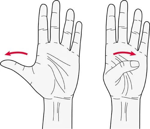
Finger-Abduction Stretch
- Lay a flat hand on a table or similar surface.
- Form a V-shape between your thumb and index finger by slowly removing your thumb from your fingers.
- Using your other hand, carefully place your thumb over your palm with your right to deepen the stretch.
- After holding the stretch for 15 to 30 seconds, let go.
- After two or three repetitions, move to the other hand.
Thumb Flexion Stretch
- Stretch out your right arm so that the palm faces up.
- Using your left hand, slowly bend your right thumb across your palm as if you were trying to touch the base of your pinky finger.
- Use your left hand to exert light pressure to lengthen your adductor pollicis.
- Take a 15–30-second hold, then let go.
- Repeat two or three times, then go on to the opposite hand.
Strengthening Exercise Adductor Pollicis Muscles
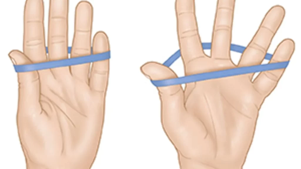
Thumb Adduction with Rubber Band
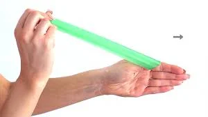
- Gently wrap a little rubber band around your hand, encircling your fingers and thumb.
- Start by separating your thumb from your palm.
- Slowly bring the rubber band closer to your index finger while applying a little pressure with your thumb against its resistance.
- Hold the thumb there for a short while, and then slowly release it.
- Do 2-3 sets of 10–15 repetitions on each hand.
Thumb Adduction with a Soft Ball
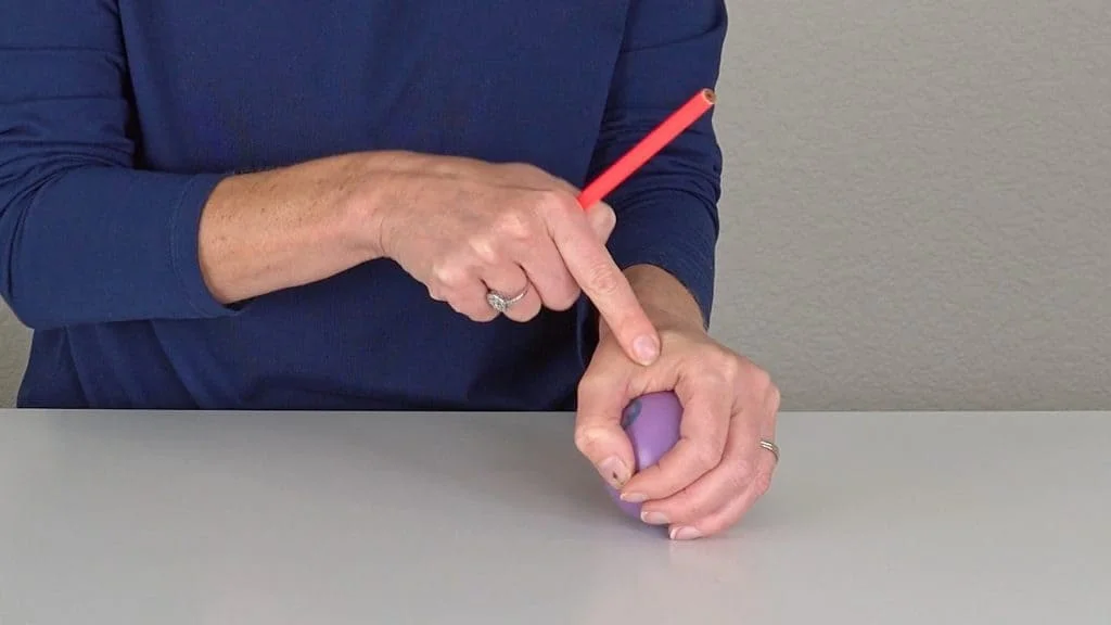
- Use a stress ball or a small, soft ball that is easy to hold in one hand.
- Squeeze the ball with your thumb toward your index finger while holding it with your other fingers.
- Squeeze the ball with your thumb while applying light yet strong pressure.
- Hold for one to three seconds, then let go.
- Do 2-3 sets of 10–15 repetitions on each hand.
Pinch Grip Exercise
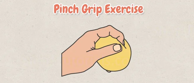
- Use a little object, such as a clothespin or small clip, to practice pinch grips.
- Put your index finger on one side of the object and your thumb on the other.
- Using your thumb and index finger, squeeze the object, focusing on the movement and pressure from your thumb.
- Squeeze for three to five seconds, then let go.
- Do 2-3 sets of 10–15 repetitions on each hand.
Finger Adduction with Putty or Clay

- Roll a piece of modeling clay or therapeutic putty into a smooth round.
- After placing the putty on a level surface, keep your other fingers around the Putty’s corners and press your thumb into the center.
- Try to bring the putty toward your palm by applying pressure with your thumb.
- Hold for a brief moment before releasing your grip.
- Do 2-3 sets of 10–15 repetitions on each hand.
Conclusion
The adductor pollicis muscles, which are necessary for certain vital activities requiring grip strength, coordination, and fine motor skills, enable thumb adduction. These hand muscles are required for some tasks, including gripping pinching, and playing an instrument.
FAQ
What is the adductor pollicis muscle used for?
The adductor pollicis’ primary function is to add the thumb. Adduction in this context refers to moving the thumb into an opposing position at the palm’s center due to the thumb’s modified plane with the palm and other digits.
Is adductor pollicis a thenar muscle?
When talking about thumb movements, the adductor pollicis which is not a member of the thenar muscle group occasionally included alongside the thenar muscles. It is a palm muscle that is fan-shaped, short, and broad, and it arises from the oblique and transverse muscle heads.
What is the origin of the adductor pollicis?
Twenty human adductor pollicis muscles were grossly dissected, and the results showed that the transverse and oblique heads have a wide origin. The capitate, trapezoid, and trapezium bones, as well as the second, third, and fourth metacarpal bones, were among previous generations.
What are the attachments of adductor pollicis?
comes from the neighboring posterior surfaces of the ulna and radius as well as the interosseous membrane. It affixes to the first metacarpal’s lateral side at the base
How do you test adductor pollicis?
The adductor pollicis muscle’s function is evaluated using the Froment’s sign. If the adductor pollicis muscle is weak, the thumb IP joint will flex when the thumb and index finger are brought together against resistance.
How do you treat adductor pollicis?
The majority of these injuries can be resolved with a simple plaster that immobilizes the thumb, allowing the injured ligament to recover. If there is a large or complete tear in the ligament, therapy, and surgery are needed to fix it and make the thumb more stable.
What nerve is abductor pollicis?
The posterior interosseous nerve (C7, C8), a continuation of the radial nerve’s deep branch, innervates the abductor pollicis longus. A branch of the brachial plexus’s posterior chord is the radial nerve.
What is abductor pollicis muscle pain?
localized pain and swelling across the tendons on the wrist’s thumb side. It’s possible to feel a sudden stinging pain when you lift your child or even just pots and pans. Sometimes, after getting worse, it can continue to pain or pulse.
How do you palpate adductor pollicis?
Find the muscle on the palm side of the hand, close to the base of the thumb, to palpate the adductor pollicis. Put your index finger on the dorsal side of the first metacarpal bone and your thumb on its palm side. Gently squeeze while feeling the contraction of the muscle beneath your fingers.
What does the adductor pollicis muscle do?
The main function of the adductor pollicis is to adduct the thumb. Because of the thumb’s altered plane concerning the palm and other digits, adduction in this context refers to putting the thumb into an opposite position at the center of the palm.
What is normal adductor pollicis?
The adductor pollicis is the intrinsic muscle of the hand. It is triangular and has two heads. The radial artery runs anteriorly across the space between the two heads, forming the deep palmar arch.
References
- Adductor pollicis muscle. (2024, May 4). In Wikipedia. https://en.wikipedia.org/wiki/Adductor_pollicis_muscle
- Acosta, J. R., Graefe, S. B., & Varacallo, M. (2023, August 8). Anatomy, Shoulder and Upper Limb, Hand Adductor Pollicis. StatPearls – NCBI Bookshelf. https://www.ncbi.nlm.nih.gov/books/NBK526059/