Electrical stimulation (ES)
What is Electrical Stimulation?
Electrical stimulation (ES) is a therapy that uses electrical currents to deliver tiny pulses to nerves or muscles. These pulses can cause muscles to contract, similar to how the brain sends signals to muscles during voluntary movement.
The idea is that employing an electrical current develops muscles, decreases pain signals, and enhances the circulation of blood.
If you have an accident or condition that causes discomfort or stops you from moving freely, your physical therapist may employ electrical stimulation, or e-stim, as part of your rehabilitation program.
Why is e-stim applied?
Electrical stimulation is utilized for a variety of purposes in physical therapy. It can be applied to:
- Provide medicine for inflammation.
- Improve the muscles that are weak or not working properly.
- Help reduce pain or spasms.
- If you are experiencing discomfort, spasms, inflammation, or muscle weakness, your physical therapist may prescribe this therapy.
E-stim can be used to treat the following medical conditions:
- Lower back pain
- Post-operative pain
- Muscle weakness or impaired motor control
- Tendonitis
- Bursitis
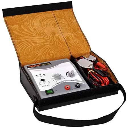
There are several types of electrical stimulation used in physical therapy:-
Physical therapy can employ a variety of electrical stimulation (e-stim) techniques. These therapies can relieve pain and inflammation, increase circulation, and help your muscles contract appropriately.
The different types include:
- Transcutaneous electrical nerve stimulation (TENS).
- Russian Current.
- Neuromuscular electrical stimulation (NMES).
- Inferential current (IFC)
- High-voltage electrical stimulation
- Iontophoresis
- Faradic-type current
- Interrupted direct/galvanic current
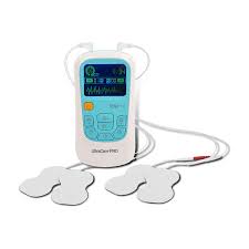
Transcutaneous electrical nerve stimulation (TENS)
Transcutaneous electrical nerve stimulation (TENS) is a form of electrical stimulation that can help relieve acute and chronic pain.TENS works by modifying or blocking the painful signals sent from wounded tissues to the brain. It involves placing electrodes on the painful portion of your body and using electricity to send signals for discomfort to your brain.
Advantages:
The TENS device is lightweight, making it simple to transport and use on the move. It may usually be kept in your pocket or fastened to your belt.TENS is hypothesized to break the pain cycle by sending a non-painful feeling to the nerves around the target region, hence lowering pain signals sent to the brain. The electrical impulses may also cause the body to release endorphins, which work as natural pain relievers.
Uses:
TENS can treat chronic (long-term) and acute (short-term) pain and muscular cramping caused by a variety of disorders, including:
- Arthritis
- Fibromyalgia
- Knee discomfort.
- Backache.
- Neck pain.
- Diabetic neuropathy
- Pelvic discomfort due to periods or endometriosis
Russian Stimulation
Russian stimulation is a high-voltage electrical wave stimulation technique that employs electricity to construct muscular tissues. You may experience muscular weakness following an accident or surgery. Muscles are frequently inhibited after an injury, preventing them from contracting forcefully. Russian stimulation is used to help your muscles contract more efficiently.
Neuromuscular Electrical Stimulation (NMES)
Neuromuscular electrical stimulation (NMES) is used the same as Russian stimulation. Your physical therapist may use NMES to help your muscles contract appropriately following an injury or surgery. This type of muscle re-education can help you get back to normal function rapidly after an injury or surgery.
NMES can also assist you in doing functional activities. Small switches in the unit may be put on your body to regulate whether the stimulation is turned on or off. When executing a job, such as walking, the switch may activate stimulation when your leg muscles are intended to contract and then turn it off when they are supposed to relax.
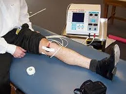
NMES Parameters for Foot Drop:
If you have a drop foot, your physical therapist may utilize NMES to enhance muscle contraction. It is a disorder characterized by anterior tibialis muscle weakening or paralysis.
Application of NMES in Physical Therapy:-
When using NMES, your physical therapist will most likely do some simple procedures.
Here’s the fundamental protocol:
- Expose the anterior tibialis muscle in the front of your shin.
- Your Physical Therapist will place little adhesive electrodes on the front of your shin.
- The electrodes will be attached to an electrical stimulation machine.
- Your Physical Therapist will then switch on the equipment and raise the level of stimulation.
- You will most likely experience a tingling feeling on the front of your shin.
- Increase the effort until the anterior tibialis muscle contracts visibly. Your ankle will automatically flex up.
- While the NMES unit flexes your ankle, try to bring your toes and ankle up higher.
Your physical therapist will program the NMES device to cycle on and off. Typically, it will turn on for 15 to 20 seconds before turning off for the same amount of time. - While the machine is flexing your foot, you should also flex your foot; when the unit turns off, rest your anterior tibialis muscle.
- NMES for foot drop is often used in physical therapy clinics for 15 to 20 minutes. Following the therapy, the electrodes will be removed.
- After removing the electrodes, you should continue to develop your anterior tibialis muscle with specific routines.
Interferential current (IFC)
Your physical therapist may employ inferential current (IFC) electrical stimulation to assist relieve pain and enhance circulation to damaged tissues. The IFC operates similarly to TENS, but the current may be readily shifted and changed to target the most painful location of damage.
Benefits of Interferential Therapy:
- Decrease pain and swelling.
- Enhance blood circulation.
- Causes vasodilation. Removes waste materials from the affected region.
- Enhances metabolic rate.
- Lowers blood pressure.
- Treat persistent ligamentous lesions.
- Treats edema and hematoma.
- Enhances restricted joint motions.
- Enhance muscular stimulation.
- Rebuild lost muscular mobility.
High Voltage Stimulation
Your physical therapist may use high-voltage electrical stimulation as one type of electrical stimulation to improve circulation or reduce pain. It is also infrequently used to promote wound healing. High voltage stimulation is supposed to help modify the sort of cells around your wound, perhaps speeding up healing.
Iontophoresis
Iontophoresis is a type of electrical stimulation that delivers drugs to your body via the skin. For example, Dexamethasone can help reduce inflammation, localized edema, and muscular spasms. Some medications used in iontophoresis can also assist in reducing calcium deposits and regulating scar tissue.
Common Uses:
Iontophoresis is commonly used for several purposes.
This includes, but is not limited to:
- Decrease inflammation.
- Reduces pain and muscular spasms.
- Reduces swelling and edema.
- Reduce calcium accumulation in the body and control tissue scarring.
Faradic-type current:
It is a short-lived, interrupted direct current. Pulse duration is 0.1 to 1 ms, with a frequency of 50 to 100 Hz.
The term “faradism” was originally used to characterize the type of current generated by an induction coil, or faradic coil.
The current produced by coils was unevenly alternating.
Each cycle is composed of two unequal stages.
The first was of modest intensity and lasted a long time.
The second is of great intensity but brief duration.
The frequency was at 50 Hz, and the second phase, which was the most potent, lasted around 1 ms.
Electronic Simulators:
Faradic coils have been replaced with electronic simulators.
These provide currents generate the same physiological consequences as the original faradic currents, but with significantly different waveforms.
The physiological effects are produced by repeating shocks with durations ranging from 0.1 to 1 ms at a rate of 50 to 100 times per second.
Electronic simulators for the generation of faradic-type current operate on the same principles as those for the interrupted.
However, the resistances governing the length of the impulses and the intervals between them are extremely low, allowing for the needed duration and repetition rate.
Modified faradic current:
Unmodified current refers to the use of faradic currents for therapy purposes, resulting in near-normal muscular contraction and relaxation.
Surged current means that the current is surged in such a way that the strength of succeeding impulses steadily increases, with each impulse reaching a peak value larger than the previous one before falling, either abruptly or gradually.
The current in initial faradic coils was generated by hand, while newer stimulators employ electrical devices.
The circuit may be modified to produce surges of varying durations, frequencies, and waveforms.
The duration of the surges and the interval between them should be controlled separately to get the most satisfactory muscular contractions and relaxation periods for each patient.
Nerve transmission involves electrical activity:
Because of the difference in ion concentrations inside and outside the plasma membrane, there is a potential differential between the inside and outside of the nerve.
The resting nerve has a positive outside and a negative inside, and the plasma membrane is not permeable to sodium ions.
This is referred to as the membrane’s polarising phases.
When a neuron is activated, the potential difference across the plasma membrane decreases.
The membrane’s permeability to sodium ions changes when this decline reaches a threshold level.
As a result of this change in ion concentrations within and outside, the PD continues to decline until polarity is revealed. presently, the membrane is positive inside as well as negative outside.
Following this action, the sodium ions are pushed out again, and that portion of the neuron returns to its resting condition.
The potential difference between the active and resting sections of the nerve results in local electron flow between the active and neighboring regions of the nerve.
Now, the current passes through the membrane in the opposite direction of the potential difference [PD] between the fibers.
The fibers function as a barrier to the current, lowering the PD and making the membrane permeable to sodium ions, which reverses the PD as previously.
These alterations then spread throughout the length of the nerve fibers.
This wave of change of polarised state represents the passage of an impulse along the nerve.
Electrical stimulation of nerves:
Electrical stimulation can trigger nerve impulses.
To do this, a changing current of appropriate intensity must be applied.
The plasma membrane of nerve fibers produces resistance in series with the other tissues, resulting in PD as the current travels across it.
The surface of the membrane closest to the cathode turns negative in comparison to the opposite surface.
The surface of the membrane closest to the anode becomes positive in comparison to the opposite surface.
If the PD falls down the level at which the membrane becomes permeable to sodium ions, these ions enter the axon and start the chain of events described above, resulting in the initiation of a nerve impulse.
If the PD is sufficient to pass any region of the plasma membrane of the nerve cells or fibers, an impulse is triggered.
Because the current travels throughout the tissues, the current density is lower on the distant surface of the nerve fiber than on the nearer one, resulting in the anode being less efficient than the cathode in generating impulses.
Effect of nerve stimulation:
Nerve stimulation can cause nerve impulses to travel in both directions from the point of stimulation, unlike when they originate at nerve cells or end organs.
When a sensory neuron is activated, downward-traveling impulses have no impact, but upward-traveling impulses are appreciated once they reach the conscious regions of the brain.
When impulses of varying durations are delivered with the same current, it is discovered that the sensory stimulation experienced by wires is proportional to the impulse duration.
Long-length impulses cause a painful, stabbing feeling, but this reduces as the duration of the impulses decreases, and with impulses of 1 ms or less, just a light prickling sensation occurs.
When a motor nerve becomes active, the upward-traveling impulses cannot reach the first synapse because they are traveling in the wrong direction. Still, the downward-traveling impulses travel to the muscles the nerve supplies, causing them to contract.
When a stimulus is delivered to a motor nerve trunk, impulses are sent to all of the muscles supplied by the nerve below the site of stimulation, causing them to contract.
When a current is applied directly to an innervated muscle, the nerve fibers within the muscle are activated in the same way.
The maximal reaction is produced either by stimulating the motor point, which is the point at which the major nerve enters the muscle, or, in the case of deeply located muscles, at the sites where the muscle emerges from underneath the more superficial ones.
Effects of stimulation frequency:
A single stimulation sends impulses to several motor units, resulting in a rapid contraction followed by instantaneous relaxation.
If a series of stimuli are applied at relatively long intervals, such as one stimulus per second, each produces an isolated muscle contraction with time for complete relaxation until frequencies above 20 Hz, at which point there is no time for complete relaxation between contractions, resulting in partial tetanus.
Further frequency increases result in a reduction of nearly 60 Hz, no discernible relaxation, and a complete large contraction.
Strength of contraction:
Contraction strength varies based on motor unit activation and current rate.
If the current’s intensity suddenly increases, there is little time for accommodations, resulting in a muscle contract.
If the current increases more slowly, as with trapezoidal, triangular, and saw-tooth impulses, some accommodation occurs, and a higher current intensity is required to achieve a contraction.
Physiological effects of faradic type current:
The physiological effects resulting from faradic-type electricity include the ability of biological tissue to carry electric current due to the presence of ions that act as conductors.
As a result, the current going through the body is a two-way movement of ions, and the conductivity of the various tissues changes depending on how much fluid they contain.
Muscle equals good conductor.
Fat equals a lousy conductor.
The current prefers to pass through tissues with low resistance, although it is not always feasible to avoid high-resistance layers.
The epidermis has a high resistance of 1000 or more since it contains little fluid and the surface layers do not readily absorb moisture.
When performing electrical therapies, the current must flow through the epidermis, which is reduced in resistance by proper methods.
The passage of electricity may cause chemical changes, which can be dangerous in particular therapies.
Sensory nerve stimulation:
it involves when applying a faradic current to the body, resulting in a minor prickling sensation.
This is caused by sensory nerve activation, which is not very noticeable because the impulses are brief.
Sensory stimulation promotes reflex vasodilatation of the superficial blood vessels, resulting in minor reddening of the skin [erythema].
Vasodilation is mainly limited to the surface tissues and has little practical significance.
Motor nerve stimulation:
it involves when faradic current stimulation, which can produce muscular contractions if the current is strong enough.
Because the impulses are repeated 50 times per second or more, the contraction is enormous.
Muscle fatigue occurs when this sort of contraction is maintained for an extended amount of time, hence the current is often increased to allow for muscle relaxation.
Contraction strength gradually increases and falls when the current explodes, similar to voluntary contraction.
Effects of muscle contraction:-
Electrical stimulation causes muscular contractions, which are very comparable to voluntary contractions.
There is increased metabolism, which increases the need for oxygen and food, as well as the emission of waste products, including metabolites.
The metabolic process causes dilation of capillaries and arterioles, resulting in a significant increase in blood flow to the muscle.
Muscle contraction and relaxation cause a pumping action on the veins and lymphatic vessels that lie inside and surrounding them.
The valves in these veins guarantee that the fluid they contain is transported to the heart.
If the muscle contractions are powerful enough to create joint movement, they also provide a pumping effect.
This leads to enhanced venous and lymphatic return.
Stimulation of denervated muscle:-
To stimulate denervated muscle, the current required to induce a contraction lasting 1 ms is typically too high for effective therapy.
As a result, the faradic current is ineffective for stimulating denervated muscles.
Chemical consequences of the faradic type of current:-
Direct current passing through an electrolyte causes chemical reactions in electrodes.
If the chemical generated comes into touch with the tissue, there is a risk of electrolytic burns, however, the risk is much lower with the intermittent direct current than with constant direct current.
When the current alternates, the ions flow one way during one phase and the other direction during the other, and if the two phases are equal, the chemicals generated during one phase are neutralized during the next.
If the reverse wave of the current is not identical to the forward wave, there may be chemical changes that result in an electrolyte burn.
The current derived from electronic apparatus may have a larger flow in one direction, but the chemical production should not be significant enough to provide a substantial burn risk.
Indications for using faradic currents:
Facilitation of muscle contraction:
Electrical stimulation can help patients who are unable to make voluntary muscular contractions.
Muscle contraction is the product of a complex integration of the neural circuit, both at the spinal level and in higher centers.
Excitation of the tiny efferent fibers leads to contraction of the intrafusal muscle fibers.
Stretching of the muscle spindle activates the main nerve ending, sending information to the big anterior horn cells and activating the extrafusal muscle fibers.
Inhibition of anterior horn cells that feed the antagonistic muscle group.
Pain inhibits the big anterior horn cells, which delays impulse transmission to the motor units.
Re-education of muscle action :
Muscle inability to contract voluntarily can be caused by disuse or inappropriate usage, such as in the intrinsic foot muscles in standing flat feet or the abductors hallucis in hallux valgus deformity.
Faradic stimulation may also be utilized to induce contractions and aid in the restoration of movement.
The brain values motions over muscle activities, hence the current should be administered in such a manner that it creates movements that the patient is unable to do.
Active contraction should be undertaken concurrently with electrical stimulation, with the therapy serving as a warm-up for active activity.
It will likely take longer to create voluntary contraction.
Training a new muscle action :
After tendon transplantation or repair, a muscle may need to be trained to execute a different movement than before.
A new movement sequence has to be created.
The muscle is stimulated with a faradic-type current, causing it to perform an uncommon motion, and the patient must concentrate on the movement while attempting to help with voluntary contraction.
Muscle action can be taught in this manner, but it will take longer than re-educating a previously performed motion.
Neurapraxia of a motor nerve :
Neurapraxia of a motor neuron occurs when signals from the brain cannot travel through the lesion to the muscles supplied by the afflicted nerve.
Voluntary power may be weakened or lost.
The nerve, however, does not degenerate, thus when stimulated with faradism below the site of the lesions, impulses flow to the muscles, causing them to contract.
Severed motor nerves:
it results in axon degeneration and impaired responsiveness to short-duration stimuli.
Muscle degeneration occurs many days to a few days following an injury.
Improved venous and lymphatic drainage through muscle contractions and joint movement.
The therapy is successful when current is administered using the faradism under pressure approach.
Prevention and loosening of adhesion :
To avoid and release adhesions caused by effusion in tissues, maintain structures moving about one other.
If enough physical exercise is not achievable electronic stimulation can be utilized as an alternative.
Muscle contractions can stretch and release previously established adhesions.
Preparation of apparatus for faradic current:
- To prepare the apparatus for faradic current, a low-frequency electronic simulator with automated surge is typically utilized.
- To test the machine, connect the leads and electrodes to the terminals, hold the two electrodes in a moistened hand, enter the core if using a smart-Bristow coil, and increase the current until a minor prickling sensation and muscle spasms occur.
- Describe to the patient the sensation you are experiencing and ensure that the patient can observe the muscular spasms that are occurring.
- If the surge is automated, the duration and frequency of the surge should be investigated.
- Check that the device is far enough away from an active short-wave treatment machine [about 2 meters] to avoid output disruption caused by radio frequency [RF] radiation.
- The active electrode can be a disc electrode or a tiny lint/sponge pad with a flat plate as the electrode.
- The latter is preferable for big muscles like the quadriceps and glutei because it is easier to mold to the surface, resulting in better contact.
- Flat plate and lint/sponge pad electrodes are utilized to complete the circuit.
- The pads are made up of at least eight layers of lint, which allows them to establish good contact with the tissues and electrodes while also absorbing any chemicals that may accumulate.
- They should be folded properly and without wrinkles, otherwise, the current will be unevenly distributed, causing discomfort.
- The pads and lint that cover the disc electrode are soaked in warm 1% saline.
- Tap water can be used, however, adding salt reduces the resistance of the wetting solution; 1% saline has a lower resistance than tissue fluids.
- Electrodes should be 1 cm smaller all around than the pads to limit the risk of them coming into contact with the skin, resulting in an unpleasant concentration of current and possibly tissue damage via chemical activities.
- The electrode corners should be rounded, as the points may bend and dig into the pads, creating current concentration.
Patient Preparation for Faradic Current:–
- Clothing is removed from the treatment region, and the patient is comfortably supported in a well-lit environment.
- The patient must be warm; otherwise, the muscles will not respond properly to stimulation.
- Muscle contraction in response to electrical stimulation is typically easier to achieve when the portion is supported in a short posture.
- Positions are adjusted as needed.
- If the goal of the treatment is to re-educate to muscular activity, the patient may be placed so that movement occurs when the muscle contracts. [For quadriceps = knee in mild flexion, such that extension occurs when the muscle is activated.]
- In rare circumstances, mobility can be accomplished by supporting the limb in slings during the therapy.
- Joint movement should be avoided if it causes discomfort, as this inhibits muscle function.
- Skin has a high electrical resistance, which may be decreased by washing with soap and water to remove natural oils and moistening with saline before applying the pads.
- To protect the pads against damaged skin, apply a tiny amount of petroleum jelly and cover it with a small piece of non-absorbing cotton wool.
- The indifferent pad should be large enough to keep the current density to a minimum.
- The indifferent electrode can be bandaged, secured with a rubber strap, or held in place by body weight.
Interrupted direct current/galvanic current:-
Interrupted direct current, also known as galvanic current, occurs when the current flows intermittently at regular intervals.
The peak and fall in intensity might be rapid or gradual.
The length and frequency of impulses may be changed; typically, 100 ms are employed, but this can be increased to 300 or 600 ms.
The frequency required for 100 ma impulses is around 30 per minute.
If the time is extended, the frequency must decrease.
A depolarized impulse is a low-intensity reversed current produced by some equipment between impulses.
The passage of a direct current through an electrolyte induces chemical changes at the electrodes.
Because of the chemical burn, continuous direct current is no longer widely employed.
Using depolarized impulses reduces risk.
Production of the interrupted direct current:-
Interrupted direct current is created using modernized circuits with transistors and timing devices.
The duration of the electrical pulse produced may be changed by alternating the portions of the circuit through which current flows and a selector switch allows you to choose among a variety of fixed-interval pulses and frequencies.
A potentiometer is often used to apply current to the patient, and the strength of the current is gradually increased from zero.
Physiological effects of interrupted direct current:-
A contraction of the denervated muscle can be started if the present intensity and duration are appropriate.
When contractions and relaxations are slow, the motor nerve is activated.
Sensory stimulation of the nerves:
it can provide a sharp or burning feeling due to long-lasting impulses.
The superficial blood vessels dilate reflexively, resulting in cutaneous erythema.
Motor nerve stimulation:
it involves repetitive impulses that cause muscle twitching followed by rapid relaxation.
As a result, the muscles receive minimal benefit.
Indication of interrupted direct current:-
Contraction of denervated muscle.
When a muscle loses its nerve supply, it undergoes structural and functional alterations.
There is a significant loss of muscular fibers, and if the degeneration is prolonged, they tend to become fibrosed and lose their qualities of irritability, cons tractability, extensibility, and elasticity.
These alterations are slowed by the electrical stimulation of muscle fibers.
It also restores muscular mass and characteristics.
It is also used to re-educate muscles.
It produces a sufficient number of contractions.
Selection of Impulse Types:
For good contractions = rectangular impulses.
The increase of rectangular impulses is quick, but trapezoidal impulses are rather sluggish.
With the triangle slower, and the saw-tooth even slower.
Impulse duration:-
for denervated muscle contraction is 100 milliseconds.
Shorter impulses fail to constrict.
Innervated muscle contraction = 300 to 600 milliseconds.
Methods of applying interrupted direct current:
The labile method:
The labile approach involves placing one pad over the muscle group’s origin and stimulating it with an active electrode.
Active electrodes are discs or small pads that can be positioned at the lower part of the muscle’s tender belly to be stimulated or stroked softly along it.
Advantages: This approach produces the optimal contraction of each muscle.
Disadvantage: It is not practical to create a significant number of contractions of each.
Stable technique:
two disc electrodes are employed.
One electrode is placed over the origin and the other over the lower end of the muscle group.
Advantages: This approach is beneficial for deep muscles that are difficult to separate and allows for a high number of contractions.
Disadvantage: It is difficult for the operator to grasp both electrodes.
Another method:
it involves using an active pad to completely cover a muscle or set of muscles.
The circuit is finished with a big directing or indifferent electrode.
Equipment Preparation of interrupted direct current:-
To prepare equipment for interrupted direct current, use the same testing procedures as for faradic current.
Ensure that the disc electrodes and pads have a minimum of eight layers of material.
It is required in therapy due to the potential of chemical burns with long-duration pulses if the treatment is administered at the same location for an extended length of time.
Metal should not come into touch with the patient’s tissues.
Patient preparation:-
it involves cleaning and protecting the skin from abrasions for treatment.
It is feasible to use warm water because the skin’s resistance is lesser.
However, if there is a considerable loss of feeling, it is important to ensure that the water is not excessively hot.
For easier contraction = shorter posture.
Application of interrupted direct current:-
Interrupted direct current can cause muscle contractions, but it’s not necessarily necessary to connect the electrode to the anode.
Each patient should be checked to see whether the anode or cathode produces the best reaction, as well as which pole is most beneficial for the active electrode.
Different between faradic and interrupted direct current:
| No. | Faradic current | Interrupted direct current | |
| 1 | Direct | Biphasic with variable amplitude | Constant in direction & amplitude |
| 2 | Duration | Short < 1 ms ”interrupted AC” | Long 100 ms “interrupted DC” |
| 3 | Frequency | 30-100 Hz | Low < 30 Hz |
| 4 | Source | Dynamo | Batteries and accumulators |
| 5 | Stimulation of motor nerves | Mainly ++ of innervated muscle the Titanic’s contraction if >50 Hz. | +++ for denervated muscle. Brisk contractions on make and break. |
| 6 | Stimulation of sensory nerves | Pricking sensation VD [ Erythma] | Burning sensation reflex VD |
Limitations of electrical stimulation:
Keep in mind that many types of electrical stimulation are passive treatments. You do nothing while being stimulated. Some types of e-stim, such as NMES and Russian stim, need you to be active while they are in use.
Active participation in your physical therapy program, with or without electrical stimulation, produces the best outcomes. E-stim should only be used in conjunction with an intensive physical therapy program that includes targeted movements and exercises to treat your ailment.
Hazards of electrical stimulation:
Your physical therapist should describe the advantages and hazards of using electrical stimulation during rehabilitation therapy.
The risks of e-stim may include:
- Muscle tearing
- skin irritation
- Tissue burn
If You Have A Muscle Tear
If the electrical impulse is too powerful, you could have severe muscular discomfort. This may cause muscular tissue to rip. In this scenario, the e-stimulation should be discontinued immediately.
The healthcare physician should next begin treating the acute muscle damage. This may involve resting, icing, and elevating the affected body part.
If skin irritation occurs
Some kinds of electrical stimulation might irritate the skin beneath the electrode. Iontophoresis employs a direct current during application. This has been shown to irritate the skin.
People with sensitive skin may experience irritation from the electrode’s adhesive or electrical stimulation. If irritation develops, the operation should be discontinued. The affected region may then be treated with a soothing lotion.
If You Experience a Tissue Burn
If electrical stimulation is administered at an excessive intensity, tissue burns may develop. This seldom occurs. However, if you get a tissue burn, the physical therapist should immediately halt the operation. To cure the burn, offer appropriate skin care.
Your physical therapist can verify that electrical stimulation is performed correctly, which reduces the hazards associated with its usage. Understanding these dangers might help you decide whether or not to incorporate them into your rehabilitation plan.
Benefits of electrical stimulation:-
- post-stroke recovery.
- Muscle weakness during ACL reconstruction and complete knee replacement.
- Muscle weakness is connected with knee osteoarthritis.
- Treating debilitation and weakness caused by serious diseases.
- Promoting recovery of peripheral nerve damage caused by trauma, certain medical disorders, and autoimmune illnesses.
Reasons You Shouldn’t Use E-Stim:-
There are several situations where electrical stimulation should not be employed. Your physical therapist should be aware of the reasons why e-stim should be avoided.
Avoid electrical stimulation if you have:
- Changes in tissue feeling.
- impaired mental state
- The presence of an implanted electrical device. (E-stim may interact with pacemakers and implanted pain stimulators.)
- Malignant tissue
- Wounds with excessive moisture
- A damaged region near the eyes, carotid sinus, front of the neck, or above reproductive organs.
E-stim should also be avoided if:-
- Avoid using e-stim if you are pregnant or suspect you are pregnant. TENS may not be suggested early in pregnancy.
- You have epilepsy or a heart condition.
- You have a hyperactive bladder.
Alternatives to electrical stimulation:
If you are refusing or unable to get e-stim as a treatment, your physical therapist may provide alternatives. Other kinds of therapy might include:
- Stretching and flexibility exercises.
- Muscle-building workouts using weights, resistance bands, machines, or your body weight
- Heat and Ice Treatments
- Ultrasound Therapy
- Range-of-motion exercises.
- Massage Therapy
- Pain medicines
If you are in discomfort or have restricted movement, contact your physical therapist. They will help you determine whether electrical stimulation is the best therapy option for you and your individual ailment.
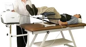
Muscle group stimulation with a faradic current:-
Faradic current stimulation can effectively re-educate the activity of muscle groups, including quadriceps, small foot muscles, and pelvic floor muscles.
Quadriceps Muscle:
Set up a plinth and the patient’s affected knee supports it in flexion.
One electrode (12 X 9 cm) is inserted on the femoral nerve in the femoral triangle and secured with a sandbag or bandage.
Use jaconet to protect clothing and bandages from dampness.
Choose an appropriate length and frequency to achieve a good contraction followed by full relaxation of the muscle.
Give several moderate contractions to help the patient adjust to the current, then gradually raise the intensity until a forceful contraction is obtained.
Instruct the patient to consciously contract the muscle as the current passes.
When the patient achieves a voluntary contraction, terminate the treatments.
Small muscle of foot :
This condition, also known as faradic foot bath, involves a little muscle in the foot.
Electrical stimulation by the faradic-type current can be used in baths; water creates ideal contact with the tissues, pads, and electrodes are not required, and prolonged socking minimizes skin resistance.
Position the patient on a plinth, back adequately supported, feet on a stool covered with a plastic sheet.
This posture may need to be adjusted for elderly people or those with a history of dizziness.
Place the patient’s foot in a bath with enough warm water to cover their toes.
Lumbrical muscles :
To activate the lumbrical muscles, insert two electrodes transversely across the bottom of the bath, one under the heel and another obliquely under the metatarsal heads.
Interossei muscles :
To activate the planter interossei muscles, put one electrode on each side of the foot, near the metatarsal shafts.
Abductor hallucis :
To stimulate the abductor hallucis muscle, place one electrode under the heel and apply a button electrode to the motor point.
In three ways, pick an appropriate duration and frequency to achieve a satisfactory contraction followed by full muscle relaxation.
Give several moderate contractions to help the patient adjust to the current, then gradually raise the intensity until a forceful contraction is obtained.
Instruct the patient to consciously contract the muscle as the current passes.
It may be impossible to activate the tiny muscle groups of the foot in water if the patient has a foot infection, pes cavus, or an open unhealed cut.
Muscle of the pelvic floor :
Electrical stimulation can help re-educate pelvic floor muscles in cases of early prolapse and stress incontinence (Bladder Bowl Incontinence).
There are different methods for administering electricity, but a good muscular contraction is required, and vaginal electrodes are frequently the most satisfying technique for doing this.
Voluntary contractions must be undertaken concurrently with electrical stimulation; electrotherapy is an adjunct to the exercises, which are a crucial component of therapy.
Male patients with incontinence after prostatectomy can be addressed utilizing a rectal electrode.
Position the patients on their side, laying with a cushion between their lower legs.
A big electrode is implanted and fastened to the lumbosacral area.
Sterilized lubricant Jelly is smeared onto the rectal electrode.
Choose an appropriate length and frequency to achieve a strong contraction followed by full muscle relaxation.
FAQs
Which conditions can electrical muscle stimulation handle?
Physical therapists utilize EMS to treat muscular weakness and poor motor coordination. Lower back discomfort, tendinitis, bursitis, and post-surgical pain are all medical disorders that benefit from e-stim therapy.
Is it possible to grow muscle using electric muscle stimulation?
Maybe, but not in the manner it is commonly advertised. According to research, EMS can improve muscle mass and function. However, the study was conducted on persons who had a muscular injury or atrophy. After six weeks of therapy three times per week, the muscular mass grew by just 1%. Muscle function enhanced by 10-15%.
Is electrical muscle stimulation safe?
Conclusion: EMS appears to be a safe and appropriate method of developing physical fitness in healthy people.
What are the four benefits of electrical stimulation?
Relieve discomfort and suffering. Reduce muscular spasms. Restore muscular tone. Rehabilitate some areas of the body.
Is electrical stimulation healthy for the nerves?
According to clinical research, electrical stimulation promotes axon development during nerve healing and speeds up sensorimotor recovery.
How long does electrical stimulation work?
According to research, TENS works best for chronic pain when administered for at least 30 minutes while exercising.
What is one example of electrical stimulation?
If you suffer brain or spinal cord damage, functional electrical stimulation can help you move your muscles more freely. For example, FES can cause your muscles to contract to raise your foot or elevate your arm. The electrical impulse can interfere with signals sent to your brain that indicate pain.
References
- Inverarity, L. (2023, November 9). How Electrical Stimulation Is Used in Physical Therapy. Verywell Health. https://www.verywellhealth.com/electrical-stimulation-2696122
- Geng, C. (2022, July 22). Some of the top medications for muscle pain. https://www.medicalnewstoday.com/articles/best-medication-for-muscle-pain
- Pt, B. S. (2023, February 16). Types of Electrical Stimulation Used in Physical Therapy. Verywell Health. https://www.verywellhealth.com/estim-use-in-physical-therapy-2696490
- Ladva, V. (2023, July 20). Electrical Stimulation Machine – Mobile Physiotherapy Clinic. Mobile Physiotherapy Clinic. https://mobilephysiotherapyclinic.in/electrical-stimulation-machine/#Muscle_of_facial_nerve

5 Comments