Elbow Joint
The elbow joint is a complex hinge-type synovial joint that connects the upper arm to the forearm. It plays a critical role in the movement and function of the arm, enabling activities such as lifting, pushing, pulling, and fine motor skills.
This essential joint supports the range of motions required for daily tasks, involving bending the forearm (pronation and supination) and bending and straightening the arm (extension).
The arm’s ulna, radius, and humerus.
The elbow joint is classified as a synovial joint in terms of structure.
Introduction
These joints, which unite the upper arm to the forearm, are called elbows.
The skeletal system includes every joint in your body, including your elbows.
Elbow joints fall under the structural category of synovial joints.
This vital joint allows the perform various motions necessary for daily tasks, such as rotating the forearm (pronation and supination) and bending and straightening the arm (extension).
The Anatomy of elbow joint
The elbow joint, located in the upper portion of your arm, is responsible for the movement of the forearm and upper arm joints.

Distal End of Humerus
- Although the capitellum and humeral condyle articulate near the distal end of the humerus, it begins at the anterior trochlear.
- This cartilage-infused condyle is given stability by the concave superior layer of the radius head and the trochlear notch of the ulna.
- The axis of the humerus forms a 40° angle with the capitellum and trochlea’s anterior articular surfaces.
- When the elbow is bent, the trochlea, the humerus’ distal medial articulating end, rotates the ulnar trochlear notch like a pulley.
- She joined to the joint’s radial head.
- Moreover, it helps the front part of the ulna and the coronoid muscle.
- The radial head also called the radial fossa, is joined to the body by the smaller lateral fossa, which encourages bending.
- At the trochlear notch, it starts and removes in a C at the proximal end of the ulna.
- Some specific muscles and ligaments follow the ridges and epicondyles.
- Extensor muscles in the hands and arms wrapped develop the lateral epicondyle, while flexor muscles produce the medial epicondyle.
Ulna
- The olecranon process makes the trochal notch expansion, an articular cartilage near the apex of the ulna, possible.
- A trochal notch develops as articular cartilage grooves into the olecranon process.
- The shaft comes next, even farther away.
- The radius head is later shown in the coronoid process’s radial notch.
Radius
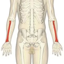
- The elbow joint’s radial head is its primary structural component.
- The elbow joint’s superior articulation has an impact on the hinge.
- The radius head’s medial circumferential area is where radioulnar region articulation starts.
- The neck can bend laterally due to the protection given by the skull’s covering bone.
- Its radial tuberosity is its thickest area.
- The shaft follows the radial tuberosity distally.
Type of the joint
Joints are divided by healthcare providers using the following rules:
- A synovial joint is the elbow.
- Smooth, silky hyaline cartilage surrounds the ends of the bones that make up an elbow.
- This extra cushioning allows for the least friction when moving through synovial joints.
- It has a pivot joint as well.
- Pivot joints remain in their original configuration when rotating in place.
- Your elbow’s swivel allows you to effortlessly elevate or lower your palm by bending your forearm.
Structure of elbow joint
What constitutes your elbow joints is:
- Bone
- Cartilage
- Ligament
- Muscle
- Nerves
- Blood supply
Articulating Surfaces
- The humerus’s and the ulna’s trochal notches
- The radius head and the humerus capitulum
Several studies have referred to the proximal radioulnar joint, a significant part of the elbow’s joint capsule, as an isolated articulation.
- Joint type: hinge joint
- Bones: Humerus, radius, ulna
- Various mnemonics exist Trochlear = ULnar, Capitulum = RAdius, and Crazy TULips.
- CUTER (Radial Trochlea, Ulnar Capitulum)
- The ulnar, intermediate, and radial arteries are collateral at the elbow joint.
- Flexion involves the brachii, brachialis, and brachioradialis muscles.
- Muscle extension: Triceps brachii
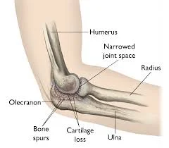
- Common illnesses include fractures, arthritis, venipunctures, and epicondylitis.
Osteology
Bone in elbow joint
Your elbow joint consists of three bones:
- Upper arm bone: humerus.
- Your forearm’s longest bone is the ulna.
- Radius, the shorter forearm bone.
From these bones, three joints are formed:
Humeroulnar joint
- The proximal ulna’s trochlear notch and the medial part of the distal end of the humerus’s trochlea form the shoulder and humeroulnar joint.
- The glenohumeral joint can be handled by two movements: flexion and extension.
- It articulates with the humerus’s greater trochlea using the proximal ulna’s sigmoid arch.
Humeroradial joint
- The radiocapitellar junction appears above the capitulum on the lateral side of the humerus distal end.
- The radiocapitellar joint, however, defines how the humerus and radius move.
- They could effectively give it both pronation and supination.
- Spherical capitalism is seen on the concave surface of the proximal radial head on the lateral distal humerus.
Proximal Radioulnar Joint
- Pronation and supination of the forearm remain regulated by the proximal radioulnar joint, also known as the trochoid joint.
- Both the ulna’s radial notch and the radial head’s borders articulate.
The humerolateral and humeroradial joints give the elbow its characteristic hinge-like characteristics.
The ulna’s concave trochlear notch and radius head surfaces are connected to the humerus’s round trochlea and capitulum surfaces.
The radius of the ulna can rotate due to the proximal radioulnar joint’s pivot.
Cartilage in the elbow joint
- It serves as a body-wide shock absorber.
- Hyaline cartilage borders your elbows.
- Your joints’ hyaline cartilage is a smooth substance that facilitates easy bone-to-bone movement.
- Hyaline cartilage lines the surfaces of your humerus, ulna, and radius where they are in contact with one another.
Carrying Angle
- The carrying angle can be found by fully extending and supinating the elbows.
- The ulnar and humeral long planes are utilized to measure that angle, which is approximately 13 to 16° for females and 11 to 14° for men.
- It is aptly titled since it permits carrying goods and lets our arms pass over our hips while we walk.
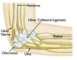
Ligaments of the elbow joint
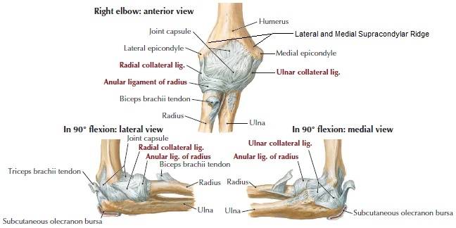
The two relevant bones in the articulation are attached by the ligaments of the humeroulnar and humeroradial joints.
The collateral ligaments include radial and ulnar.
The humerus’ medial epicondyle and the ulna’s coronoid process define the terminal region of the ulnar collateral ligament.
This ligament connects the lateral epicondyle of the humerus to the radial collateral.
The annular ligament, which forms the radial head, the extensor carpi radialis brevis, and the supinator muscle fibers, accompany the distal fibers.
At this point, the quadrate ligament is strained when the forearm is pronated or supinated.
Anterior, posterior, and inferior layers come together to create a triangle.
Your elbows include three major ligaments:
- The medial collateral ligament provides stability to the joint between the ulna and the inside margin of the humerus.
- Your ulna is connected to your humerus head by an annular ligament.
Musculature
The posterior tricep, medial flexor-pronator, lateral extensor-supinator, and anterior bicep are the four substantial muscle groups the ability to support the elbow.
Elbow joint compression happens as any muscle group relaxes.
- Primary Elbow Flexors
- Brachialis
- Biceps brachii
- Brachioradialis
- Secondary Elbow Flexors
- Pronator teres
- Extensor carpi radialis longus
- Flexor carpi radialis
- Primary Elbow Extensors
- Triceps
- Anconeus
- Secondary extensors
- Flexor Carpi ulnaris
- Extensor carpi ulnaris
- Pronation
- Pronator teres
- Pronator quadratus
- Supination
- Mainly Biceps
- Assistance from supinator
- Lesser degree finger and wrist extensors
Bursae of Elbow Joint
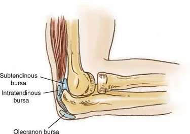
- Intratendinous: Found inside the triceps brachii tendon.
- Subtendinous: located between the triceps brachii tendon and the olecranon.
- Subcutaneous: The connective tissue layer beneath the subcutaneous layer envelops the olecranon.
Nerve supply of elbow joint
Musculocutaneous nerve
- Origin: the brachial plexus’s lateral cord
- Laterally, it appears after separating from the biceps tendon.
- Deep within the cephalic vein, it will end in the LABC or forearm.
- Innervation at elbow
- It provides the biceps and brachialis.
- Between these muscles is a nerve.
Radial nerve
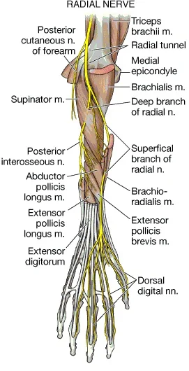
- Origin: the brachial plexus posterior cord
- The teres major, the triceps’ long head and the humeral shaft form the last triangle.
- A spiral indentation 13 cm superior to the trochlea.
- This usually happens where the distal and middle thirds of the humerus meet.
- Together, the brachialis and brachioradialis are located.
- The radiocapitellar joint is situated at a distance near the joint capsule.
Median nerve
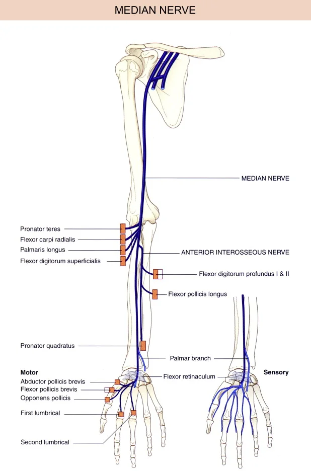
- Origin: Brachial plexus lateral and medial cords
- The brachial artery operates from its medial to lateral branches.
- Innervation at the elbow joint.
- It provides elbow joint branches.
- There are none on its upper arm.
Ulnar nerve
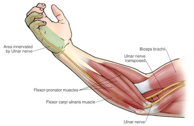
- Origin: Brachial plexus medial cord
- It reaches the Struthers arcade level and proceeds medially to the brachial artery, passing through the medial intermuscular septum to enter the posterior compartment.
- The medial epicondyle uses the cubital tunnel to assist in its posterior migration.
- Innervation at elbow
- The elbow joint receives branches from it.
- There are none on its upper arm.
- The FCU’s initial motor branch and the elbow joint are far apart.
- The radial, ulnar, and musculocutaneous nerves forward blood to the elbow joint.
Blood supply and innervation
The brachial, profunda brachii, radial, and ulnar arteries donate blood to the elbow joint through collateral and recurrent branches. These branches arise from numerous periarticular anastomoses.
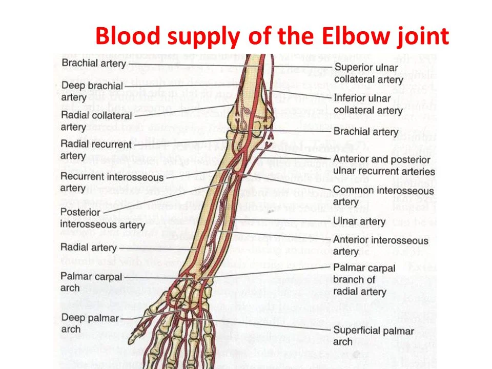
These two arteries were around the elbow joint. They support and round the anastomotic loop, which supplies blood to the joint.
The arm’s brachial and deep brachial arteries are attached above the elbow.
Blood Supply of Elbow
Brachial artery
- It is still located on the upper arm’s medial side.
- Laterally, it enters the cubital fossa.
- The median nerve, medial biceps tendon, & brachial artery
- Lateral border–brachoradialis
- Medial border–pronator teres
- Proximal border –distal humerus
- The elbow is the base of the radial and ulnar arteries.
Function of the elbow joint
Arm bending is achieved by flexing your elbow. It can travel in four directions:
- Extension: Finding forward for something with your arm further from your body.
- Flexion is a kind of extension; it includes extending your lower arm inward toward your body.
- Supination: Raising your hand’s palm.
- Pronation: Downward your hand’s palm.
Muscle of the elbow joint
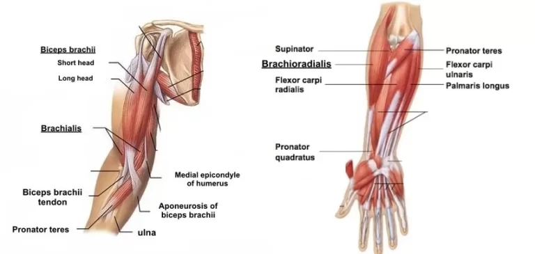
- The biceps brachii, brachialis, and brachioradialis are the muscles that most frequently stimulate the elbow joint during flexion.
- Most biceps brachii flexion and supination are done primarily by the elbow.
- The biceps brachii are particularly unusual, as they have a more laterally oriented tendon that inserts on the proximal radius and a medially directed aponeurosis that extends into the fascia of the proximal forearm.
- The triceps brachii contract primarily to extend the elbow joint, with the anconeus muscle providing little assistance.
- The pronator teres and quadratus pronate the forearm to the elbow, where as the biceps brachii and supinator supinate it.
Movements of elbow joint
- One way to lessen the angle that the arm and forearm make is to bend the forearm at the elbow joint.
- As the arm and forearm extend, they make a wider angle.
- Together, the arm’s anterior and posterior muscular groups do those functions.
Elbow joint Flexion
Flexor muscles are primarily found in the anterior part of the arms.
This compartment contains two elbow-flexing muscles.
- The brachial bicep muscle has two heads.
- The scapula’s supraglenoid tubercle is where the long-head tendon starts.
- It travels via the bicipital groove on the humerus’ anterior surface and the shoulder joint’s capsule.
- The biceps brachii muscle’s short head arises into the coracoid process of the scapula.
- The biceps brachii’s muscular belly grows by joining these two heads.
- The radial tuberosity found distal to the elbow joint, identifies the point at which the muscle connects to a single tendon.
- The bicipital aponeurosis, a flattened sheath of connective tissue wrapping the forearm, is where the tendon originates.
- It is positioned far inside the biceps’ brachial muscle.
- Although the brachioradialis muscle aids forearm flexion, the brachialis and biceps brachii are the main elbow flexors.
- On the distal humerus, the brachioradialis muscle rises atop the lateral epicondyle.
- This muscle works on the elbow joint because it crosses it, even though its primary location is in the forearm. The radial nerve innervates it.
Mnemonic
It becomes easy if you tie learning the muscles that bend the elbow to a mnemonic, like the one below.
3 Bs flex their elbows
- Biceps
- Brachialis
- Brachioradialis
Extension of Elbow Joint
The rise in elbow angle to return the forearm from a flexed posture to its natural position is known as an extension of the forearm at the elbow joint.
- The triceps brachii contain three heads.
- The medial head begins at the lateral aspect of the humerus, the lateral head ends at the scapula’s infra glenoid tubercle, and the long head commences at this location.
- The tendon passes through the olecranon of the ulna once the three heads connect.
- The lateral and medial heads of the muscle originate a radial groove to let the radial nerve, which depends on the muscle, reach the arm.
- While the elbow joint can only move in the flexion and extension directions, the proximal radioulnar joint, which supports the elbow joint, can also move.
- We refer to pronation and supination as movements at this joint.
Pronation antebrachial, or pronation of the forearm;
- A supinated forearm looks upward, while a pronated forearm appears to be tilted downward.
- The forearm is anatomically situated in the supine position.
- The raised forearm that faces the palm posteriorly is known anatomically as pronation.
Pronation and Supination
- The radiocapitellar and proximal radiolunar joints regulate pronation and supination movements.
- The usual range of motion is 180 degrees, which consists of 80 to 90 degrees of pronation and 90 degrees of supination.
- One can use shoulder abduction to compensate for decreased pronation range of motion.
- A loss of motion may be more incapacitating than a loss of pronation range of motion due to the absence of a balancing action connected to supination.
Clinical significance
The elbow joint is a crucial anatomical structure with several clinical significances due to its complexity and functional importance in upper limb movement. The following are some of the most significant problems related to the elbow joint:
Bursitis
- Constant pressure and friction may allow the bursa to get irritated in subcutaneous bursitis. Due to its relatively superficial location, this bursa is susceptible to infection (e.g., skin laceration following a fall on the elbow).
- Subtendinous bursitis: Usually diagnosed in assembly line workers, this ailment comes on by repetitive forearm flexion and extension. Flexion typically causes more pain because it puts extra strain on the bursa.
Dislocation
- A young child who falls on a hand with their elbow flexed most often dislocates their elbow.
- The most common injuries are to the ulnar nerve and ulnar collateral ligament, though there may be additional effects as well.
- Given that most elbow dislocations occur in the posterior region, this is quite important.
Epicondylitis (Tennis elbow or Golfer’s elbow)
- Tendinous tissue is the basis for almost all of the forearm’s flexor and extensor muscles.
- A common tendon overuse strain may happen in athletes, resulting in pain and inflammation near the injured epicondyle.
- A common complaint among golfers with the same flexor origin is soreness in the medial epicondyle.
Supracondylar Fracture
- Although a direct blow to the flexed elbow can also result in a fracture, a child’s outstretched hand is the most common location for supracondylar fractures (95%).
- The most common type of fracture is transverse, extending between the two epicondyles in the weak epicondylar region of the distal humerus, which is made up of the opposing olecranon and coronoid fossas.
- The brachial artery, which supplies blood to the forearm, can become clogged by direct injury or edema.
- Because the flexor muscles become fibrotic and short, the ensuing ischemia may result in Volkmann’s ischemic contracture, which is an uncontrollable flexion of the hand.
- Further possible consequences include injury to the medial, ulnar, or radial nerves.
- Therefore, it is crucial to assess and record the neurovascular status of every patient who presents with these injuries.
- Acute disruption of the blood flow can occasionally result in a “pale, pulseless” leg, generally in a youngster, necessitating emergency surgery.
Understanding these clinical significances helps healthcare providers diagnose and treat various elbow joint conditions effectively, often requiring a multidisciplinary approach involving orthopedic surgeons, physiotherapists, and occupational therapists.
FAQs
Is the elbow a pivot joint?
The elbow joint gives the arm’s capacity for bending, stretching, and rotating. A connective system involving muscles, tendons, and ligaments connects the distal humerus to the upper arm bone, joining the ulna, radius, and forearm bones.
Which one is referred to as an elbow?
Forearm and hand movement in both directions with the body is made easier by the elbow joint. Normal hinge joints that distinguish the arm from the forearm involve the humerus in the upper arm and the radius and ulna at the wrist.
The elbow joint is situated where?
Your elbow joint, which is situated in the middle of your arm, controls how much your forearm and upper arm can bend.
What blood supply goes into the elbow joint?
The brachial artery’s branches form a surrounding network of vessels that supply the elbow joint with a rich arterial supply.
What is the elbow’s functional anatomy?
Different functional structures exist for the elbow joint complex in terms of both orientation and structure. The humeroradial, humeroulnar, inferior radioulnar, and superior radioulnar joints develop by the joint cartilage of the ulna, radius, and humerus.
Which are the three most important elbow muscles?
These upper extremity movements may result in the connecting of muscles that pass through the elbow joint.
Biceps Brachii.
The biceps brachii muscle attaches to the elbow and shoulder joints and is primarily responsible for elbow flexion.
Triceps Brachii.
Brachioradialis.
Brachialis.
References
- Professional, C. C. M. (n.d.-b). Elbow Joint. Cleveland Clinic. https://my.clevelandclinic.org/health/body/elbow-joint
- Dhameliya, N. (2024, February 26). Elbow Joint -. Samarpan Physiotherapy Clinic. https://samarpanphysioclinic.com/elbow-joint/
- Physiotherapist, N. P. (2023, December 30). Elbow Joint: Anatomy and Function – Clinic for Mobile Physiotherapy. A mobile clinic for physical therapy. https://mobilephysiotherapyclinic.in/elbow-joint/
10 Comments