Shoulder muscles
Introduction
The shoulder joint, also known as the glenohumeral joint, is a ball and socket joint with the widest range of motion in the human anatomy. The shoulder muscles perform a variety of functions, including abduction, adduction, flexion, extension, internal and external rotation.
The scapula is the shoulder’s central bony structure and the site of all muscle interaction. The glenoid cavity, or articular surface of the glenohumeral joint, is located on the lateral aspect of the scapula. The glenoid labrum, shoulder joint capsule, supporting ligaments, and the rotator cuff muscles’ myotendinous attachments surround and reinforce the glenoid cavity.
The shoulder muscles maintain the greatest range of motion of any joint in your body. Because of its flexibility, the shoulder can become unstable or sustain an injury. The humerus (upper arm), clavicle (collar bone), and shoulder blade (scapula) are the attachment points for about eight shoulder muscles. Many other muscles help to stabilise and guide the shoulder’s movements.
These are the largest shoulder muscles:
- Trapezius
- Deltoid
- Pectoralis major
- Serratus anterior
- Rhomboid major
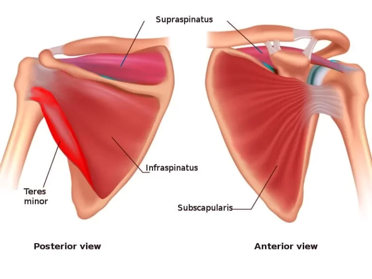
Another four shoulder rotator cuff muscles:
- Supraspinatus
- Infraspinatus
- Teres minor
- Subscapularis
Other shoulder muscles include:
- Pectoralis minor
- Latissimus dorsi,
- Biceps brachii, or biceps,
- Triceps
Trapezius muscle
Origin
- Medial one-third of the superior nuchal line
- External occipital protuberance
- Ligament nuchae
- C7 spine
- T1-T12 spines
- Correspondings supraspinous ligaments
Insertion
- Upper fibres into the lateral third of the clavicle’s posterior border
- Middle fibres into the upper lip of the crest of the scapula’s spine and the medial border of the acromion
- Lower fibres, on the apex of the triangular area at the medial end of the spine, with a bursa intervening
Nerve supply
- Spinal part of accessory nerve(XI)
- Branches from C3,C4
Actions
Upper fibers act with levetor scapulae, and elevate the scapula, as in shrugging. The upper fibres of both sides extend the neck.
Middle fibres act with the rhomboid, and retract the scapula,
Upper and lower fibres act with the serratus anterior, and rotate the scapula forwards around the chest wall thus playing an important role in the abduction of the arm beyond 90 degrees.
Deltoid muscle
The acromial part of the deltoid is an example of a multipennate muscle. many fibres arise from the septa of origin that are attached above the acromian. the fibers converge onto three septa of insertion which are attached to the deltoid tuberosity.
Origin
- the front edge and surrounding surface of the clavicle’s lateral third.
- The lateral border of the acromion where three septa of origin are attached.
- the bottom lip of the scapular spine’s crest.
Insertion
the humerus’s deltoid tuberosity, to which three insertional septa are connected.
Nerve supply
Actions
- The multipennate acromial fibres are powerful abductors of the arm at the shoulder joint from the beginning to 90 degrees.
- The anterior fibres are the flexors and medial rotators of the arm.
- The arm’s lateral rotators and extensors are the posterior fibres.
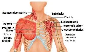
Pectoralis major muscle
Origin
- the medial two-thirds of the clavicle’s anterior surface
- Half the breadth of the anterior surface of the manubrium and sternum up to 6th costal cartilage
- Second to sixth costal cartilages, sternal end of 6th rib
- A abdominal external oblique muscle aponeurosis
Insertion
- It is inserted by a bilaminar tendon on the lateral lip of the bicipital groove in the form of “U”
- The inferior surface of the two laminae is continuous with one another.
Nerve supply
Medial and lateral pectoral nerves
Actions
- The muscle as a whole results in the following actions: Adduction and medial rotation of the arm’s shoulder joint.
- The clavicular region results in Arm flexion
- The sternocostal part is used in the extension of the flexed arm against resistance, Climbing
- Acts as an accessory muscle during inspiration when the humerus is fixed in abduction
Serratus anterior muscle
Another name for the serratus anterior is the boxer’s muscle.
Origin
The fascia covering the intervening intercostal muscles and the upper eight ribs in the mid-axillary plane give rise to the eight digitations that make up the serratus anterior muscle. The first digitation appears in the posterior triangle of the neck. It arises from the outer border of the 1st rib and a rough impression on the 2nd rib. Also, 5th-8th digitations interdigitate with the costal origin of the external oblique muscle of the abdomen.
Insertion
All 8 digitations pass backwards around the chest wall.
- Along the medial border of the scapula, the muscle inserts into the costal surface.
- Starting from the superior angle and moving towards the spine’s root is the first digitation.
- The subsequent two or three digits are placed on the medial border, lower down.
- Over the inferior angle, the lower five or four digitations are inserted into a sizable triangle.
Nerve supply
Long thoracic nerve(C5,C6.C7)
Actions
- Pushing and punching movement
- The fibres inserted into the inferior angle of the scapula pull it forward and rotate the scapula so that the glenoid cavity is turned upwards. The trapezius assists the serratus anterior in this action by pulling the acromion both upward and backwards.
- The muscle steadies the scapula during weight-carrying
- It helps in forced inspiration
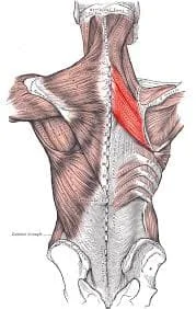
Rhomboid major muscle
Origin
- Spines of T2-T5
- Supraspinous ligaments
Insertion
The medial border of the scapula below the root of the spine
Nerve supply
Dorsal scapular nerve (C5)
Actions
Retraction of scapula
Supraspinatus muscle
Origin
- the middle two-thirds of the scapula’s supraspinous fossa. The muscles merge with the shoulder joint capsule by passing a tendon laterally beneath the coracoacromial arch.
- The subacromial bursa divides the tendon from the arch.
Insertion
upper impression on the humerus’s larger tubercle
Nerve supply
Suprascapular nerve(C5,C6)
Actions
It stabilises the head of the humerus during arm movements, along with other short scapular muscles. its action as abductors of the shoulder joint from 0-15 degrees is controversial. The deltoid and supraspinatus are both involved in the beginning and end of abduction.
Infraspinatus muscle
Origin
the middle two-thirds of the scapula’s infraspinous fossa
Insertion
middle impression on the humerus’s larger tubercle
Nerve supply
Suprascapular nerve (C5,C6)
Actions
Lateral rotator of arm
Teres minor muscle
Origin
the top two-thirds of the lateral scapular border’s dorsal surface as two slips
Insertion
The largest tubercle of the humerus’s the lowest impression
Nerve supply
Axillary nerve (C5, C6)
Actions
Lateral rotator of arm
Subscapularis muscle
Origin
Medial two-thirds of the subscapular fossa
Insertion
Lesser tubercle of the humerus
Nerve supply
Upper and lower subscapular nerves (C5, C6)
Actions
Medial rotator and adductor of arm
Pectoralis minor muscle
Origin
- 3,4,5 ribs, near the costochondral junction
- Intervening fascia covering external intercostal muscles
Insertion
The coracoid process’s upper surface and medial border
Nerve supply
Medial and lateral pectoral nerve
Actions
- Draws the scapula forward
- Depresses the point of the shoulder
Latissimus dorsi muscle
Origin
- one-third of the iliac crest’s posterior outer lip
- Posterior layer of lumber fascia; thus attaching the muscle of the lumber and sacral spines
- Spines of T7-T12, lower four ribs
- Inferior angle of scapula
Insertion
- The muscle winds around the lower border of the teres major and forms the posterior fold of the axilla
- The tendon is twisted upside down and inserted into the floor of the intertubercular sulcus
Nerve supply
Thoracodorsal nerve (C6-C8)
Actions
- When swimming, rowing, climbing, pulling, folding the arm behind the back, and scratching the opposite scapula, the shoulder’s adduction, extension, and medial rotation are all involved.
- Hold the inferior angle of the scapula in place
Biceps brachii muscle
Origin
It has two heads of origin:
- From the tip of the coracoid process, the short head emerges with the coracobrachialis.
- The glenoidal labrum and the supraglenoid tubercle of the scapula give rise to the long head.
Insertion
The posterior rough part of the radial tuberosity. the tendon is twisted; the anterior fibres become medial. A bursa separates the tendon from the anterior portion of the tuberosity.
Nerve supply
Musculocutaneous nerve (C5,C6)
Actions
- The arm’s flexor is the short head.
- The humerus’s head cannot move upward due to its long head.
Triceps muscle
Origin
The Triceps muscle arises from the following three heads:
- The long head arises from the infra glenoid tubercle of the scapula; it is the longest of the three heads.
- The lateral lip of the radial groove, which corresponds to an oblique ridge on the upper portion of the posterior surface of the humerus, is where the lateral head originates.
- The medial and lateral intra-muscular septa, as well as a sizable triangular region on the humerus’ posterior surface beneath the radial groove, are the sources of the medial head. The medial head is medial to the lateral head at the level of the radial groove.
Insertion
A superficial flattened tendon covering the medial head and inserted into the posterior region of the olecranon process’s superficial surface is formed by the convergence and fusion of the long and lateral heads. Part of the medial head is inserted into the olecranon and part of it into the superficial tendon.
A tiny bursa divides the medial head from the elbow joint capsule, but some of its fibres are inserted into this area of the capsule to keep the capsule from nipping during arm extension. These fibres are referred to as the articularis cubiti or the sub anconeus.
Nerve supply
The radial nerve gives each head its branch (C7, C8). The radial groove and the axilla are where the branches emerge.
Actions
An effective active elbow extensor is the triceps. When the arm is abducted, the long head stabilises the humeral head. Gravity extends the elbow passively.
Shoulder Muscles Functions
The muscles of the shoulder perform a variety of functions, including
- Shoulder and Arm Movement: The shoulder muscles move the upper arm and shoulder girdle through flexion, extension, abduction, adduction, internal and external rotation, elevation and depression, protraction, and retraction.
- Connecting the upper limb to the trunk: transmitting forces from the hands and arms to the central body.
- Shoulder Stabilisation: The shoulder’s rotator cuff muscles help to deepen the shoulder socket and draw the head of the humerus into the socket for increased stability.
- Stabilising The Scapula: The posterior shoulder muscles control and allow smooth movement of the shoulder blade, helping to protect the shoulder.
- Preventing Joint Dislocation: The muscles of the shoulder play an important role in preventing the joints from moving out of place.
- Tightening the Joint Capsule: Some of the muscle feeds into the joint capsule, which helps to improve joint stability and reduce friction.
Embryology
The embryologic development of the limbs begins at the end of the fourth week of foetal development. By the sixth week, the foetus has developed hand and foot plates. Mesenchymal cells condensate and differentiate into chondrocytes during limb development, eventually forming the upper and lower extremity’s bones and cartilage.
The upper and lower extremities have very similar embryological development patterns. Limb musculature is first visible around the seventh week. Mesenchyme migrates from the dorsolateral cells of somites to the limb, where it differentiates into muscle cells. The upper extremity’s distal muscles develop later than the shoulder muscles.
Blood Supply
The upper extremities receive blood supply from the subclavian artery. These vessels are located on both sides of the body and supply blood to the upper extremities. The blood supply for both arteries comes from the aortic arch. The vertebral artery, internal thoracic artery, thyrocervical trunk, and dorsal scapular artery are among the branches of the subclavian artery that run parallel to each other on both sides of the body.
When the subclavian artery reaches the first rib’s lateral border, it becomes the axillary artery.
The axillary artery is divided into three parts, each of which contains arterial branches that supply the shoulder muscles. The superior thoracic artery, thoracoacromial artery, circumflex humeral artery, and lateral thoracic artery all branch off of the axillary artery. The subscapular artery is a branch of the third segment of the axillary artery. The subscapular artery separates into the circumflex scapular artery and the thoracodorsal artery. In general, the shoulder muscles are supplied by arteries that are named after the muscles they supply.
Lymphatic drainage
Efferent lymphatic vessels originate in the distal upper extremity and travel through the shoulder. Furthermore, axillary lymph nodes supply efferent lymphatic vessels in the shoulder region and pass proximally through the shoulder. Deep lymphatic vessels exist alongside superficial lymphatic vessels. The deep lymphatic vessels transport lymph from the joint capsule, tendons, and nerves. The subclavian lymphatic trunk drains lymphatics from the shoulder and axillary region. The right lymphatic duct receives drainage from the subclavian trunk. On the left, the subclavian trunk empties into the thoracic duct.
Shoulder muscles injury
Because the shoulder joint is extremely flexible, the muscles and soft tissues surrounding it are subjected to a great deal of wear and tear. This makes the shoulder muscles prone to injuries and degenerative conditions, including:
- Adhesive capsulitis, also known as frozen shoulder, occurs when the capsule around the shoulder joint thickens and stiffens. It can cause spasms, pain, and extreme stiffness in the shoulder muscles.
- Shoulder bursitis is an inflammation of the bursae (tiny fluid-filled sacs) in your shoulders. Inflammation can make it difficult to move your shoulder joint and cause muscle irritation.
- Rotator cuff injury: Rotator cuff injuries, such as tears, typically affect tendons but can also involve muscles.
- Shoulder impingement syndrome occurs when your shoulder muscles or tendons rub against bones excessively, causing the soft tissues to become painful and inflamed.
- Strain: A shoulder strain is caused by overstretched muscle fibres. A muscle strain, also known as a pulled muscle, is determined by how much muscle stretches or tears. Muscle strains are divided into three grades: Grade 1 (mild)—A few muscle fibres have been torn, resulting in shoulder pain and tenderness the day after exercise.
Grade 2 (moderate): Approximately half of the muscle fibres are torn, resulting in significant pain, swelling, and weakness after activity.
Grade 3 (severe)—Complete muscle rupture, resulting in severe pain, swelling, and loss of strength. - Dislocation. The top of your arm may come out of its socket if you rotate or pull your shoulder back too forcefully. Your shoulder will be painful and weak. You may also experience swelling, numbness, or bruising.
- Separation. This injury impacts the joint where your shoulder blade and collarbone meet. It’s known as the acromioclavicular (AC) joint. A fall or hard blow tears the ligaments that keep it together. If your collarbone is pushed out of position, you will develop a bump on the top of your shoulder.
- Fracture. If you fall or take a hard hit, your bone may break or crack. The clavicle (collarbone) and humerus (arm bone closest to the shoulder) are the most commonly broken bones. You will feel a lot of pain and may bruise. If your collarbone is broken, your shoulder will sag and you may be unable to lift your arm.
Shoulder muscles exercises
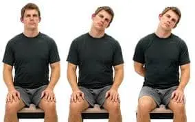
Shoulder rolls
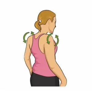
- Stand with your feet hip-width apart.
- Lean the arms at your sides to hang loosely.
- Inhale deeply and lift your shoulders to your ears.
- Squeeze the shoulder blades together by repositioning the shoulders.
- Exhale and drop your shoulders back.
- Move the elbows forward and feel the stretch in the back of the shoulders.
- Repeat this ten times.
Cross-body shoulder stretch.
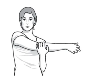
- Stand with your feet hip-width apart.
- Stretch your right arm out straight.
- Bring the right arm across the body, with the hand pointing to the floor on the opposite side of the left leg.
- Bend your left arm at the elbow.
- Hook the left forearm under the right arm to support it above the elbow.
- Stretch the back of the right shoulder by pulling the right arm further in and across the body with your left forearm.
- Hold for 20 seconds, then repeat the stretch on the opposite side.
Child’s Pose.
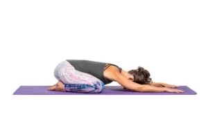
- Kneel on the floor or a mat.
- Touch your big toes together.
- Spread the knees apart.
- Sit up straight.
- Breathe out and raise your arms above your head.
- Exhale and bow forward, towards the floor, with arms extended in front.
- Touch the ground with your palms.
- Bring both elbows to the ground.
- Sit back, bringing the bottom of the back to the heels.
- As you stretch the back of your shoulders, feel it.
- Breathe deeply and hold the position for one minute or more.
Cow Face Pose
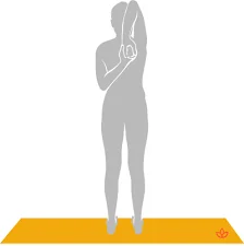
- Stand with your feet hip-width apart.
- Elevate your right arm vertically towards the heavens.
- Bend your right arm at the elbow.
- Keeping the elbow raised, extend the right hand over the head and down the back.
- Stretch your left arm down.
- Reach your left hand behind and up the back.
- Bring the left and right hands together, clasping them if it is comfortable.
- Take three to four deep breaths.
- Give up the stretch and repeat on the other side.
Arm lifts (standing)
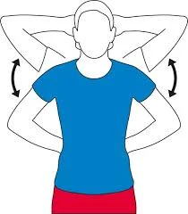
- Place your hands behind your head, elbows pointing to the sides, and press back as far as you can. Hold for 5 seconds.
- Then, place your hands behind your back, elbows pointing out, and pressed as far back as possible. Hold for 5 seconds.
- Repeat each movement five times.
Shoulder circle
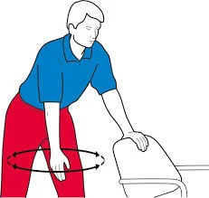
Stand with one hand on the chair. Let your other arm hang down and gently swing it back and forth in a circle. Repeat this motion five times and aim to do it two or three times per day. This can be an effective warm-up exercise.
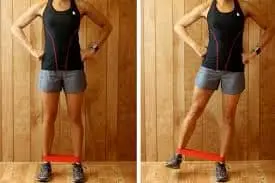
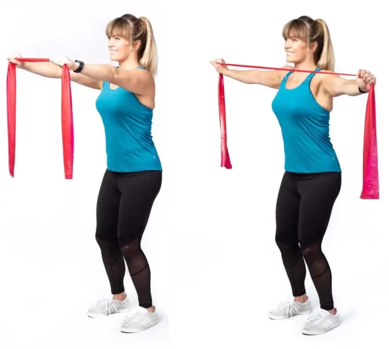
Stretch your shoulders with a resistance band.
- Hold a yellow or red elasticated resistance band in your hands, fingers curled around it, facing inward. Your elbows should be bent at waist height, just above your hips, with your arms and hands aligned with your shoulders.
- Keeping your elbows at your sides, extend the band as far as is comfortable. Hold for 10 counts. Gently bring your hands back into line with your shoulders. Repeat this ten times. Try doing this exercise three times per day.
Summary
The shoulder joint, also known as the glenohumeral joint, is a ball and socket joint with the widest range of motion in human anatomy. It is composed of eight muscles, including the trapezius, deltoid, pectoralis major, serratus anterior, and rhomboid major. The largest shoulder muscles are the trapezius, deltoid, pectoralis major, serratus anterior, and rhomboid major. These muscles maintain the greatest range of motion but can become unstable or sustain an injury due to their flexibility.
Shoulder muscles are responsible for various functions, including shoulder movement, connecting the upper limb to the trunk, stabilizing the shoulder socket, preventing joint dislocation, and tightening the joint capsule. They also play a crucial role in blood supply, with the subclavian artery providing blood to the upper extremities and the axillary artery supplying blood to the lower extremities.
Shoulder muscle injury is common due to the flexibility of the shoulder joint, making it susceptible to wear and tear. Examples include adhesive capsulitis, shoulder bursitis, rotator cuff injuries, and shoulder impingement syndrome.
Various shoulder muscle exercises, such as shoulder rolls, cross-body shoulder stretches, child’s poses, cow face poses, arm lifts, shoulder circles, and resistance band stretching, can help maintain the shoulder’s flexibility and prevent injuries.
FAQs
What is the primary muscle in your shoulder?
The primary skeletal muscles of the shoulder are: The rotator cuff is a group of four muscles—the supraspinatus, infraspinatus, teres minor, and subscapularis—that work together to stabilize the humeral head.
What kind of joint is the shoulder?
The shoulder joint (glenohumeral joint) is the articulation between the scapula and the humerus. It is a ball and socket synovial joint, one of the most mobile in the human body.
How many major muscles are in the shoulder?
This joint is supported by approximately eight muscles in your shoulder. They lend it strength, stability, and shape. Your shoulder muscles are skeletal. Tendons connect them to bones.
Which muscle rotates the arm?
During arm movements, the rotator muscles contract to prevent the head of the humerus from sliding, allowing a full range of motion and stability. Rotator cuff muscles also contribute to shoulder joint mobility by allowing for abduction, medial rotation, and lateral rotation.
What is the most frequently injured muscle in the shoulder?
Rotator cuff tears.
The muscles and tendons that hold your shoulder bones together are known as the rotator cuff. A rotator cuff tear is a common shoulder injury that causes a partial or complete tear in this group of muscles and tendons.
References:
- Chaurasia, B. D. (2019, June 30). Bd Chaurasia’s Human Anatomy, Volume 1. CBS Publishers & Distributors Pvt Limited, India. http://books.google.ie/books?id=F7cRyAEACAAJ&dq=BD+chaurasias&hl=&cd=1&source=gbs_api
- McCausland, C., Sawyer, E., Eovaldi, B. J., & Varacallo, M. (2023, August 8). Anatomy, Shoulder and Upper Limb, Shoulder Muscles. StatPearls – NCBI Bookshelf. https://www.ncbi.nlm.nih.gov/books/NBK534836/
- Hecht, M. (2020, April 17). Anatomy of the Shoulder Muscles Explained. Healthline. https://www.healthline.com/health/shoulder-muscles
- Muscles Of The Shoulder: Anatomy, Function & Common Injuries. (n.d.). Shoulder-Pain-Explained.com. https://www.shoulder-pain-explained.com/muscles-of-the-shoulder.html
- Shoulder Pain. (2023, May 8). WebMD. https://www.webmd.com/pain-management/why-does-my-shoulder-hurt
- Professional, C. C. M. (n.d.). Shoulder Muscles. Cleveland Clinic. https://my.clevelandclinic.org/health/body/21798-shoulder-muscles
- Dpt, J. F. P. (n.d.). Shoulder Muscle Anatomy: How to Strengthen and Avoid Injury. Hospital for Special Surgery. https://www.hss.edu/article_shoulder-muscle-anatomy.asp
- Burgess, L. (2024, March 18). Top 10 stretches for shoulder tightness. https://www.medicalnewstoday.com/articles/324647
- Exercises for the shoulders. (n.d.). Versus Arthritis. https://versusarthritis.org/about-arthritis/exercising-with-arthritis/exercises-for-healthy-joints/exercises-for-the-shoulders/