Oculomotor Nerve
Introduction
The oculomotor nerve is the third cranial nerve (CN III), and its name indicates its function. The oculomotor nerve will innervate muscles that move the eye or its components, as implied by the name alone. The nerve’s ability to move makes it an effective indicator of brain injury.
It is a mixed nerve with motor, parasympathetic, and sympathetic fibres. It originates in the midbrain and travels through the cavernous sinus to the orbit, where it controls the movements of four of the six extraocular muscles (superior, medial, inferior rectus, and inferior oblique), as well as the levator palpebrae superioris and superior tarsal muscle.
It performs somatic motor (general somatic efferent) and visceral motor (general visceral efferent-parasympathetic) functions. Essentially, the oculomotor nerve has three major functions, including:
Innervation of eye muscles for gaze fixation and eye tracking (somatic motor).
Innervation of the lens and pupil (autonomic parasympathetic)
Innervation of the upper eyelid (somatic motor)
Anatomical Course of Oculomotor nerve
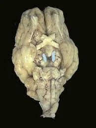
The oculomotor nerve originates in the oculomotor nucleus, which is located in the midbrain of the brainstem, ventral to the cerebral aqueduct. It emerges from the anterior aspect of the midbrain and connects to the posterior cerebral artery and the superior cerebellar artery.
The nerve then passes through the dura mater and into the lateral aspect of the cavernous sinus. The cavernous sinus receives sympathetic branches from the internal carotid plexus. These fibres do not connect with the oculomotor nerve; rather, they travel within its sheath.
The superior orbital fissure is where the nerve leaves the cranial cavity. It now splits into superior and inferior branches at this point:
The levator palpabrae superioris and superior rectus muscles are innervated by the superior branch.
Sympathetic fibres connect to the superior branch and innervate the superior tarsal muscle.
The inferior branch provides motor innervation to the inferior rectus, medial rectus, and inferior oblique.
It also provides pre-ganglionic parasympathetic fibres to the ciliary ganglion, which eventually innervates the sphincter pupillae and ciliary muscles.
Embryology
The oculomotor nerve and its associated cranial nerve nuclei are located within the midbrain. The midbrain develops from the mesencephalon. Neuroblasts from the basal plates form the tegmentum. The Edinger-Westphal nuclei, oculomotor nuclei, trochlear nuclei, red nuclei, reticular nuclei, and cranial nerves III and IV make up the tegmentum.
Brainstem nerve III is involved in both autonomic and somatic processes. Spinal nerves and somatic nerves have similar ventral roots. The inferior rectus, medial rectus, superior rectus, and inferior oblique muscles are innervated by them; they originate from the basal plate. The first preoptic myotome is the source of these muscles.
Most studies on the ciliary ganglion and the development of parasympathetic nerves have been done on chickens. The caudal midbrain and rostral hindbrain neural crest cells provide the parasympathetic nerve supply for the oculomotor nerves.
Some cells may also arise from the ectodermal placode caudal to the eye. These cells move ventrolaterally and rostrally towards the optic vesicle. Axons from the differentiating neurons in the ciliary ganglion innervate the choroid plexus’s blood vessels as well as the striated muscles of the iris and ciliary body.
Blood Supply and Lymphatics
The oculomotor nerve’s somatic and autonomic components have different vascular supplies. The inner somatic (voluntary) nerve fibres are supplied by the vasa vasorum, whereas the outer autonomic nerve fibres are supplied by pia mater blood vessels.
The lymphatic drainage of the eye’s orbit is still poorly understood. Lymphatic structures in the arachnoid and lacrimal gland have been identified morphologically; other unique lymphatic structures in the orbit are unknown.
The oculomotor nucleus and Edinger-Westphal nucleus are both found in the medial midbrain, which is supplied by the paramedian branches of the upper basilar artery and the proximal posterior cerebral artery.
Muscles
The oculomotor nerve regulates several muscles:
- Levator palpebrae superioris: raises the upper eyelid.
- The superior rectus muscle rotates the eyeball backwards, “looking up”
- the muscle of the medial rectus conjugates the eye,” looking towards the nose”.
- The inferior rectus muscle rotates the eyeball forwards, “looking down”.
- The inferior oblique muscle rotates the eyeball backward when it is adducted.
- The ciliary muscle controls lens shape to focus on close-up objects.
- Sphincter pupillae: constricts the pupil.
Function of Oculomotor nerve
Motor
The oculomotor nerve is the primary motor nerve for the ocular and extraocular muscles. Except for the superior oblique and lateral rectus, the oculomotor nervous system supplies all extraocular muscles with somatic motor fibres. The levator palpebrae superioris, which raises the upper eyelid, and the superior rectus muscle, which raises the eyeball, are supplied by the superior branch.
The inferior branch innervates three muscles: the inferior rectus, which depresses the eyeball, and the inferior oblique, which elevates, abducts, and laterally rotates the eyeball. The trochlear and abducens nerves supply the superior oblique and lateral rectus muscles, respectively.
Parasympathetic
The autonomic parasympathetic (involuntary) oculomotor nerve serves two primary functions. When stimulating the smooth muscle (sphincter pupillae) related to the pupil, it constricts the pupil (miosis). It also stimulates the ciliary muscles. The sphincter pupillae narrow the pupil to prevent diverging light rays from the corneal periphery from producing a blurred image. The ciliary muscle alters the shape of the lens during accommodation.
Sympathetic
The oculomotor nerve has no main function, but sympathetic fibres control together with it to stimulate the superior tarsal muscle (all of which takes the eyelid).
While this may appear to be a minor task, lens curvature, and pupil constriction are essential to vision and enable the following actions:
- Accommodation is focusing on objects as they move closer or farther away from you.
- Light reflex is the process of changing the pupil’s size to let the right amount of light into the eye for optimal vision.
- Near focus allows you to focus on and see close-up objects.
Oculomotor nerve palsy
Oculomotor nerve dysfunction can affect the third cranial nerve. This is caused by a group of disorders that cause paralysis of the third cranial nerve. When this happens, it’s called third nerve palsy.
Third nerve palsy is defined as partial or complete paralysis of the oculomotor nerves. Palsy has varying degrees and can affect different functions from person to person.
Oculomotor nerve palsy can be congenital (present at birth) or acquired (developed later in life).
Causes of Oculomotor nerve palsy
Oculomotor nerve palsy may be idiopathic. This indicates that doctors are unsure of the cause.
Possible causes of congenital third nerve palsy are:
- Aplasia: The oculomotor nucleus has disappeared or did not develop properly.
- Hypoplasia refers to a small or poorly developed oculomotor nucleus.
- Birth trauma: the moulding forces used during labour can affect the skull.
- Intrauterine trauma is an injury or stress to the foetus during pregnancy.
- Infection – an infection, such as meningitis, can cause third nerve palsy, but it is rare.
Acquired third nerve palsy can be caused by the following:
- The most common cause of poor blood flow is diabetes and high blood pressure.
- Trauma on the head or face
- An aneurysm, high intracranial pressure, or a brain tumour can all result in abnormal pressure on the oculomotor nerve.
- Trauma or aneurysm can cause brain bleeding.
- Infections, including Lyme disease and HIV
- Multiple sclerosis is a type of autoimmune disease.
- Migraine
Symptoms of Oculomotor nerve palsy
Symptoms associated with the oculomotor nerve include:
- Trouble moving one or both eyes.
- Ptosis, or drooping of the eyelid.
- Eye misalignment (strabismus).
- Double vision (diplopia).
- Blurred vision.
- Eye strain and migraines.
- Mydriasis is a condition in which your eye’s pupil is unusually large and responds slowly or never to light.
Assessment
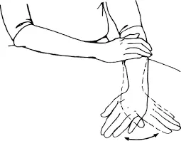
Using your finger or the tip of a pen, draw the letter “H”; instruct the patient to follow it with their gaze. During this procedure, the patient’s head should remain motionless, and the patient should report any blurring or double vision (diplopia).
Total oculomotor nerve palsy refers to the involvement of all muscles innervated by the oculomotor nerve, including pupillary involvement. Movement will be restricted in all fields of view except abduction and intorsion. Incomplete palsy refers to either partial limitation (paresis) of elevation, depression, or abduction, or even total mobility in one or more of these directions.
Diagnosis
CT or MRI are required. If a patient has a dilated pupil and a sudden, severe headache (suggesting a ruptured aneurysm) or becomes increasingly unresponsive (suggesting herniation), neuroimaging (CT or, if available, MRI) is performed right away.
If a ruptured aneurysm is suspected and CT (or MRI) scans do not reveal blood or are not available quickly, other tests, such as lumbar puncture, magnetic resonance angiography, CT angiography, or cerebral angiography, are recommended. Cavernous sinus disease and orbital mucormycosis necessitate immediate MRI imaging for proper treatment.
Treatment
To treat oculomotor nerve palsy, medical management of systemic predisposing factors and conservative symptom relief are the first steps in treatment. If these measures are ineffective, surgical intervention is the next step.
During the acute phase, conservative management is typically considered. Instruments like an eye patch, opaque contact lens, or blurred lens to shield the affected eye may be helpful interventions in the case of diplopia.
In other cases, botulinum toxin may be used to treat partial third nerve palsy with an adduction component, particularly when there is medial rectus involvement. If there is a deficit in vertical deviation, prism glasses may help to correct the abnormality.
After six months of conservative treatment with no significant results, surgical intervention is usually recommended. Strabismus surgery is primarily used to treat this condition. Strabismus surgery corrects misalignment in the extraocular muscles.
After failed pharmacological treatment, physical therapy can be an effective treatment of choice in patients with cranial nerve III and VI damage. Oculomotor nerve palsy is treated with physical therapy, which includes electrical stimulation, kinesiotaping, LASER therapy, trigger point therapy, and a neuromuscular exercise programme.
Prevention
Taking care of your oculomotor nerve health is similar to caring for your entire body. You can do the following things:
- Attend yearly examinations and routine eye exams; these can often identify eye and health problems before symptoms manifest.
- Give up using nicotine-containing products (like vaping, smokeless tobacco, and cigarettes), or refrain from starting to use them. For resources on quitting smoking, ask your provider.
- To avoid head injuries, use safety gear such as helmets and seatbelts.
- Manage chronic conditions such as type 2 diabetes and high blood pressure.
Summary
The oculomotor nerve (CN III) innervates the muscles that move the eye and its components. It is a mixed nerve with motor, parasympathetic, and sympathetic fibers and originates in the midbrain. It travels through the cavernous sinus to the orbit, controlling the movements of four of the six extraocular muscles, as well as the levator palpebrae superioris and superior tarsal muscle.
The oculomotor nerve performs somatic motor and visceral motor functions, with three major functions: innervation of eye muscles for gaze fixation and eye tracking (somatic motor), innervation of the lens and pupil (autonomic parasympathetic), and innervation of the upper eyelid (somatic motor).
The oculomotor nerve’s somatic and autonomic components have different vascular supplies, with the inner somatic nerve fibers supplied by the vasa vasorum and the outer autonomic nerve fibers by pia mater blood vessels.
Oculomotor nerve palsy is a condition affecting the third cranial nerve, causing partial or complete paralysis. It can be congenital or acquired and can be caused by various factors such as aplasia, hypoplasia, birth trauma, intrauterine trauma, infection, diabetes, high blood pressure, head or face trauma, brain tumours, aneurysms, infections, multiple sclerosis, and migraines. Symptoms include difficulty moving eyes, ptosis, eye misalignment, double vision, blurred vision, eye strain, migraines, and mydriasis.
Diagnosis involves CT or MRI scans. Treatment involves medical management, conservative measures, and surgical intervention if symptoms persist. Preventative measures include regular eye exams, quitting smoking, using safety gear, and managing chronic conditions.
FAQs
What is the blood supply to the oculomotor nerve?
The paramedian branches of the upper basilar artery and the proximal posterior cerebral artery supply the medial midbrain, which is the location of the oculomotor nucleus and the Edinger-Westphal nucleus.
What is the meaning of oculomotor weakness?
Oculomotor nerve palsy is a neurological condition that impairs your vision. It can cause double vision and make it difficult to use both eyes together. This condition is caused by a weakness in the oculomotor nerves, which leads to a loss of control over important eye muscles.
Is oculomotor a mixed nerve?
The oculomotor nerve is considered a mixed nerve because it contains both motor and parasympathetic fibres.
What disease affects the oculomotor nerve?
Third cranial nerve disorders can affect ocular motility, pupillary function, or both. Diplopia, ptosis, and paresis of eye adduction and upward and downward gaze are some of the symptoms and signs. The pupil becomes dilated, and light reflexes are impaired.
Is the oculomotor nerve purely motor?
This motor nerve communicates with all extraocular muscles except the lateral rectus and superior obliques.
What number of branches does the oculomotor nerve have?
The oculomotor nerve divides into two branches either upon entering the orbit or shortly before. It is divided into two branches: superior (cephalic branch) and inferior (caudal branch). The nasociliary nerve (a branch of the ophthalmic nerve) is located between the two divisions.
What symptoms indicate oculomotor damage?
Diplopia, ptosis, and paresis of eye adduction and upward and downward gaze are some of the symptoms and signs. The pupil becomes dilated, and light reflexes are impaired.
What is the origin of the oculomotor nerve?
The oculomotor nerve emerges from the midbrain’s tegmentum near the superior colliculus, ventral to the periaqueductal grey matter.
References
- Joyce, C., Le, P. H., & Peterson, D. C. (2023, March 27). Neuroanatomy, Cranial Nerve 3 (Oculomotor). StatPearls – NCBI Bookshelf. https://www.ncbi.nlm.nih.gov/books/NBK537126/
- The Oculomotor Nerve (CN III) – Course – Motor – TeachMeAnatomy. (2022, December 21). TeachMeAnatomy. https://teachmeanatomy.info/head/cranial-nerves/oculomotor/
- Oculomotor Nerve. (n.d.). Physiopedia. https://www.physio-pedia.com/Oculomotor_Nerve
- Sprabary, A. (2022, October 12). Oculomotor nerve. All About Vision. https://www.allaboutvision.com/eye-care/eye-anatomy/oculomotor-nerve/
- Professional, C. C. M. (n.d.). Oculomotor Nerve (CN III). Cleveland Clinic. https://my.clevelandclinic.org/health/body/21708-oculomotor-nerve
- Rubin, M. (2024, February 21). Third Cranial (Oculomotor) Nerve Disorders. MSD Manual Professional Edition. https://www.msdmanuals.com/en-in/professional/neurologic-disorders/neuro-ophthalmologic-and-cranial-nerve-disorders/third-cranial-oculomotor-nerve-disorders

One Comment