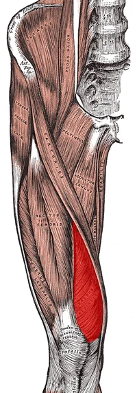
V M O Muscle (Vastus Medialis Oblique)
What is a V M O Muscle (Vastus Medialis Oblique)?
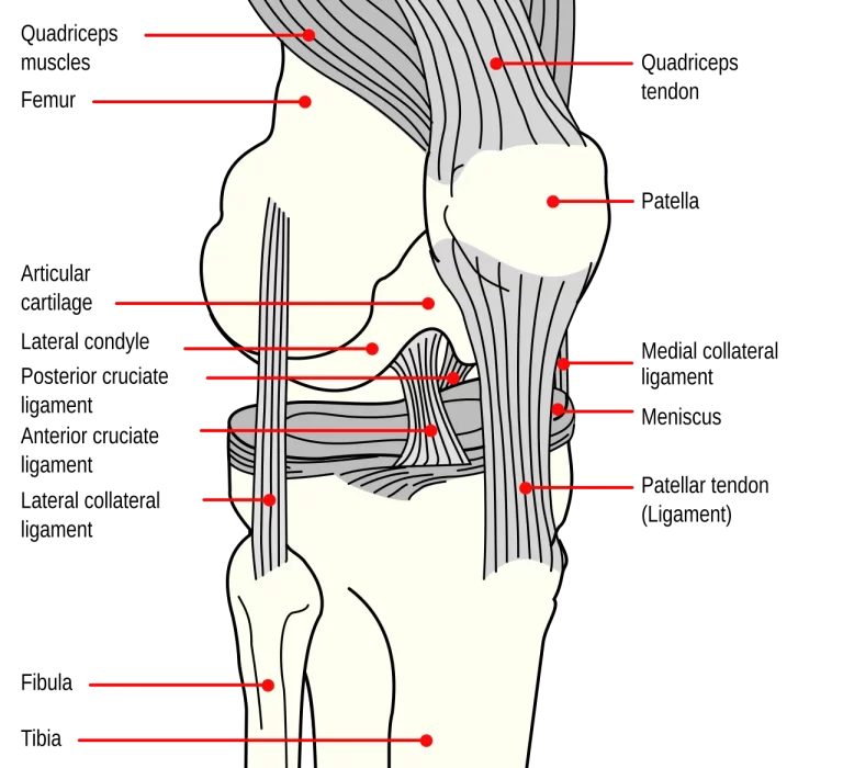
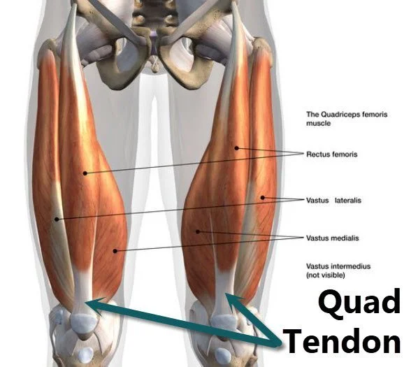
The V M O Muscle (Vastus Medialis Oblique) is a quadriceps muscle in the thigh. The quadriceps femoris is a group consisting of four muscles on the front of the thigh, including the VMO. The quadriceps have the responsibility of extending the knee and play an important role in activities like walking, running, as well as jumping.
The VMO is a teardrop-shaped muscle found on the inner side of your thigh, just above the kneecap. It is considered a part of the quadriceps group’s most medial (interior) muscle, the vastus medialis. The VMO is frequently mentioned when discussing knee health, particularly concerning the patella (kneecap).
Anatomical documents show that the muscle fiber pennation points of the vastus medialis oblique (VMO) as well as vastus medialis (VM) differ, resulting in functional differences between the two. While VM travels farther longitudinally and contributes more to knee extension, VMO runs obliquely assisting in medial patellar translation.
Strengthening the VMO is often emphasized in rehabilitation as well as injury prevention programs, especially for illnesses like patellofemoral pain syndrome as well as patellar tracking abnormalities. Exercises that target the VMO include leg extensions, terminal knee extensions, and additional isolation exercises focusing on the inner part of the quadriceps.
It’s interesting to note that the VMO works in tandem with the other quadriceps muscle tissue and that the knee’s stability and functionality are determined by the quadriceps’ overall strength.
In this blog post, we’ll talk about the VMO muscle, why it’s important, how to strengthen it, as well as how you can incorporate VMO exercises into your daily routine. Most people don’t give much thought to their VMO muscles, but it’s essential to keep your body sound and looking good.
Anatomy of V M O Muscle (Vastus Medialis Oblique)
Origin
The VMO has several points of origin. The muscle fibers primarily originate at the pubic point along the Adductor Magnus tendon. The other points of origin are the medial lip of linea aspera as well as the medial supracondylar line.
Insertion
VMO attaches to what is called the medial border of the patella as well as the knee joint capsule. It additionally includes a small area that directly connects to the patella tendon.
Nerve Supply
VMO’s nerve supply comes from the Femoral Nerve branches.
Blood Supply
The VMO muscle receives its blood supply from the femoral artery.
Function
The VMO muscle functions as follows:
- The VMO muscle holds the patella in the femoral groove while the knee is extended.
- It helps to keep the patella tracking straight inside the femoral groove throughout knee movement.
- Arrangements to help extend and straighten the knee joint.
- It is also beneficial to abduct (going away from the midline) as well as medially rotate (turning inward) the thigh at the hip joint.
The VMO muscle aids in knee extension by producing an eccentric force that slows the patella as it goes up the femoral groove.
So those are the functions of the VMO muscle.
Relations
The rectus femoris partially covers the vastus medialis. The vastus medialis’ superficial surface is also crossed by the sartorius muscle. The vastus medialis muscle in the middle third of the thigh serves as the lateral wall of the adductor canal (Hunter’s canal).
This canal is completed by the adductor longus as well as adductor magnus posterior muscles, as well as the sartorius medially. It delivers the femoral artery, femoral vein, saphenous nerve, and nerve to vastus medialis (both of which are femoral nerve branches).
Assessment
VMO assessment: The VMO muscle may be assessed by touching its contraction, assessing its volume, or performing surface electromyography (EMG). These methods can aid in the diagnosis of VMO weakness or disorders, as well as monitoring the progression of rehabilitation.
Examination of the VMO muscle.
- First, ensure that your VMO contracts are correct. VMO dysfunction is a common cause of chronic conditions such as patellofemoral knee pain.
- Sit with the legs extended in between you and a rolled-up towel under the injured knee to evaluate the VMO contraction.
- Place your fingers on the inside of your thigh’s VMO area to contract the muscle. To remove the foot from the sofa, press the knee into the towel and straighten the leg.
- The muscles under your fingertips must be firmly contracted.
- If the muscle does not contract, repeat the exercise with light pressure on the muscle, focusing on getting the muscle fibers under your fingertips.
Clinical Importance
- The vastus medialis oblique (VMO) muscle is required to maintain proper kneecap tracking. In simple terms, it stabilizes the patella as it moves.
- In pain-free normal individuals, the VMO fibers are active all through the entire range of motion.
- People who suffer from chondromalacia patella or patellofemoral knee pain experience irregular muscle contractions. They thus get tired easily.
- This can cause the patella to misalign or shift out of its groove, causing instabilities, pain, and damage to nearby structures. VMO weakness can result from damage, disuse, and poor neuromuscular control. If the muscle contracts continue the strengthening routine.
- The VMO is the largest as well as most powerful muscle in your quadriceps. It helps to stabilize your knee and extend (erect) your leg.
- A strong VMO helps to prevent knee pain and injuries like patellofemoral pain syndrome (PFPS) as well as patellar tendinitis. Strong VMO muscles can boost your performance and lower your risk of injury.
- The VMO muscle (Vastus Medialis Oblique) is a teardrop-shaped muscle found on the inner side of the thighs. It is one of four quadriceps muscles, which are accountable for straightening the leg at the knee.
Here’s why the VMO muscle is important:
The VMO muscle stabilizes and tracks the knee joint, preventing injuries like patellar subluxation as well as patellofemoral pain syndrome (runner’s knee).
It helps to keep the patella (kneecap) tracking straight inside the femoral groove during knee movement.
Knee Extension:
The VMO contracts to straighten the knee joint.
It generates an eccentric force that slows the patella as it shifts up the femoral groove during knee extension.
A strong VMO helps prevent knee pain and injuries, such as patellofemoral pain syndrome (PFPS) as well as patellar tendinitis.
Athletes, particularly those participating in sports that require running, jumping, and quick changes of direction (such as basketball as well as soccer), profit from well-developed VMO muscles.
The VMO is the strongest and largest quadriceps muscle.
It is essential for knee stability and keeping the patella in proper alignment during leg extension.
Patellofemoral taping
Tapping your patella may be beneficial if it is not tracking properly or if you are experiencing pain while performing VMO exercises.
The patella is taped, and support strips are used to direct the tape out of sore spots. As a result, you may be able to perform strengthening exercises without experiencing pain.
Patella Taping & Q-Angle
The VMO’s specific role is to stabilize the patella inside the patella groove as well as to control patella tracking when the knee is bent and straight.
Misfiring deficiencies in the VMO cause mal tracking of the patella, resulting in injuries to the surrounding structures as well as aching pain.
Is my VMO muscle contracting normally?
First, check that your VMO is properly contracted. Long-term injuries, particularly patellofemoral knee pain, frequently occur due to VMO malfunction.
To assess VMO contraction, sit with your legs extended in front with a rolled-up towel on the injured knee.
Place your fingers on the VMO muscle on the inside of your thigh and contract it. The knee ought to sink into the towel while the leg straightens, lifting the foot off the couch.
You ought to sense a strong contraction from the muscle beneath your fingertips.
If the muscle cannot contract, keep going to practice by gently pressing down on it and concentrating on contracting the muscle fibers beneath your fingertips.
If the muscle contracts continue with the strengthening exercises.
What occurs If Your VMO Muscles Are Weak?
If your VMO muscle is weak, it can result in patellofemoral pain syndrome (PFPS). PFPS is a condition characterized by pain near the kneecap. The kneecap is located in a groove at the tip of the femur. The VMO muscle helps to keep the kneecap in this groove. If the VMO muscle is weak, the kneecap may track improperly as well as rub against the bone, resulting in pain.
PFPS is a common condition that can affect people of any age. It is more prevalent in women than in men and usually affects people aged 30 to 50.
How do you strengthen the VMO muscle?
The exercises listed below help to strengthen the VMO muscle. They are as follows.
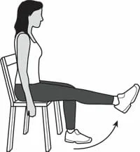
Seated Leg Extension:
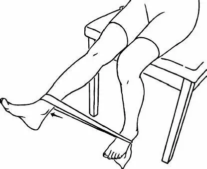
This exercise can be performed at the gym or at home using a resistance band. Sit on a chair or bench, legs extended toward you. Cover the band near your ankles, then slowly raise your leg while maintaining your knee straight. Lower the leg and repeat. For example, if you’re using a 10-pound weight, begin with three sets of ten repetitions.
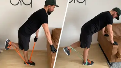
Hamstring Curl with a Resistance Band:
This exercise may be done at the gym or at home using a resistance band. Anchor the band to a sturdy post and lie face down. Wrap the band near your ankles and curl your legs up to your buttocks while keeping your hips as well as your upper body still. Lower your legs as well as repeat.
Squats:
Squats improve leg strength and target the VMO muscle. For a squat, stand shoulder-width apart and bend your knees. Keep your knees behind your toes as well as your buttocks out as you lower down. Return to standing, and repeat.
Lunges:
Lunges are a great exercise for increasing leg strength. To do a lunge, stand with your toes approximately shoulder-width apart then take a big step forward using one leg. Lower the body after both knees form a 90-degree angle. Make sure that your front knee doesn’t cross over the toes and your back is straight. Return to standing and continue with the other leg.
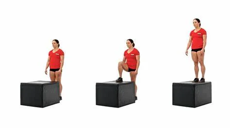
Step-Ups:
This exercise can be done in the gym or at home using a step stool or box. Stand in front of the step and rest your entire foot on top of it. Press down using your heel to raise your body, then bring your other leg up to stand on top of the step. Step back and then repeat with the opposite leg.
Leg Press:
This exercise is commonly performed at the gym, but it can also be done at home with a resistance band. Sit on the ground, legs extended in front of you, as well as cover the band around your ankles. To lift your body, press your legs down into the ground. Lower back and then repeat.
Consistently performing these exercises will help to strengthen the muscles while enhancing overall leg strength. For each exercise, attempt three to four sets of 10-15 repetitions. Add weight or resistance as needed to make the exercises difficult but not impossible.
Conclusion:
It can be concluded that the VMO muscle is essential for knee function as well as injury prevention. To keep the knees healthy and injury-free, it is recommended that you keep strong VMO muscles. You can perform VMO-targeted exercises like squats and lunges. To avoid knee injuries, exercise with proper form and wear supportive shoes.
If you have any questions about how to do these exercises properly, consult a physical therapist or a certified personal trainer. Physical therapy helps patients recover from pain.
Heel Drop:
Once you can maintain the contraction described above, begin to incorporate it into useful exercises like lunges as well as heel drops. To perform a heel drop, stand on a step and move your heel forward off the step while bending your knee slightly. Take only what you need to feel the vastus medialis obliquely constrict. Keeping a straight knee while controlling the hips is critical. When executing this exercise, many therapists recommend taping the patella to ensure proper tracking.
Knee Extension with a Resistance Band:
Position yourself on the border of a table or bench, securing the back of your knee as well as a fixed point with a resistance band. Stretch the knee against the band’s resistance, focusing on your quadriceps’ inner regions. Do not lock the knee at the end of the extending; rather, shift carefully.
VMO Squeeze: Squeeze a large ball, such as a football, between your knees. Because the adductor magnus tendon gives rise to the VMO, this promotes VMO contraction as well as adductor muscle activation. Gradually progress to 5-second holds as well as 20 repetitions after holding for 3 seconds as well as repeating 10 times.
Conditions that affect the Vastus Medialis.
A variety of conditions, including surgery, trauma, and athletic injuries, can impair the function of the vastus medialis. Muscle injuries can cause weakness, alter the way your knee moves, and impair your capacity to walk and run.
- Patellofemoral Stress Syndrome(PFSS): Patellofemoral stress syndrome happens when the kneecap does not track correctly in the femoral groove. This causes pain around your kneecap, making it difficult to walk, jump, or run. Weakness in the vastus medialis, which is a major stabilizer of your kneecap, could be the cause of PFSS.
- Femoral Nerve Injury: Your femoral nerve originates in the lower lumbar spine. An injury to it can result in paralysis (lack of movement) and paresis (partial absence of movement) in the quads and vastus medialis. Arthritis is a herniated disc, and spinal tumors are all possible causes of nerve injury. The resultant weakness may make it difficult or impossible to straighten your knee. Your ability to walk, get out of a chair, or ascend stairs may be affected.
- VMO Weakness Following Surgery or Injury: If you’ve had knee surgery, you will most likely experience swelling around the knee joint. The swelling can irritate the nerves providing muscles, including the vastus medialis, causing weakness. These symptoms typically optimize as the swelling subsides as well as the injury heals.
- Patellar dislocation or subluxation: When you have a patellar dislocation, the vastus medialis could be injured or torn. This injury may result in pain, weakness of the muscles, as well as trouble walking or running.
- Vastus Strain Because of Trauma: A sudden blow to the thigh can strain the vastus medialis, resulting in pain, swelling, as well as muscle weakness.
- Plica syndrome: A plica is a small collapse of tissue that covers a portion of the kneecap. This tissue can become pinched between the kneecap and the femoral groove, causing pain. The vastus medialis stabilizes your kneecap and keeps it in place, preventing the plica from becoming pinched. If you’re experiencing knee pain and weakness, see your doctor. It may refer you to a physical therapist who will assist you in your recovery.
Rehabilitation
- Injuries to the knee and vastus medialis muscle can limit function. The rehabilitation process will be customized for your specific injury as well as your needs.
- For an acute vastus medialis injury, it is usually suggested that you rest for a few days before gradually resuming movement.
- Exercises that increase muscle strength and flexibility can help you regain full mobility while also preventing future problems.
- Most quadriceps injuries require a minimum of six to eight weeks to recover.3 The length of recovery depends on the severity of the tear as well as whether surgery is required.
Vastus Medialis Strain:
When you tear or strain your vastus medialis, the first therapy is to rest. You may require a brace for your knee or compression sleeves to support your knee and manage swelling while you recover.
After a full week or so of rest, start with moderate exercises including heel slides, quad sets, as well as erect leg raises. Stretching your quadriceps gently extends the muscle.
VMO Weakness Due to Femoral Nerve Injury:
If your femoral nerve is pinched by arthritis and a bulging disc within your back, it may result in vastus medialis weakness as well as impair your ability to walk.
The initial phase in therapy is to relieve pressure on the femoral nerve as well as restore normal nerve function using the vastus medialis. Once the nerve is free, you can use strengthening exercises to restore normal knee function.
Patellofemoral Stress Syndrome:
Weakness in the vastus medialis muscle, which is a major knee stabilizing agent, can cause PFSS.
PFSS is treated by increasing vastus strength through thigh sets, erect leg raises, as well as patellar tracking exercises.
The hip muscles (gluteus medius) control the position of your knee. Treatment for PFSS may also include strengthening the muscles of the hip and vastus medialis.
How Hip Weakness Causes Knee Pain and Vastus Inhibition after Injury or Surgery:
Swelling is common after a knee injury as well as surgery, which may affect the function of both your quad and vastus medialis.
Heat or ice may be employed to reduce swelling. Exercises such as heel slides and stationary bike riding can also be beneficial.
As a component of a long-term recovery, you may want to see a physical therapist to improve the vastus medialis function.
Physical therapists may use particular kinds of neuromuscular electrical stimulation (NMES) that allows the vastus to contract properly and help restore normal muscle function.
FAQ
What is the vastus medialis oblique (VMO)?
The Vastus Medialis Oblique (or VMO) is a muscle responsible for knee extension, knee stability, and optimal patella tracking. The exercises listed below are arranged in order of difficulty. Please keep in mind that you may need to start with easier exercises and work your way up to more difficult ones.
How do you do a VMO exercise?
Hold the position for three seconds, then gently lower the foot. You must then repeat with the opposite leg. Perform the VMO exercise for the knee ten times, gradually increasing to 25 repetitions. You must lie on your back with your knees bent for this exercise.
What is the vastus medialis oblique (VMO)?
The vastus medialis oblique (VMO) is one of the four quadriceps muscles located on the front of your thigh. It allows you to extend your knee while also stabilizing it. Learn more about this important muscle and how it can be injured.
What is a VMO muscle & how does it work?
VMO connects with the medial limit of the patella as well as the knee joint capsule. It also has a small area where it directly connects to the patella tendon. The primary function of the VMO muscle is to pull the patella medially. The horizontal alignment of the muscle fibers makes it the patella’s primary medial stabilizer.
What is the clinical relevance of VMO?
VMO’s primary function limits its clinical relevance to knee function as well as stability.Patellar stability: VMO muscle weakness or any change in muscle activity can cause mal-tracking of the patella, resulting in instability and subsequent damage to surrounding structures, as well as pain in the area.
REFERENCES
- Clinic, M. P. (2023, December 13). V M O Muscle (Vastus Medialis Obliquus) – Origin, Exercise. Mobile Physiotherapy Clinic. https://mobilephysiotherapyclinic.in/v-m-o-muscle-vastus-medialis-obliquus/
- Vastus Medialis Oblique. (n.d.). Physiopedia. https://www.physio-pedia.com/Vastus_Medialis_Oblique
- Pt, B. S. (2023, January 8). The Anatomy of the Vastus Medialis. Verywell Health. https://www.verywellhealth.com/vastus-medialis-anatomy-4691789
- Walden, M. (2024, February 13). VMO Muscle & Knee Rehabilitation. Sportsinjuryclinic.net. https://www.sportsinjuryclinic.net/rehabilitation-exercises/knee-hamstring-thigh-exercises/vmo-rehab
- M. (2023, January 9). 12 VMO Exercises To Strengthen Your knee. Posture Direct. https://www.posturedirect.com/vmo-exercises/


3 Comments