Lumbrical Muscles of the Hand
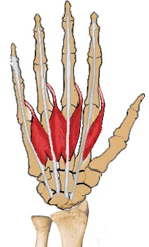
The lumbrical muscles of the hand are a group of four small, intrinsic muscles located within the palm. Despite their diminutive size, they play a crucial role in hand function, particularly in fine motor movements and dexterity.
The lumbrical muscles are located at the base of the palm. The proximal and middle phalanges of the fingers are where these muscles attach, and they begin with the FDP muscles.
What Are The Lumbricals Muscle?
The lumbrical muscles are a group of four intrinsic hand muscles attached to the digits’ distal extensor and the flexor digitorum profundus (FDP) muscle proximally. These muscles are worm-shaped and cylindrical, like those of the genus Lumbricus, which includes earthworms. The proximal interphalangeal (PIP) and distal interphalangeal (DIP) joints are extended, while all four lumbricals flex the metacarpophalangeal (MCP) joints.
The lumbricals assist in controlling the little and difficult movements of the fingers by working with other hand muscles. These muscles are necessary for typing, holding things, and completing other effective hand actions. This is a summary of the lumbricals’ anatomy and clinical significance.
Structure Of The Lumbrical Muscles
On each hand, the lumbricals are four tiny, worm-like muscles. However, they attach proximally to the flexor digitorum profundus muscles and distally to the extensor expansions. While the third and fourth lumbricals are bipennate, the first and second lumbricals are unipennate.
Function Of The Lumbrical Muscles
Most lumbrical muscles’ attachment locations are highly movable since they arise from muscle and are inserted into the extensor expansions rather than bone structures. This indicates that the muscles may do two different tasks. These are the proximal and distal interphalangeal joints’ extension and the metacarpophalangeal joints’ flexion.
The reasons for different activities are because the muscle layers insert distally at the dorsal side of the finger, although passing the metacarpophalangeal joints on the palmar side. These coordinated motions contribute to the hand’s overall ability and are involved in complex hand movements, such as holding a pen.
Origin
- First: It originates from the most radial tendon of the flexor digitorum pro fundus, which attaches to the index finger, on the radial side.
- Second: It originates from the same place as the middle finger, on the radial side of the second-most radial tendon of the flexor digitorum profundus.
- Third: The flexor digitorum profundus muscle matches the ring finger on the radial side, whereas the other head originates on the ulnar side of the tendon for the middle finger.
- Fourth: One head begins at the flexor digitorum profundus muscle in the little finger, whereas the second head originates on the ulnar side in the tendon for the ring finger.
Insertion
- First: It inserts on the extensor extension close to the metacarpophalangeal joint after moving posteriorly along the radial side of the index finger.
- Second: It inserts on the extensor expansions close to the metacarpophalangeal joint after moving posteriorly along the radial side of the middle finger.
- Third: The ring finger’s radial side is where the muscle inserts after passing posteriorly along the extensor.
- Fourth: The muscle inserts on the little finger’s extension of the extensor after passing posteriorly along the radial side of the finger.
Nerve Supply
The first and second lumbricals are innervated by the digital branches of the median nerve. The ulnar nerve’s deep branch supplies the third and fourth lumbricals. Damage to these neurons causes problems in motor and sensory function in several hand regions.
Action
Extend interphalangeal joints and flex metacarpophalangeal joints
Blood Supply
The superficial palmar arch is the most significant of the four veins that supply the lumbricals’ arterial circulation. The dorsal digital arteries, palmar digital arteries, and deep palmar arch are additional arteries providing support to the anastomotic network in this area. These arteries are connected by similar veins that discharge into the basilic vein on the medial aspect of the hand and the cephalic vein on the lateral side.
Lymphatic Drainage
The upper limbs have two kinds of lymphatic drainage: superficial and deep. For example, the palmar and dorsal lymphatic channels develop into the cubital and axillary lymph nodes along the basilic and cephalic veins. The deep lymphatic channels are complete in the humeral lymph nodes, after which are the primary deep veins.
Related Muscles
The palm’s deepest part contains the lumbrical muscles. These muscles attach to the extensor expansions of the fingers, which are attached to the proximal and middle phalanges, and their origins start from the FDP muscle. Except for the thumb, which is connected to only the first lumbrical muscle, each finger is attached to two lumbricals.
The fingers’ fine motor control and precise grasp depend on the lumbricals. Together with other hand muscles, these muscles flex the MCP joints and lengthen the PIP and DIP joints.
Anatomical Variations
The ulnar nerve innervates the medial 2 lumbricals while the median nerve supplies the lateral 2 lumbricals in the most frequent innervation pattern of the lumbricals. There is a 50–60% prevalence for this pattern. In contrast, the median nerve innervates the lateral three lumbricals in 20–30% of people, but only the first lumbrical in the other individuals.
A variation was found in cadaveric research where the third lumbrical had dual innervation from the ulnar and median nerves. The median nerve always supplies the first lumbrical, and the deep branch of the ulnar nerve innervates the fourth lumbrical; however, the middle lumbricals may not always have the same innervation.
The arrangement and structure of the lumbricals vary amongst individuals as well. There have previously been reports of a bipennate 2nd lumbrical, a double 1st lumbrical, and an absent 3rd lumbrical.
Variations can also be found in the distal lumbrical insertion sites, but these vary more from the lateral to the medial lumbricals. There have been reports of insertions on the volar face of the MCP joint, the proximal phalanx, the extensor hood, and combinations of these. The first lumbrical is more likely to have additional insertions on the extensor hood, while the fourth lumbrical is more likely to have attachment to the volar plate and bone.
Surgical Importance
Neuromas in continuity and carpal tunnel syndrome are two disorders affecting the median nerve and are treated with lumber flaps. This method was used in a case study where the exposed median nerve was covered by the first lumbrical. Despite the patient’s hand infection, soft tissue repair was not possible because of the diabetes-related inflammatory condition’s severity.
The patient whose right median nerve was constricted by significant scarring was treated using the same procedure, according to the same study.
Clinical Importance
Compression of the median nerve can result from aberrant lumbrical construction, such as when the muscle originates proximal. Additionally, lumbrical muscle hypertrophy, which can happen if the muscle has an abnormal origin and is prone to be overused, can cause carpal tunnel syndrome.
The ulnar nerve is captivity-induced compression neuropathy, which occurs second the most frequently. Usually, nerve captivity at the elbow, forearm, or wrist, brachial plexus compression, or nerve root impingement causes this disease.
By asking the patient to flex the MCP joints while maintaining supine palm and extended interphalangeal (IP) joints, a physician can evaluate the strength of the lumbrical muscles. The physician then administers a counterforce individually to digits 2 through 5 along the palmar surface of the proximal phalanx.
When the flexor digitorum profundus muscle is distal to the lumbrical origin points, a stimulating event occurs, causing the fingers to anticipate being closed.
The lumbricals now function as the flexor digitorum pro fundus’s new attachment surface following the distal muscle ‘separation. This indicates that the lumbricals are truly moved when the flexor muscle is carefully activated. Additionally, the planned fist closure paradoxically results in an extension of the fingers because the flexor digitorum profundus and the lumbrical are antagonists at the proximal and distal interphalangeal joints. The lumbrical-plus finger is the clinical term for this anomaly, which can develop following trauma or the cut.
Crush injuries
Crush injuries involving the hand can occasionally cause damage to the lumbrical muscle. Adhesions between the interosseous and lumbrical muscles may form after these injuries. Adhesions distal to the inter-palmar plate ligament restrict the proximal movement of the interosseous and lumbrical muscles because the lumbrical passes volar to the ligament while the interosseous muscle passes dorsal. When the patient makes a fist, the abnormality creates intermetcarpal pain. Usually, the affected muscles can be released to treat it. It has also been demonstrated that the lumbrical muscles play a role in carpal tunnel syndrome.
Assessment
To assess lumbrical muscle strength, the patient is requested to flex the metacarpophalangeal joints with the palm supine and the interphalangeal joints extended.
The physician then applies a counterforce along the palmar surface of the proximal phalanx of each of the fingers 2 through 5.
Applying individual resistance to the dorsal side of the middle and distal phalanges of digits 2–5 is a popular technique to measure the degree of flexion maintained in the metacarpophalangeal joints.
Exercises of Lumbrical Muscles of the Hand
Stretching Exercise
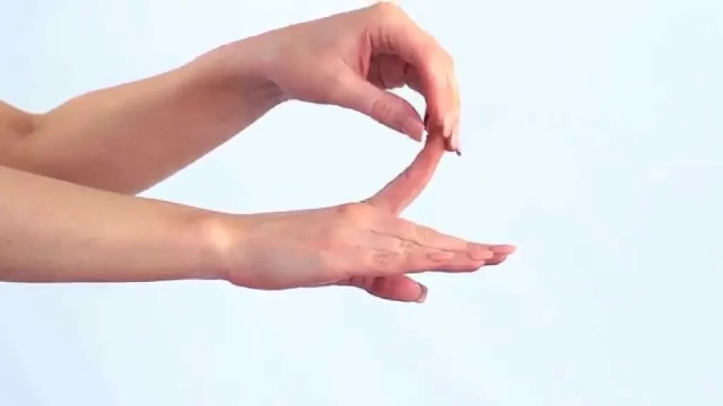
Finger Extension Stretch
- First, take a comfortable seat or stand with proper alignment.
- With your palm facing down, extend your arm shoulder-height in front of you.
- Press the fingers of your outstretched hand gently back towards your body with your other hand.
- For 15 to 30 seconds, hold the stretch with light pressure.
- Using the other hand, perform the same stretch.
Finger Spread Stretch

- With your fingers stretched apart, hold out your hand to begin.
- Stretch the space between each finger by gently pushing it apart from the others.
- Stretch for 15-30 seconds.
- Repeat the stretch multiple times, alternating between extending all fingers immediately and concentrating on each finger individually.
Wrist Flexor Stretch
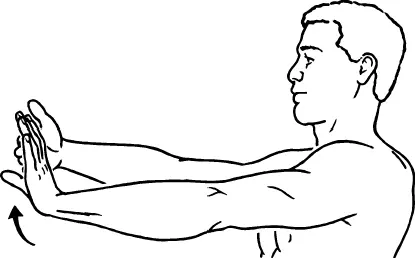
- With your palm facing up, extend your arm shoulder-height in front of you.
- Feel a stretch down the underside of your wrist and forearm as you slowly bring your fingers back towards your body with your other hand.
- Stretch for 15-30 seconds.
- Using the other hand, perform the same stretch.
Wrist Extensor Stretch
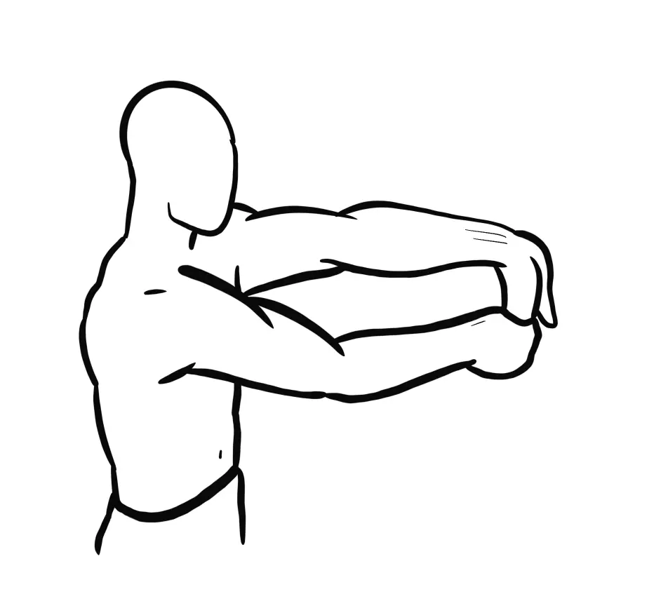
- With your palm facing down, extend your arm shoulder-height in front of you.
- With your other hand, gently press your fingers downward toward the floor until you feel a stretch along the top of your wrist and forearm.
- Stretch for 15-30 seconds.
- Using the other hand, perform the same stretch.
Strengthening Exercises
Finger Flexion with Rubber Band

- Put a rubber band around one hand’s five fingers.
- Start by separating your fingers and wrapping the rubber band around them.
- Carefully press your fingers together, applying pressure against the rubber band’s resistance.
- Squeeze for a short, then release to return to the initial position.
- Do this ten to fifteen times.
Finger Extension Exercise
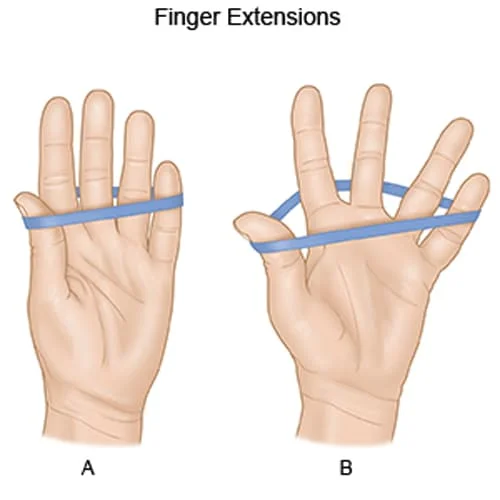
- Place your forearm, palm down, on a table while you sit or stand.
- Put something little in your palm, such as a rolled-up towel or a small ball.
- To release it and straighten your fingers as much as possible, curl them around the object first and then carefully push them apart.
- After a few seconds of holding the stretched position, curl your fingers back toward the object.
- Do this ten to fifteen times.
Thumb Opposition Exercise
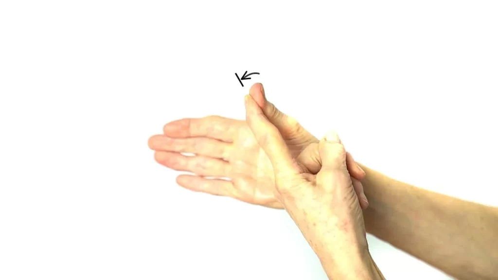
- Extend your hand and hold it palm up.
- Beginning with your index finger and working your way down to your little finger, touch the tips of each finger with your thumb.
- Repeat this action ten to fifteen times, being careful of your movements and maintaining your composure.
Grip Strengthening Exercise with Hand Gripper
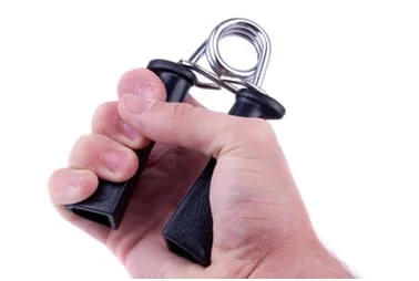
- With one hand, grasp a hand gripper by encircling the handles with your fingers.
- Using any of your hand and finger muscles, firmly squeeze the hand gripper.
- Squeeze for a short while, then slowly let go.
- Repeat ten to fifteen times for each hand.
Conclusion
Strengthening the lumbrical muscles is necessary to maintain hand flexibility, grip strength, and general hand function. The control and mobility of the fingers depend on these tiny hand muscles. The lumbrical muscles can be strengthened with exercises that focus on grip strength, thumb opposition, and finger flexion and extension. To differentiate and strengthen these muscles, use resistance training with bands or hand grippers combined with finger and thumb opposition exercises.
FAQ
What are the lumbrical muscles?
The lumbrical muscles are located in the deepest region of the palm. These muscles attach to the proximal and middle phalangeal extensor expansions of the fingers and are derived from the FDP muscle. Except for the thumb, which is only connected with the first lumbrical muscle, each finger is associated with two lumbricals.
What is the function of the lumbrical muscles?
The origin and insertion of the lumbrical muscles are unique as they are located on muscle. As deflectors of the proximal interphalangeal joint, the lumbricals aid in the flexion of the metacarpophalangeal joint as they contribute to the extension of the interphalangeal joint.
What nerves are in the lumbrical muscles?
The ulnar nerve innervates the third and fourth lumbricals, while the median nerve generally innervates the first and second lumbricals.
How do you test for lumbrical strength?
The following procedures must be followed by the lumbrical muscle tightness test for it to be able to participate in the FDP: When the patient reaches the end of their range of flexion, ask them to tuck their fingertips more tightly inside the range of motion. The DIP joints should receive special attention if the patient pulls during end-range flexion.
What is the blood supply of the lumbricals?
It was discovered that the superficial palmar arch (SPA), dorsal digital artery, deep palmar arch (DPA), and common palmar digital artery are the 4 different blood supply origins for each muscle.
What is the clinical importance of lumbricals?
The lumbricals assist in controlling the minuscule and delicate movements of the fingers by collaborating with other hand muscles. These muscles are necessary for typing, holding things, and executing other dexterous hand tasks.
What is Lumbrical muscle pain in the hand?
Lumbrical muscle injuries result when a finger (middle or ring finger) is forcefully straightened while the other fingers are actively gripping or bending. The most vulnerable group to lumbrical muscle rips is rock climbers.
References:
- Valenzuela, M., Launico, M. V., & Varacallo, M. (2023, November 17). Anatomy, Shoulder and Upper Limb, Hand Lumbrical Muscles. StatPearls – NCBI Bookshelf. https://www.ncbi.nlm.nih.gov/books/NBK534876/
- Hirpara, D. (2023, December 13). Lumbrical muscles of the hand -Origin, Insertion, Function, Exercise. Mobile Physiotherapy Clinic. https://mobilephysiotherapyclinic.in/lumbrical-muscles-of-the-hand/
- Lumbricals of the hand. (2024, May 4). In Wikipedia. https://en.wikipedia.org/wiki/Lumbricals_of_the_hand
- Lumbricals of the Hand. (n.d.). Physiopedia. https://www.physio-pedia.com/Lumbricals_of_the_Hand

