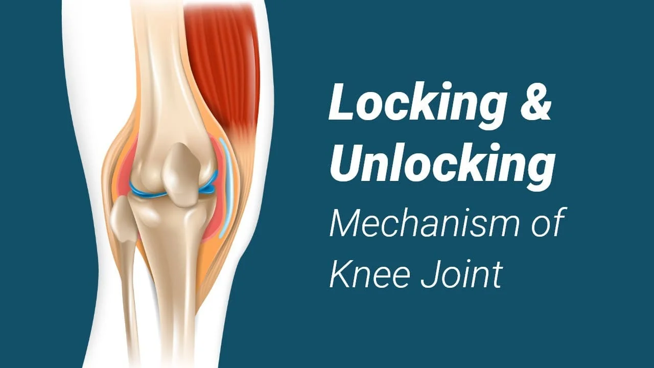
Locking and Unlocking of Knee Joint
Introduction:
A critical procedure that is necessary for the knee’s ultimate extension is the locking and unlocking mechanism of the joint, often known as the “Screw home mechanism (SHM)”.
An individual with a locked knee may find it difficult to bend or straighten their leg for extended periods. This ailment might be chronic or uncommon.
Knees may bend in both directions and can even rotate somewhat. If your knee is unable to carry out these tasks, it may compromise your range of motion and your capacity to sit, stand, squat, and kneel comfortably.
A pseudo-locked knee occurs when discomfort makes it difficult to move the knee, whereas a genuine locked knee occurs when the knee joint is physically locked into place and cannot move.
Knee anatomy:
The joint between the knees can mainly be used for both flexion and extension because it is a hinge-type synovial joint (with a limited amount of medial and lateral rotation as well). The formulations of the patella, femur, and tibia generate it.
This piece will address the knee joint’s articulating surfaces, ligaments, and neurovascular supply.
Articulating Surfaces:
The patellofemoral and tibiofemoral articulations together comprise the knee joint. Hyaline cartilage lines the joint surfaces, which are contained in a single joint cavity.
Tibiofemoral: One articulates each tibial condyle and the medial and lateral femur condyles. It is the part of the knee joint that bears weight.
The anterior portion of the distal femur articulates with the patella in the patellofemoral joint. Allowing the quadriceps femoris tendon, also referred to as the knee extensor, to be placed straight across the knee, it increases the muscle’s effectiveness.
The patella functions as a stabilizing structure that lessens the frictional stresses put on the femoral condyles and acts as a fulcrum to boost the power of the knee extensor because it is both created and lives within the quadriceps femoris tendon.
Nerve Supply
The genicular branches of the femoral and popliteal arteries send blood to the genicular anastomoses surrounding the knee, which in turn supply blood to the knee joint.
Menisci
Within the knee joint are C-shaped rings made of fibrocartilage called the medial and lateral menisci. They fulfill two primary purposes:
Increases the stability of the joint by deepening the articular surface of the tibia.
Increases the surface area of the joint and lowers the forces that are passed throughout it, acting as a shock absorber.
They are attached to the extremities of the tibia at the intercondylar area. The medial meniscus is furthermore attached to the joint capsule and the medial collateral ligament. Corresponding ligament injury is often associated with a medial meniscal rupture.
Its reduced size and lack of additional attachments make the lateral meniscus more flexible.
Bursae
Little amounts of synovial fluid are stored in a structure called a bursa, which resembles a sac. Its purpose is to lessen the friction that occurs during movement between tendons, bones, and skin. The knee joint contains four primary bursae:
Situated amidst the femur and the quadriceps femoris lies the suprapatellar bursa.
There are two types of infrapatellar bursa: superficial and deep. Positioned between the patella ligament and the tibia is the deep bursa.
The semimembranosus bursa is situated between the medial head of the gastrocnemius and the semimembranosus muscle, posterior to the knee joint.
Ligaments
The following are the main knee ligaments:
Distal to the patella, the quadriceps femoris tendon continues as the patellar ligament.
Two strap-like ligaments are called collateral ligaments. They function to stabilize the knee’s hinge action, limiting excessive lateral or medial movement.
The broad, flat medial collateral ligament is located on the medial side of the joint. It connects distally to the medial condyle of the tibia and proximally to the medial epicondyle of the femur.
The lateral collateral ligament is rounder and thinner than the medial collateral ligament. The attachment point is situated on the lateral side of the fibular head, close to the lateral epicondyle of the femur. It is situated distally.
Cruciate Ligaments: These two critical ligaments connect the tibia and femur. In Latin, they are called “cruciate” because of the crossing.
The anterior cruciate ligament joins the medial meniscus at the anterior intercondylar area of the tibia. It climbs posteriorly through the intercondylar fossa to join the femur. It keeps the bone of the tibia coming from displacing anteriorly against the femur.
From its attachment at the posterior intercondylar portion of the tibia, the posterior cruciate ligament travels anteriorly to connect to the anteromedial femoral condyle. It stops the tibia from posteriorly dislocating onto the femur.
Movements:
Movements The knee joint allows for the following four primary movements:
Extension – The quadriceps femoris, which inserts into the tibial tuberosity, produces extension.
Flexion is produced by the popliteus, sartorius, hamstrings, and gracilis muscles.
Femur biceps contractions cause lateral rotation.
medial rotation
Mechanism of locking system:
When a person is standing up, normal knee locking occurs in the latter stages of extension. The lateral condyle of the femur uses up space at the tibia’s lateral condyle during extension, which causes the femur to rotate medially. As a result, the medial femoral condyle rotates backward on the lateral femoral condyle’s axis. This movement is made possible by the quadriceps muscles contracting and the knee ligaments pulling obliquely. The ligaments tense up when the knee joint is locked in place when standing.
Different Knee Locking Types:
There are two basic types of knee locking: true and pseudo. When a section of your knee joint stalls out and remains immobile in a specific posture, it results in a true knee lock. The knee joint may rotate and bend both upward and downward by design. The knee may lock and become immobile if something is obstructing its range of motion. At times, this phenomenon can be extremely painful.
When a person experiences pseudo knee locking, they may feel completely immobile due to the intense discomfort. But in this instance, there isn’t anything stopping the person from moving their legs. Usually, a muscle spasm that produces pain in or around the knee region is the source of this.
The causes of a locked knee:
- Depending on the kind, locked knees might have different causes.
Reasons for knee pseudo-locking:
Some possible causes of pseudo-locked knees include the following:
- pain resulting from a knee injury
- inflammation resulting from damage or degenerative joint disease (osteoarthritis)
- plica syndrome, or inflammation of the tissue surrounding the knee joint
- incorrect kneecap motion
Causes of a truly locked knee:
A truly locked knee can be caused by several things, including:
Meniscal tear
- Two pieces of cartilage with a “c” shape that rest on either side of the knee joint are called menisci. Between the thigh and shin bones, they act as a cushion.
- If a meniscus tear occurs, a fragment may break off and become stuck in the knee, locking the joint.
- Severe knee rotation or twisting may cause a meniscal tear. Overuse and degenerative changes to the knee are additional factors.
bodies loosened in the knee
Similar to cartilage, bone shards can lodge in the knee joint and cause it to lock.
Following an accident or developing osteoarthritis cartilage as well as bone fragments may become loose.
Dislocation of the patella
A few different types of knee injuries can cause the patella, or kneecap, to move. We refer to this as patella dislocation. It may cause the knee to become rigid during extension.
inflammation of the knee joint
If the structures inside the knee joint expand and become inflamed, it may be difficult to extend the knee. Swelling can be brought on by injury, osteoarthritis, or overuse.
Acute Osteochondritis Dissecans
Osteochondritis dissecans (OCD) is a joint disorder that arises from inadequate blood flow behind the protective cartilage at the ends of bones. These bone layers begin to erode, separating them from the main bone and removing the cartilage along the way. Children and teenagers are more likely to suffer from this condition.
The knee joint locking:
extension of the knee flexion from a 30-degree angle in the closed kinematic chain.
When the smaller lateral side halted, the larger medial femoral condyle continued to roll and glide posteriorly. In the final five degrees of extension, this causes the femur to rotate medially on the tibia. Known as “automatic” or even “terminal rotation,” a medial rotation of the femur near the end of the extension is not voluntary or caused by muscle action.
A joint is brought into the closed, packed, or locked position by rotation within the knee joint.
Lateral rotation of the tibia on the femur is known as an open kinematic chain.
Unlocking the knee joint:
The knee needs to be unlocked to begin flexion.
The flexion force will inevitably cause the femur to rotate laterally.
Because of this, the longer lateral condyle will shift after the larger medial condyle.
The main muscle used to unlock the knee is the popliteus.
Features of a locked knee:
- Having unsteadiness in the knees
- discomfort when bending the knee
- stiffness in the knee joint
- swelling around and in the knee joint when injured
- The sensation of catching when extending the knee
- challenges with sprinting, leaping, walking, and climbing stairs.
Diagnosis:
A physician will initially enquire about any soreness and edema. Subsequently, a physical examination of the knee will be performed to look for indications of discomfort, swelling, and bruising.
During the examination, the doctor may exert force or apply pressure to the knee joint to evaluate it. They can then ask the person to cross the room to evaluate their mobility.
Sometimes a doctor will suggest further examination of the knee through diagnostic imaging. These tests include, for example:
- CT (computerized tomography) scan and magnetic resonance imaging (MRI) scan
- Ultrasound
- X-ray
With the use of extra testing, the physician can search for signs of inflammation or infection. These consist of arthrocentesis procedures and blood testing. During this operation, a knee fluid sample is taken.
Treatment:
Treatments for locked knees different according to the kind of condition and its underlying cause.
Meniscal tears:
The location and degree of a meniscal tear determine the kind of treatment that is needed.
The RICE treatment may be advised by the doctor if the tear is tiny and located on the outer portion of the meniscus. RICE, as an acronym, stands for:
- Rest: Give up on the painful activity.
- Ice: Several times daily, administer a 15–20 minute application of a wrapped ice pack.
- Compression: use an elastic compression bandage.
- Elevation: Raise your knees above your heart. This aids in removing extra blood from the knee, which lowers swelling.
One nonsteroidal anti-inflammatory medication (NSAID) that can help lessen inflammation and discomfort is ibuprofen.
In certain situations, a physician could advise physical therapy to aid in the patient’s recovery of knee mobility.
Loose body in the knee:
A person may need surgery to remove a loose bone or fragment of cartilage if it is causing their knee to lock.
Additional reasons:
Other causes, such as plica syndrome or patellar maltracking, typically call for physical treatment in addition to rest, ice, and anti-inflammatory drugs.
Prevention:
Sometimes there is no way to avoid the ailments and injuries that can result in a locked knee.
On the other hand, there are steps that people can do to better safeguard their knees. Among them are:
- Stay away from contact sports
- stopping the activities that cause knee pain
- exercising within your physical capabilities
- supporting the leg muscles and maintaining the flexibility of the core
- exercising caution in locations that are unstable or slippery to help prevent falls
Summary
True and pseudo-locked knees are the two varieties. The knee joint is immovable while it remains firmly in place. A person with a pseudo-locked knee can move their knee physically, but they are unable to do so because of excruciating discomfort or swelling.
For both kinds of locked knees, rest, ice, and painkillers are usually necessary. Physical therapy may also be suggested by a physician to assist the knee joint regain its range of motion. If these remedies prove ineffective, surgery may be required.
For a diagnosis, a person experiencing a locked knee should visit their physician as soon as feasible.
FAQs
When is a knee locked or unlocked?
It is called a locked knee whenever the knee cannot be bent or straightened. Two varieties of locked knees exist. When the knee joint is genuinely locked into position and is immobile, it is said to have a truly locked knee. A pseudo-locked knee occurs when pain limits the knee joint’s natural range of motion.
Why does this locking and unlocking thing happen to my knee?
The most prevalent reason for knee locking, particularly in the elderly, is frequently arthritis. The knee joints sustain a great deal of wear and strain since they bear weight. A joint’s inflammation may make it difficult to move the knee properly. This inflammation may appear following a previous sports injury or knee fracture
Which muscles unlock the knee?
Due to its necessity in “unlocking” the knee, which results in the femur on the tibia bending and rotating laterally, popliteus muscle contraction is referred to as the “key” to the knee joints.
Can you walk while your knee is locked?
It varies. If a meniscus tear does not impede joint movement, you might be able to walk on a locked knee.
Can one repair lock knees?
When a child’s growth is complete, osteotomy surgery can be used to address more severe abnormalities or knock knees that do not heal on their own.
Is it typical for knees to lock?
Knee locking can occur for non-traumatic reasons as well. For example, teenagers who have osteochondritis dissecans are more likely to develop loose bodies down the road. However, the majority of knee-locking events are more frequently caused by trauma.
How can I prevent the locking in my knee?
Rest: Give your knees time to heal by avoiding strenuous activities like running and leaping.
Stretching: The knee’s supporting muscles can be strengthened and the joint stabilized with easy at-home stretching.
Ice packs: Ice packs can be used to lessen pain, edema, and inflammation.
References
- Joint locking (medicine). (2023, November 3). Wikipedia. https://en.wikipedia.org/wiki/Joint_locking_(medicine)#:~:text=The%20ligaments%20are%20pulled%20taut,the%20lateral%20rotation%20of%20femur.
- 7 Main Causes of Locked Knees | OrthoNeuro | Columbus, OH. (n.d.). OrthoNeuro. https://orthoneuro.com/knee/locked-knees/
- Locked knee: Causes and what to do. (2019b, November 2). https://www.medicalnewstoday.com/articles/326877#summary
- Prajapati, N. (2022, July 19). Knee locking and unlocking mechanism – Screw home mechanism. Samarpan Physiotherapy Clinic. https://samarpanphysioclinic.com/knee-locking-and-unlocking-mechanism/
- Knee Locking: Causes & Treatment For A Locked Knee. (2021, December 8). Knee-Pain-Explained.com. https://www.knee-pain-explained.com/knee-locking.html
- DrBurkeWP. (2022, November 1). Locked Knee: Causes, Treatments and When to Seek Help – Dr. Burke Orthopedics. Dr. Burke Orthopedics. https://drburkeortho.com/locked-knee-causes-treatments-and-when-to-seek-help/