Hamstring vs Quadriceps
Introduction
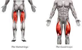
The hamstring vs quadriceps are two of the most significant muscle groups in the human body, which are essential for both movement and sports performance. The hamstrings are three muscles on the back of the thigh, and the quadriceps are four muscles on the front of the thigh. Both are found in the upper leg region.
Gaining strength in your quads and hamstrings is essential for exercises requiring quickness, power, and agility. Many lower-body actions, such as sprinting, jumping, and direction changes, rely heavily on these muscular groups to produce force. They are crucial in preventing common injuries and stabilizing the knee joint.
On the other hand, there are a few widespread myths regarding hamstring and quadriceps exercise. Some people assume that training with your quadriceps can cause muscular imbalances and increase your chance of injury. In contrast, others advise against training your hamstrings if you have ever suffered a strain.
An extensive guide to comprehending the hamstrings and quadriceps can be found in this article. It will go over the structure and purpose of these muscle groups and practical training methods for building stronger hamstrings and quadriceps. It will also dispel some common misconceptions about training the quads and hamstrings so you may create a well-rounded, scientifically supported lower-body training regimen.
Anatomy of the Hamstring
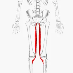
The semitendinosus
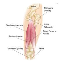
It is a fusiform muscle located in the thigh’s posterior compartment. It is a subpart of the hamstring muscle group, which also contains the semimembranosus and the long head of the biceps femoris. These three muscles are similar to each other. Along the way, they traverse the hip and knee joints and exert their influence on them. They all attach between the leg bones and the ischial tuberosity of the pelvis. The sciatic nerve’s tibial division innervates individuals.
The semitendinosus is an excellent mover that flexes and internally turns the leg as well as extends and rotates the thigh. Additionally, it stabilizes the pelvic girdle in a postural capacity.
From where the semitendinosus muscle originates and ends: The semitendinosus muscle connects itself between the proximal end of the tibia and the ischial tuberosity of the pelvis. This muscle is unique in that a rounded tendon (-tendinosis) makes up nearly half of its mass (semi-). The long head of the biceps femoris and the semitendinosus have equal roots. A posteromedial impression on the upper section of the ischial tuberosity is where both muscles get their shared tendon origin.
The muscle fibers then descend to the posterior thigh, where they are replaced by a tendon that resembles a cord in the middle of the thigh. Following an inferomedial path, the semitendinosus tendon passes laterally to the medial condyle of the tibia and posteriorly to the medial condyle of the femur.
It goes via the medial condyle. It produces a common insertion tendon known as the pes anserinus by partially blending with the tendons of the gracilis and sartorius muscles. The proximal end of the tibia’s medial surface is where the pes anserinus inserts.
Relationships: Semitendinosus, along with semimembranosus, forms the superomedial boundary of the popliteal fossa. When the knee is flexed against the opposition, as in a squat, its tendon can be felt as the most posterior and lateral tendon of the superomedial border.
The semitendinosus has an ischial connection deep within the gluteus maximus muscle. The muscular belly and its tendon run posteriorly to the adductor magnus and semimembranosus in the thigh, in the same plane as the biceps femoris. There are two intriguing relationships about tibial attachment. The semitendinosus inserts into the pes anserinus in a vertical line that runs posteriorly to the sartorius and postero inferiorly to the gracilis.
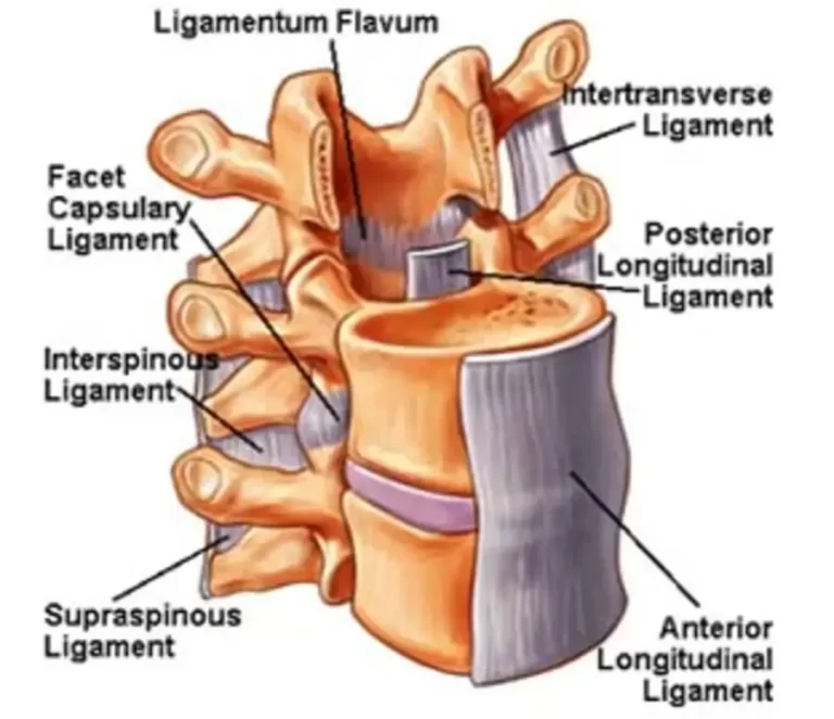
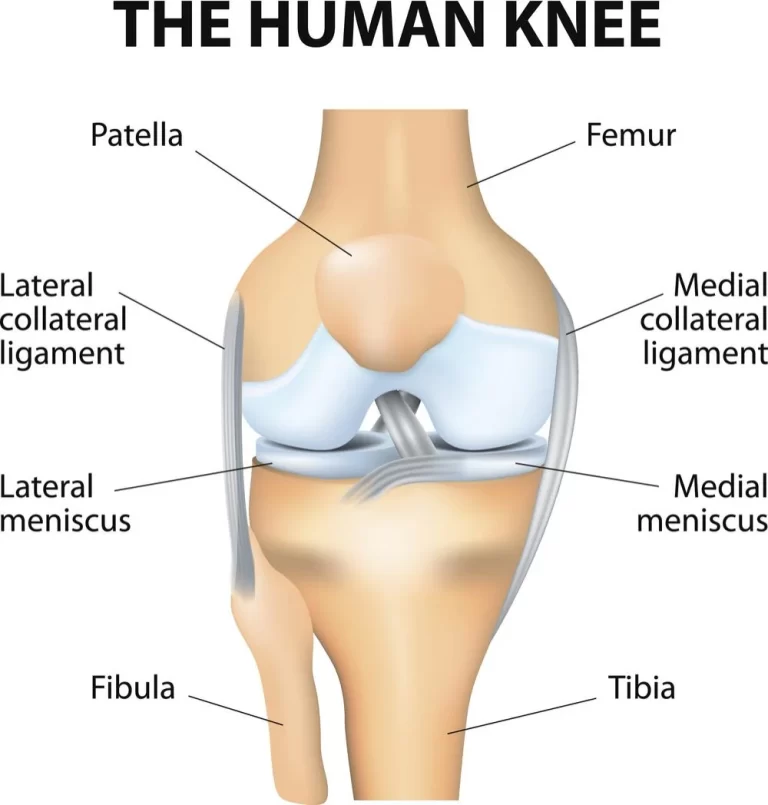
A bursa divides the three muscles that make up the pes anserinus from one another. Additionally, the anserine bursa divides the entire pes anserinus from the knee’s medial collateral ligament.
The nerve supply: The sciatic nerve’s tibial division innervates the semitendinosus (L5-S2).
Blood supply: Three major artery sources supply branches that supply this muscle;
Femoral artery via the medial femoral circumflex artery, deep femoral artery, and its first perforating branch
inferior gluteal artery to the internal iliac artery
via The inferior medial geniculate artery to the popliteal artery.
Function: The semitendinosus, which attaches between the hip and leg, moves the hip and knee joints, causing thigh extension, internal rotation, leg flexion, and pelvic stabilization (knee joint).The position of the body parts that a muscle moves and which of its attachments is fixed determine which way the muscle works origin or insertion. When its tibial connection is secured, the semitendinosus serves two purposes relating to the hip joint: The thigh rotates internally when the body is in the anatomical posture.
The thigh is extended by this muscle when the trunk is flexed anteriorly.
When the lower limb is in the correct anatomical posture, the semitendinosus flexes the leg. When the knee is semiflexed, it internally rotates the leg in concert with all of the hamstring muscles. This is the situation when the ischial attachment is fixed. The semitendinosus stabilizes the knee in addition to the pelvis and all of the short hip muscles that attach between the pelvis and proximal femur. As a matter of fact, because of where their insertion points are, all hamstrings serve as an additional set of knee stabilizers, assisting the medial collateral ligament in its role.
It is a lengthy muscle located on the back of the thigh. It creates the group of muscles comprehended as the hamstrings, along with the semitendinosus and semimembranosus muscles.
The muscle comprehended as the biceps femoris extends from the ischial tuberosity to the proximal part of the fibula. The hip joint and the knee joint are struck by the muscle in this action. The biceps femoris performs a variety of vital actions concurrently on these joints, including flexion and external rotation at the knee joint and extension and external rotation at the hip joint. This muscle, as its name implies, has two heads, one deep about the other. Though they share the same insertion, each head has a unique origin and innervation.
Beginning and ending: The most lateral hamstring muscle in the posterior thigh is the biceps femoris. This muscle has two heads—long and short—as its name would imply. Despite their disparate beginnings, these have one insertion in common.
The medial aspect (inferomedial impression) of the ischial tuberosity, which is superior to the adductor magnus muscle origin and medial to the semimembranosus muscle origin, is where the long head of the biceps femoris muscle begins. It is crucial to emphasize that the sacrotuberous ligament and the semitendinosus muscle share this tendon. Before splitting into two separate muscles, the tendons of the semitendinosus and biceps femoris travel together for a considerable distance.
Originating from the lateral lip of the inferior third of the linea aspera and the supracondylar ridge of the femur, the short head is located relatively distant from the long head. This origin is located laterally to the adductor magnus muscle and medially to the vastus lateralis muscle.
The long head of the biceps femoris persists as an aponeurosis close to the muscle’s insertion. The aponeurotic sheet, which includes the circular common tendon that attaches to the lateral aspect of the fibula head, is formed by the muscle fibers originating from the short head. The tendon separates into two slips just before implantation, passing on either side of the fibular collateral ligament. A few fibers spread to the nearby tibial condyle, while others adhere to the ligament.
The posterolateral aspect of the knee makes it easy to palpate the biceps femoris tendon when the knee is flexed.
links: The biceps femoris lies superficially in the posterolateral thigh for the bulk of its length, only delving deep into the coatings of skin, fat, and fascia. The gluteus maximus muscle covers it at its superior aspect, the only exception to this rule. The biceps femoris travels over the semimembranosus, adductor magnus, and lateral head of the gastrocnemius muscles as it descends from the pelvis into the posterior thigh region.
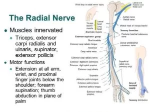
It also rescues the sciatic nerve by being superficial to it along the path. The common fibular nerve, the terminal branch of the sciatic nerve, is addressed close to the insertion of the biceps femoris. The nerve tracks the tendon as it travels shortly along the medial edge of the biceps femoris. This is a crucial clinical relationship when considering injuries or doing surgeries in this area.
Innervation: The sciatic nerve’s terminal branches supply the biceps femoris muscle. The common fibular division innervates the short head, whereas the tibial division innervates the long head. The root values of the spinal neurons L5, S1, and S2 are the same for the tibial and common fibular divisions.
blood supply: The medial circumflex femoral artery and perforating arteries of the deep femoral artery provide the majority of the blood supply for the biceps femoris. The superior lateral genicular and inferior gluteal arteries provide additional supplies.
Role: The biceps femoris muscle generally affects the hip and knee joints. The long head of this muscle functions on both joints, whereas the short head solely acts on the knee joint because of its attachments.
Hip extension is produced by the biceps femoris when it contracts the hip joint. When the trunk is bowed forward and brought into an upright position, this action is at its strongest. It’s also occasionally said that the biceps femoris helps in external rotation, which occurs when the hip joint is extended. Flexion of the leg is the primary action of the biceps femoris muscle when it acts at the knee joint.
When the lower limb is positioned anatomically, this happens. On the other hand, the biceps femoris produces external rotation of the leg at the knee when the knee is semiflexed. The biceps femoris, together with other hamstring muscles, stabilizes the pelvis, particularly as the trunk bends forward. It therefore plays a significant part in the gait cycle. The quadriceps femoris muscle, which is almost three times stronger than the hamstrings, is its main opponent.
Semimembranosus
It is one of the four muscles of the posterior thigh that extends the hip. The biceps femoris, gluteus maximus, and semitendinosus are the remaining three muscles in the hip extensor group. The term “hamstring muscles” refers to the semimembranosus, semitendinosus, and biceps femoris muscles combined. The medial aspect of the posterior thigh is occupied by the semimembranosus and semitendinosus. It is a quite lengthy muscle that extends from the hip to the knee throughout the entire length of the leg.
Beginning and ending: The somewhat big muscle known as the semimembranosus arises from a tiny facet on the abrasive superolateral side of the ischial tuberosity. Beginning at the level of the mid-thigh, the semimembranosus tendon travels caudally to the point of implantation at the medial condyle of the tibia.
Structure: The semimembranosus has some intriguing structural characteristics that aid in the general identification of the muscle. About halfway down the leg, the muscle develops a meaty belly from its flat, membranous beginnings. The fibers are directed inferomedially, and the fleshy component is medially connected to its tendon. In reality, the tendon splits distally to provide. the primary portion that attaches to the medial tibial condyle, the popliteal fascia-fusing portion in second place, and the oblique popliteal ligament-forming portion in third place. The fact that the lateral border of the semimembranosus creates the superomedial wall of the popliteal fossa is another significant characteristic.
Relationships: Numerous nearby muscular and neurovascular structures can be found along the semimembranosus muscle’s length. Throughout its whole length, the semimembranosus is medial to the biceps femoris, superficial to the adductor magnus, and deep to the semitendinosus. The gluteus maximus and adductor minimus cover the proximal portion of the muscle. Before insertion on the medial tibial condyle, the semimembranosus passes across and becomes medially connected to the gastrocnemius medial head distally. The adductor canal, which houses the lower limb’s arteries, is medial to the distal part of the semimembranosus.
The semimembranosus tendon is encased in a U-shaped bursa. It divides the tendon from the semitendinosus, medial head of the gastrocnemius, medial tibial plateau, and medial cruciate ligament. The sciatic nerve, the biggest nerve in the human body, extends laterally to the semimembranosus and ends at the top of the popliteal fossa. The popliteal artery and vein are now lateral to the semimembranosus and partially covered by it. Remember to test your knowledge of the semimembranosus and other thigh muscles to ensure you have a thorough understanding of them.
Innervation: The nerve roots of the L5, S1, and S2 innervate the semimembranosus. The sciatic nerve’s tibial division provides these fibers with access to the muscle.
Distribution of blood: The semimembranosus muscle is supplied by deep perforating branches of the femoral and popliteal arteries. The proximal portion of the muscle is occasionally supplied by the inferior gluteal artery.
The function: Due to its extension over the hip and knee joints, the semimembranosus is in charge of a variety of joint movements. To accomplish its purpose, the semimembranosus, however, collaborates with the other hamstring muscles. Semimembranosus produces hip extension when the feet are firmly planted on the ground, which pushes the upper body into an upright posture. When the hip is fully extended, the semimembranosus and semitendinosus muscles can also result in internal rotation of the thigh. Internal rotation of the leg on the thigh and flexion of the knee occurs when the legs are suspended off the ground.
When a person stands erect, their semimembranosus and the other posterior thigh muscles are at rest. But if the person leans forward too much,
By opposing the forward motion, the semimembranosus is triggered, stabilizing the hip.
Exercises specific to the hamstrings
The hamstrings react favorably to:
Deadlifts: This type of complex exercise works the hamstrings among other muscles. When deadlifts are performed correctly, the biceps femoris and the other two hamstring muscles are worked.
Deadlifts and Variations: The hamstrings during hip extension are the primary target of conventional and Romanian deadlifts. Single-leg and stiff-leg versions work quite well as well.
Hamstring Bends: By concentrating on hamstring flexion localized at the knee joint, seated and lying curls minimize the engagement of the glutes and lower back muscles.
Glute-Ham elevation: This strenuous bodyweight exercise develops hip extension strength in the hamstrings and glutes. It trains the core and doesn’t require any equipment.
Stretch your hamstrings while lying down by wrapping the Theraband Resistance Band around your foot’s arch. Adopt a flat back position. Draw the band’s two ends slowly toward the direction of your sternum.
As you pull, you should feel your hamstrings stretch. To increase resistance and stretch, pull your leg farther back. For 15 to 20 seconds, hold. Restart after lowering your leg slowly to its initial position until you feel your muscle has warmed up.
The Quadriceps muscle’s anatomy
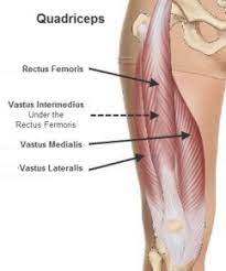
Rectus femoris
Location of origin and insertion: The rectus femoris is a fusiform muscle with two heads. It originates from two sites on the ilium: the supraacetabular groove (reflected head) and the anterior inferior iliac spine (straight head). The two heads merge to develop a single muscular belly that drops the thigh nearly vertically and covers the anterior side of this province. A thick tendon made of convergent muscle fibers penetrates the base of the patella. Infrequently, the rectus femoris can have a third head that arises from the iliofemoral ligament.
Connections: Deep to the proximal part of the rectus femoris muscle are the sartorius, iliacus, and tensor fasciae latae muscles. Up to the rectus femoris, the anterior compartment of the thigh is filled. These consist of the hip joint capsule, the anterior borders of the vastus lateralis, the lateral circumflex femoral artery, the vastus intermedialis, and the periodic femoral nerve branches.
Blood supply: The rectus femoris muscle receives blood from the quadriceps artery, which can come from the lateral circumflex femoral, deep femoral, or femoral arteries. The rectus femoris also receives blood supply via the superficial circumflex iliac and lateral circumflex femoral arteries but to a lesser extent.
Movement: flexion of the hip joint at the thighs
Lengthening of the leg at the knee joint
The vastus medialis
Location of the insertion and origin: The landmarks from which the vastus medialis muscle originates are the pectineal line of the femur, the medial lip of the linea aspera, the inferior part of the intertrochanteric line, and the proximal half of the medial supracondylar line. As the muscle passes obliquely through the thigh, its fibers wind around the long axis of the muscle. The fibers at the base of the structure are arranged in an almost horizontal plane and give rise to a noticeable protrusion above the medial side of the patella. This particular area of the vastus medialis is referred to as the vastus obliquus by some writers. Last but not least, the muscle might enter the patella’s base thanks to the quadriceps femoris tendon.
As they descend, some of their tendinous fibers penetrate the medial condyle of the tibia.
Connection: The vastus medialis is situated medial to the rectus femoris, and is partially covered by it. The sartorius muscle also crosses the superficial surface of the vastus medialis. The vastus medialis in the middle portion of the thigh forms the lateral wall of the adductor canal, also referred to as Hunter’s canal. This canal is completed posteriorly by the adductor longus and magnus, and medially by the sartorius. It transports the femoral vein, femoral artery, nerve, and saphenous nerve—both of which are branches of the femoral nerve—to the vastus medialis.
Blood transfer: The vastus medialis obtains blood from three muscle departments of the femoral artery. It also obtains minor donations from the deep femoral and descending genicular arteries.
Action: Leg extension at the knee joint
Vastus lateralis
Location of origin and insertion: The vastus lateralis is the largest of the four quadriceps femoris muscles. A large aponeurosis gives birth to it at many locations on the femur, including the proximal half of the intertrochanteric line, the anterior and inferior borders of the greater trochanter, the lateral lip of the tuberosity of the gluteus, and the proximal half of the lateral lip of linea aspera.
The deep surface of the aponeurosis, which spans the upper three-quarters of the muscle, is the source of several muscle fibers.
By means of the quadriceps tendon, the muscle inserts at the base of the patella after dropping the lateral portion of the anterior thigh compartment. Some of the tendinous fibers descend to the proximal tibia, where they insert into the lateral condyle of the tibia and combine with the iliotibial tract.
Connection: The vastus lateralis muscle, which is situated superficially to the biceps femoris muscle, is divided by the lateral intermuscular septum. The lateral portion of the muscle is covered by the tensor fasciae latae and gluteus maximus. vastus lateralis’s medial surface hooks the vastus lateralis to the vastus intermedius, from which the medial surface of the vastus lateralis is divided by the femoral nerve branches and the lateral femoral circumflex artery.
Blood supply: The vastus lateralis receives its blood flow from the superior medial artery, a branch of the lateral circumflex femoral artery. The inferior medial artery is a branch of the quadriceps artery. The first perforator of the deep femoral artery is the lateral artery.
Action: The knee joint is extended.
Vastus Intermedialis
Location of origin and insertion: The vastus intermedius muscle is located between the vastus lateralis and vastus medialis muscles. It begins at the anterior surface of the bone, or the proximal two-thirds of the femur’s shaft. As the muscle falls along the front aspect of the femur, it fills the area between the vastus medialis and lateralis. At the distal femur level, it releases a large aponeurosis, which the common quadriceps tendon connects to the base of the patella. It also inserts into the lateral condyle of the tibia.
connection: The Ventus intermedius is completely encircled by the remaining quadriceps femoris muscles. The vastus lateralis and medialis occupy their lateral and medial surfaces, respectively, while the rectus femoris covers its anterior surface.
Blood supply: The quadriceps and deep femoral arteries give blood to the vastus intermedius.
The quadriceps tendon, which is situated just above the patella, is produced by the contraction of these four muscles. As the tendon descends farther, it joins to the patella’s upper surface. After that, the patella is fastened to the tibial tuberosity. a bony protrusion made by the patellar tendon that extends inferiorly on the tibia. Occasionally considered an extension of the quadriceps tendon, the patellar tendon is involved in the ultimate attachment to the tibia.
The extension of the knee joint requires the patellar tendon and the quadriceps tendon. Activities like sprinting, jumping, stair climbing, and walking heavily rely on the quadriceps tendon and accompanying muscles functioning at their best. Quadriceps tendon strains or tears can significantly impair knee function and may require medical attention to fully recover.
Exercises specific to the quadriceps
One way to train the quads is with:
Squats and Variations: These exercises work the quadriceps over a wide range of motion while activating the glutes and hamstrings. They can be performed in a frontal or back fashion.
Leg Press: This machine exercise minimizes impact on the lower back while isolating the quads through knee extension.
Step-ups and lunges are examples of functional exercises that target the quadriceps unilaterally, improving stability and reducing side-to-side muscular imbalances.
Quadriceps lying down Pull: Encircle the top of the foot arch with a theraband resistance band. On your stomach, lie flat. Draw the band’s two ends slowly in the direction of your head.
The resistance in your quad will determine how far you can extend it. For 15 to 20 seconds, hold. Return your leg to the initial position gradually and do it again as required.
The Quadriceps’ function in joint stability and mobility
The quadriceps carry out several crucial tasks:
Extensor Function: Their primary function is to straighten the leg and extend the knee joint. This produces force for actions like sprinting, jumping, and kicking.
Aid with Knee Extension and Flexion: The quadriceps assist in controlling the motion when bending and straightening the knee, even if the hamstrings carry out knee flexion.
Percentage to Total Strength of Lower Body: More strength development in other lower body muscular groups, such as the glutes and calves, is also made possible by strong quadriceps. Both injury resiliency and athletic performance depend on this.
Considerations for training your hamstrings vs. quadriceps: An Understanding of the Different Types of Muscle Fibers
Muscle fiber types are distributed differently in the hamstrings and quadriceps.
Slow-Twitch Fibers in the Hamstrings: The proportion of slow-twitch fibers is higher in the hamstrings. Because slow-twitch fibers respond better to more reps with moderate weights, this affects the amount of training required.
Fibers with Fast Twitch in the Quadriceps: The bulk of the muscle fibers in the quadriceps are fast-twitch, designed for strength and power. This encourages higher loading patterns and fewer repetitions in each set.
Comparison of hamstring vs quadriceps
| Muscle goup | Hip extension, lower leg rotation | Quadriceps |
| Muscles | Biceps femoris, semitendinosus, semimembranosus | The vastus intermedialis, The vastus lateralis, The vastus medialis, The rectus femoris |
| Location | Back of thigh | Front of thigh |
| Major task | Knee flexion | Knee extension |
| Secondary Function | Hip extension,lower leg rotation | Hip extension, lower leg rotation |
| Fiber Type | Higher ratio slow-twitch | Mostly fast-twitch |
| Main Exercises | Leg curls,gluteal-hamstring raise, Romanian deadlift | Squats, leg presses, quadriceps lying down pull,step-ups and lunges |
| Best Rep Range | 8-15+ reps (moderate load) | 3-8 reps (heavy load) |
| Priority | Endurance, stamina | Strength, power, speed |
| Benefits | Injury protection, postural balance | Athletic performance, injury resilience |
| Strength ratio | Generally weaker than quadriceps | Generally stronger than hamstrings |
| Romanian deadlift, leg curls, glute ham raise | Very high | High |
| Injury risk | Prone to tears and strains, especially in athletes | Prone to tendonitis and overuse injury |
Techniques for training hamstrings and quadriceps
Advantages of a Well-Composed Quad and Hamstring Exercise Program
It is essential to create a balanced program that works both the hamstrings and quadriceps to optimize the advantages of training. Numerous benefits arise from a well-rounded approach, such as the reduced risk of injury, increased sports performance, and better muscular symmetry and appearance. Exercises like lunges, leg lifts, and squats are great for strengthening the quadriceps. These complex exercises strengthen the entire lower body by using several different muscles.
However, focused hamstring exercises are just as crucial. Exercises that specifically target the hamstrings, such as deadlifts, hamstring curls, and glute-ham raises, can help to strengthen them and enhance performance in exercises that call for hip extension and knee flexion. Similar to quad workouts, ongoing improvement is ensured by adjusting factors including load, rep range, and exercise selection.
Gaining balanced, powerful quads and hams has various benefits
Injury Avoidance: Injury risk is decreased by having a balanced strength between the hamstrings and quadriceps. Muscle imbalances frequently cause common knee injuries such as ACL rupture.
Increased Athletic Achievement: By increasing power generation and absorption, properly training both muscle groups enhances acceleration, jumping, cutting, and agility.
Better Muscle Symmetry and Aesthetics: Fuller, symmetrical muscular development for a more appealing appearance is achieved by focusing on all quad and hams regions.
Connection between the hamstrings and quadriceps
For both the male and female groups, at all three test speeds, the torque generated by the quadriceps was consistently higher than that of the hamstrings. The results of the analysis of variance indicated that there was a significant difference (P < 0.01) between the hamstrings and quadriceps. As the workout pace rose, the variation in torque output between the muscle groups reduced. Both muscle groups produced less torque and their foot-pound disparities decreased as the test speed was increased to 300° per second, the Cybex II dynamometer’s highest rate.
This implies that the hamstring torque output may be greater than the quadriceps torque output at test speeds higher than 300′ per second. To further describe the relationship between the hamstrings and quadriceps, the ratio between the torque output of the muscle groups
was calculated. The means of these ratios and the standard deviations for both the male and female groups at the three test speeds were similar for both sexes, even though there were large differences in the actual amounts of torque output produced. In both males and females, the ratio between the hamstrings and quadriceps comes closer to unity as the velocity of the exercise accelerates. The ANOVA determined that the ratios were significantly increased ( P < 0.01) between the test speeds for both groups
The influence of hamstring and quadriceps muscle stress on anterior cruciate ligament in-situ forces and knee kinematics
The impact of co-contraction of the hamstrings and quadriceps on the anterior cruciate ligament’s in-situ forces and the kinematics of the human knee joint during a simulated isometric extension motion. Ten cadaveric human knee specimens should be evaluated utilizing the robotic/universal force–moment sensor system. Reference sites on the path of the knee’s passive flexion/extension motion were used to assess the knee’s kinematics and in-situ forces in the anterior cruciate ligament. The knee experienced internal tibial rotation concerning the femur and anterior and lateral tibial translation with an isolated 200 N quadriceps load. When the knee was flexed from full extension to 30° of flexion, both translation and rotation enhanced; when the knee was additionally flexed, these motions were reduced. At all evaluated knee flexion angles, except full extension, the addition of an 80 N antagonistic hamstring load significantly decreased both anterior and lateral tibial translation as well as internal tibial rotation. The anterior, lateral, and internal tibial translation, translation, and rotation should be significantly reduced by 18, 46, and 30%, respectively, at 30° of flexion (p<0.05). Under the quadriceps load, it was discovered that the in-situ forces in the ACL increased from full extension to a maximum at 15° of flexion and then decreased to 10 N beyond 60° of flexion. compared to other flexion angles, the in-situ force at 15° was substantially larger (p<0.05). The in-situ forces in the ACL at 15, 30, and 60° of flexion were significantly decreased by 30, 43, and 44%, respectively, upon the addition of the 80 N hamstring load (p<0.05). These findings show that the highest in situ forces in the ACL may not always correlate with maximum knee motion. Additionally, the data indicate that hamstring and quadriceps co-contraction is useful in lowering excessive forces in the ACL, especially in the range of 15 to 60° of knee flexion.
Anterior tibial translation and ACL strain (or elongation) would both increase with isolated quadriceps muscle stresses. In cadaveric knees with isolated quadriceps loads, there should be significant anterior tibial translation between 0 and 80° of knee flexion and up to 5% strain within the anterior-medial (AM) bundle of the ACL between 0 and 45° of knee flexion. how antagonistic hamstring muscle loads affect the ACL’s AM bundle’s ability to withstand strain when simultaneous quadriceps contractions occur between 0 and 30 degrees of flexion less than 40% of the forces in ACL grafts should be reduced between 15 and 60° of knee flexion when the quadriceps load was combined with a 90 N hamstring load during a simulated squat activity.
On the other hand, little is known about the in-situ forces in the ACL during muscular loads. This information is critical to improving the surgical restoration of the ACL following injury, which is necessary to return knee function to normal. In cadaveric knees during a simulated isometric extension of the knee, this study aims to measure the multiple degrees of freedom of knee kinematics and the in-situ forces in the ACL in response to isolated quadriceps load and combined quadriceps and hamstring loads. The version of hamstring burdens will greatly diminish the magnitude of joint motion as well as the in-situ forces in the ACL. To deliver isolated quadriceps or combination quadriceps and hamstring stresses to the knee specimen, the robotic/UFS test system was adopted. With this special test setup, the same test specimen may be used to evaluate the in-situ forces in the ACL under different muscle loading scenarios.
Materials and procedures
By applying different external loads to the knee specimen, the robotic universal force–moment sensor test system can measure the related changes in knee kinematics as well as in-situ forces in the ACL. A robotic manipulator is a machine that learns and replicates any preset settings of a knee specimen, allowing it to regulate the knee’s position in six degrees of freedom in space. In conjunction with the force control manner
H: Q ratio
Evaluating muscle strength is essential to determining an athlete’s level of fitness or to helping people recover from a variety of illnesses or accidents. The torque ratio between the hamstrings (H) and quadriceps (Q) (H: Q) is a crucial statistic in these applications, serving as a gauge of an imbalance in strength between the opposing muscle groups surrounding the knee joint. Finding a relative weakness in one muscle compared to the other can be used for two main purposes: first, it can be used to identify athletes who have strength imbalances during the pre-season, and then those athletes can be trained appropriately to improve their performance, and return the H: Q ratio to “normal” levels; second, it can be used to identify imbalances following injuries or other pathological conditions, and then those athletes can be trained appropriately to recover from those imbalances.
Different combinations of contraction types (concentric, eccentric, and isometric), angular velocities, torque categories peak or angle-specific torque can be used to derive the H: Q ratio. Torque values of the same contraction type for both antagonistic muscle groups are used to calculate the traditional ratio. These include the H: Q torque ratio, the eccentric H (HECC): eccentric Q, and the concentric H: concentric Q (QCON). It has been suggested that the median value in a typical population sample is represented by an isometric H: Q ratio of roughly 0.66.
This has been partially confirmed by a recent systematic review of the literature, which shows that the isokinetic conventional ratio ranges from 0.52 to 0.67 when measured at slow (12°/s) to intermediate (180°/s) angular speeds, and increases at faster rotational velocities. When the quadriceps provide force by shortening (concentric contraction), the hamstrings engage as antagonists by lengthening (eccentric contraction). A more functional method that accounts for the typical muscle actions occurring during a knee extension should be used to compute the H: Q ratio. Thus, the dynamic control or functional torque ratio (HECC/QCON) is defined as the ratio of eccentric hamstring torque over concentric quadriceps torque. The normative values of the functional ratio also vary with test angular velocity; they are roughly 0.79 at 60°/s and increase to 1 at rotational speeds greater than 240°/s.
An additional variant of the functional ratio is the combined ratio, which is calculated by dividing the HECC torque at an angular velocity of 30°/s by the QCON torque at 240°/s. Therefore, by assuming the hamstring eccentric concentric contraction torque at a gradual speed and the quadriceps contraction at a speedier speed, the resultant H: Q ratio for the typical sports population would be middle almost a value of 1.13. The main advantages of this ratio are its improved experimental validity and reduced susceptibility to inertial artifacts. This is due to eccentric torque values at a gradual angular velocity (HECC torque at 30°/s) having better validity rather than fast velocity tests, whereas the recorded torque at fast angular velocity tests reveals better experimental facts in isokinetic fast angular velocity concentric contraction trials(QCON torque at 240°/s) than in eccentric contraction trials. the relationships between various H: Q ratio types, it did not thoroughly analyze the relationship between the H: Q ratio and damage.
The monitoring of strength imbalances in patients undergoing anterior cruciate ligament (ACL) deficit or reconstruction has been a significant use of the H: Q ratio. This is based on the finding that the anterior component of the quadriceps force is opposed by the hamstrings, which co-contract at almost full extension. By preserving knee joint stability and lowering ACL load, this lessens anterior shear loading and helps achieve a physiological range of motion and safer relative position of the femur and tibia bones in the knee joint. Thus, it has been suggested that ACL injury and recurrence, particularly in female athletes, may be associated with a reduced force-generating capacity of the hamstrings in comparison to the quadriceps.
Another common risk factor for hamstring strains is an altered H: Q ratio. This is frequently explained by the hamstrings’ diminished capacity to regulate simultaneous hip flexion and knee extension during dynamic exercises like stretching or running. Strong evidence suggests that strength characteristics, such as the H: Q ratio, are not independent risk factors for hamstring injuries, according to systematic reviews. Nevertheless, the H: Q ratio in general and several additional possible risk variables and, consequently, particular problems that may affect the assessment of the H: Q ratio and its relationship to damage have yet to be considered. These include problems with the torque ratio measuring process (reliability, validity, and gravity correction) the kind of ratio (conventional, dynamic, and mixed), the calculation technique, and interpretation (cut-off value determination). These factors are crucial because the determination of the H: Q ratio is affected by methodological restrictions and problems with the experimental methodology. These restrictions are frequently disregarded, which could cause incorrect interpretation of the H: Q ratio results that are produced. the preference for one kind of ratio over another for certain uses, it is still unclear which kind of H: Q ratio works best for injury prediction or prevention. Conclusions on the efficacy of the H: Q ratio for injury prevention may also be impacted by the usage of different types of H: Q ratios in conjunction with restrictions in the collecting and analysis of injury.
Advice for strengthening your hamstrings and quadriceps
- Warm-Up: It’s important to adequately warm up your leg muscles before beginning any quadriceps or hamstring activities. Include dynamic stretches and exercises like walking lunges, high knees, and leg swings.
- Progressive Overload: Gradually raise the weights or resistance you use throughout exercises to keep your hamstrings and quadriceps harder. Strength is improved and muscular growth is stimulated by this progressive overload.
- Equilibrium Exercise: Make sure you continue to follow a well-rounded exercise regimen that works your hamstrings and quads. A weakened or neglected muscle group might result in imbalances in the muscles and a higher chance of injury.
- Rest & Recovery: Include rest days in your training regimen to help your muscles repair and strengthen. Sufficient sleep, healthy eating, and adequate water are also necessary for the best possible muscle regeneration.
- Striking a Balance: The Cooperation of the Hamstrings and Quadriceps
It’s more like a beautiful symphony that promotes the best lower body strength and movement than a conflict between the quadriceps and hamstrings. Maintaining functional movement patterns, minimizing muscular imbalances, and lowering the risk of injury all depend on the proper balance between these muscles.
Strategies for preventing injuries
The best offense is a capable defense, as is the case with most injuries. Injuries can be avoided by:
- Maintaining Fitness
- heating up before attaching in harsh work
- Extending
- ensuring that the strength of your hamstrings and quads is equivalent
- Performing targeted workouts to increase your flexibility and strength
- Theraband Resistance Bands are an ideal tool for strengthening and stretching your hamstrings and quadriceps.
The Value of Balance in Muscle
Risks Associated with Muscle Imbalance: An imbalance in the quadriceps and hamstrings can result in poor biomechanics, which raises the possibility of sprains and strains.
Redistribution of Joint Stress: Equilibrium strength in the quadriceps and hamstrings facilitates uniform force application across the knee and hip joints, hence reducing joint stress and possible deterioration.
Optimal Motion Efficiency: These muscle groups work in harmony to transmit energy efficiently, which enhances athletic performance in a variety of activities.
Techniques for improving balance
- Compound Exercises: To promote balanced muscular development, perform compound exercises such as squats, lunges, and deadlifts that work your hamstrings and quadriceps at the same time.
- Resistance Training: Include resistance training movements that specifically target the quadriceps and hamstrings, such as leg extensions and hamstring curls, to guarantee balanced strength increases.
- Flexibility training: Stretching exercises that maintain and prevent muscle tension in the hamstrings and quadriceps include seated hamstring stretches and forward bends.
- Core and Hip Stability: To build a strong foundation for quadriceps and hamstring strength during dynamic exercises, concentrate on core and hip stability exercises, such as planks and bridges.
- Rest and Recovery: To promote muscle growth and healing and lower the chance of overuse injuries, make sure you get enough rest and recovery in between sessions.
Volume of training and distribution of loads
When planning hamstring and quadriceps exercises, take into account:
Ideal Volume and Frequency of Training Ratios: Each muscle group should be worked out one to two times a week, with the quads receiving more overall volume than the hamstrings. A decent starting point is a weekly set ratio of 2:1 (hamstring: quad).
Managing Hamstring vs. Quadriceps Load in Various Training Phases: Permit periods that concentrate more on hamstring or quadriceps strength to manage fatigue and meet the demands of sports. Volume should be adjusted within ideal ratios.
Mobility and Flexibility Concerns
Programs for soft tissue work and stretching must include this:
Stretching and Mobility Exercises: After working out, make sure to perform four or five deliberate hamstring stretches. Regularly foam roll and apply massage techniques to your hip flexors and quadriceps.
Handle Tightness and Muscle Imbalances: Examine your training mechanics and posture. Focus on muscles that are more prone to dominance and tighter muscular groups. Maintain hamstring and quad balance.
What’s the H/Q ratio, and what makes it significant?
The hamstring-to-quadriceps ratio, or H/Q ratio for short, is the strength ratio between the hamstring and quadriceps muscles. We must define concentric and eccentric muscle contractions before we can go on to discuss the H/Q ratio. When a muscle contracts eccentrically, it lengthens under strain; when a muscle contracts concentrically, it lengthens under load.
An effective way to comprehend the operation of this contraction is to compare it to kicking a soccer ball. An eccentric hamstring contraction occurs when the foot strikes the soccer ball, forcing the hamstring muscles to contract in a straight leg, lengthened position, with the force of the ball. Concentric quadriceps contractions occur when the quadriceps muscles contract simultaneously while shortening (keep in mind that the primary function of the quadriceps is to straighten the knee). Because the quadriceps and hamstring muscles have an antagonistic relationship, the strength ratio between them is crucial because it influences both the likelihood of injury and knee joint function.
How is the H/Q ratio measured?
To test the H/Q ratio, an isokinetic dynamometer is typically utilized. This apparatus can be used to compute several different things, such as the forces exerted on the knee joint at various angles and speeds. Although it yields the most exact result for determining the H/Q ratio, accessing the equipment may not be simple. Thus, an alternate method would be to measure the 1RM for the hamstrings and quadriceps using a leg extension and leg curl machine.
The highest weight you can lift for one repetition is known as 1RM. The results can still be utilized to determine any strength deficiencies, even though they won’t provide as much information as an isokinetic dynamometer. Consult your reliable physiotherapist about evaluating your H/Q ratio
What is the value of normative?
Although a ratio larger than 0.6 is preferred, the general agreement indicates that a ratio of 0.5-0.8 is acceptable in a typical, healthy population. In other words, if the quadriceps can lift 60 kg at their maximum over their entire range of motion (straightening the knee), then the hamstrings should be able to lift 36 kg through the same range of motion (for a 0.6 ratio).
It is also acknowledged that an increase in movement velocity increases the H/Q ratio. When the ratio approaches 1, meaning that the strength of the hamstrings equals the strength of the quadriceps, the hamstrings can more effectively support and stabilize the knee. In the population of athletes, this knee stability may also assist in lowering the chance of injury, particularly ACL damage. The population of football players also prefers a ratio of 1.
If your ratio of H to Q is tiny, what could happen?
During a football game, players are usually expected to execute extremely forceful and quick moves. The quadriceps muscles shorten and contract to extend the knee during a leap. Then, to oppose the quadriceps action and reduce the force needed to extend the knee during a jump, the hamstrings tighten by extending. When pivoting movements like landing from a jump or making an abrupt direction change, the hamstrings and ACL help to stabilize the knee.
There are two main types of soccer injuries: contact injuries and non-contact injuries. non-contact injuries account for almost 50% of soccer-related injuries. The majority of injuries happen to athletes whose H/Q ratio is less than 0.6, and they frequently happen in the hamstring muscle group when the muscle is stretched beyond what is normally capable of being stretched.
ACL injuries are another prevalent injury that soccer players have, and they can be largely attributed to the poor H/Q ratio. As previously shown, when the knee straightens when jumping, kicking, or pivoting, the ACL and hamstring work together to stabilize the knee by keeping it from moving forward. If there is a weakness in the hamstring muscles, To stop the knee from shifting, this puts additional strain on the ACL, which over time could result in an injury. Hamstring strengthening is therefore crucial for the athlete population’s recovery from ACL injuries as well as injury prevention.
Summary
Crucial upper leg muscular groups that are essential for movement, sports performance, and injury resistance are the hamstrings and quadriceps. These muscle groups, which are found on the front and back of the thighs, respectively, cooperate to support lower body power and stability during exercises like sprinting, cutting, jumping, and more.
From a structural perspective, the quadriceps are made up of four muscles: the vastus lateralis, vastus medialis, vastus intermedius, and the rectus femoris. They strengthen the legs generally and serve as knee extensors. In contrast, the three muscles that make up the hamstring group—the biceps femoris, semitendinosus, and semimembranosus—serve as knee flexors and aid in hip extension.
The quadriceps and hamstrings offer improved athletic potential and defense against frequent ailments in training and sports when they are strengthened appropriately. However, many variables, such as the ratios of different muscle fiber types and the degree of flexibility required, vary among these groups and necessitate customized programming strategies. Because the quadriceps are mostly made up of fast-twitch fibers, they benefit from large loads and smaller rep ranges. Along with the hamstrings and glutes, the quadriceps are efficiently trained with squats, leg lifts, and lunges. On the other hand, the hamstrings adapt better to moderate loads and increased volume because of their higher proportion of slow-twitch fibers. Exercises like deadlifts, curls, and auxiliary exercises like the glute ham raise are all part of targeted hamstring training.
A balanced quad and hamstring function is facilitated for optimal performance by making sure that stretches are balanced, that the right amount of volume is applied to each muscle group, and that posture and flexibility are routinely evaluated. Monitoring development over time promotes muscular symmetry and prevents damage.
FAQS
What differentiates hamstring and quadriceps exercises from one another?
The main muscles that move your legs are the quadriceps (front muscles) and hamstrings (back muscles), which also govern your hips and knees. Whereas the hamstrings bend and stretch the hip, the quadriceps extend the knee.
Why do the hamstrings and quads hurt?
Strong Quadriceps Defeat Hamstrings, There is a lift up in the back when your quads are very tight and the pelvis is dragged down in front. “On a stretch,” the hamstring is put in this way. Additionally, this may hurt.
Does walking help hamstrings to relax?
Hamstring discomfort may be avoided by at least ten minutes of easy calisthenics, gentle running, or walking.
Does the hamstring squeeze when you walk?
There are several causes of hamstring tightness, including physical exertion. The hamstrings must be repeatedly contracted during walking, running, and running-intensive sports like tennis and soccer to slow down knee extension and bring the lower legs closer to the buttocks.
Is hamstring strain a result of excessive sitting?
After extended periods of inactivity or sitting, some people develop tight hamstrings. For instance, tightness may result from spending several hours at a desk. In other situations, the tightness may be the result of an injury, maybe a recurrent injury that increases the susceptibility of the hamstrings to tightness.
Does heat help relieve hamstring strain?
To loosen up your hamstrings before working out, apply HEAT (Circulation Boost) to them. If you still feel stiff the next day, you can apply heat again. You should keep applying heat if the stiffness persists to prevent aggravating your previous hamstring issue.
How quickly can one relieve stiff hamstrings?
hamstring stretches to release tension
With your feet flat on the floor and your knees bent, take a lying position on the ground.
Lift your right knee to your chest gradually.
Stretch the leg, bending the knee just a little bit.
Work your way up to 30 seconds of holding for 10 seconds.
How much of an impact do the hamstrings and quadriceps have on lower body strength?
Indeed, for all-around leg strength, knee stability, and injury resistance, the hamstrings and quadriceps are equally important. Both muscle groups become weaker, more painful, and less functional when neglected.
Is it possible to work the hamstrings and quadriceps on the same day?
Exercises for the quads and hamstrings combined into a single session focus on lower body strength while saving time. Just give complex exercises top priority.
How can I avoid having an imbalance in my muscles between my hamstrings and quadriceps?
assessing the mechanics of complex lifts, including unilateral workouts, stretching the quads and hamstrings, and monitoring the volume ratios between the various muscular groups to avoid imbalances.
How frequently should one train their hamstrings and quadriceps?
Depending on your level, try to work your hamstrings and quadriceps 1-2 times a week; for most people, two full lower body workouts per week are ideal.
When doing exercises for the quads and hamstrings, should you utilize machines or free weights?
Programs should be centered around barbells and dumbbells since free weights allow for increased muscular activation through stability demands. Use equipment from time to time for increased isolation or, if necessary, for injury recovery.
References
- How to Treat Quad and Hamstring Strains | Performance Health. (n.d.). https://www.performancehealth.com/articles/how-to-treat-quad-and-hamstring-strains
- In-Text Citation:(How to Treat Quad and Hamstring Strains | Performance Health, n.d.)
- Li, G., Rudy, T. E., Sakane, M., Kanamori, A., Ma, C., & Woo, S. L. (1999, April 1). The importance of quadriceps and hamstring muscle loading on knee kinematics and in-situ forces in the ACL. Journal of Biomechanics. https://doi.org/10.1016/s0021-9290(98)00181-x
- Pilates, B. P. A. (2021, September 20). Injury Prevention in Young Elite Footballers | Part 2: hamstring and quadriceps (H/Q ratio). Breathe Physio. https://www.breathephysio.com/post/injury-prevention-in-young-elite-footballers-part-2-hamstring-and-quadriceps-h-q-ratio
- Kellis, E., Sahinis, C., & Baltzopoulos, V. (2023, May 1). Is the hamstrings-to-quadriceps torque ratio useful for predicting anterior cruciate ligament and hamstring injuries? A systematic and critical review. Journal of Sport and Health Science. https://doi.org/10.1016/j.jshs.2022.01.002
- Virani, A. (2024, February 4). Quadriceps vs Hamstrings – Anatomy, Function and Exercises. Mobile Physiotherapy Clinic. https://mobilephysiotherapyclinic.in/quadriceps-vs-hamstrings/