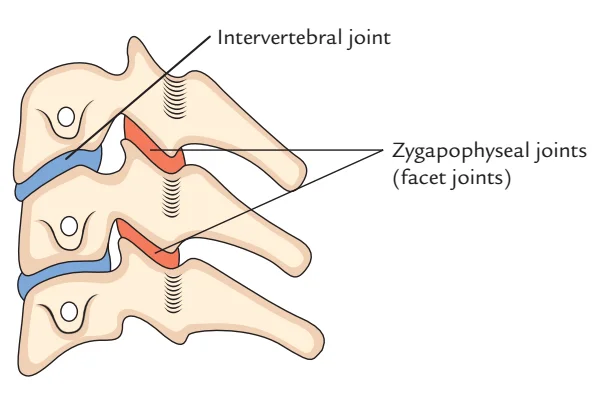
Facet Joints (Zygapophyseal Joints)
The facet joints, which are a group of synovial, plane joints between the articular processes of two neighboring vertebrae, are also known as zygapophysial joints, zygapophyseal, apophyseal, or Z-joints. Each spinal motion segment has two facet joints, and the recurrent meningeal nerves innervate each facet joint.
Introduction
This synovial connection, called the apophyseal or zygapophyseal joint, connects the superior articular process of one vertebra to the inferior articular process of the vertebra located directly above it. For every segment of spinal motion, there are two facets.
The facet joints serve as the articular pillars that give the entire vertebral column structural stability. They are located between the pedicle and lamina of the same vertebra.
The bilateral facet joints, in conjunction with the disc, transfer loads and, because of their mechanical function and geometry, guide and constrain motions in the spine.
Articulating Surfaces
The articular facet is on the superior process of the vertebra below, and the articular facet is on the inferior process of the vertebra above. The superior facet of the inferior vertebra is more convex in the lumbar area and relatively flat in the cervical and thoracic regions.
The superior vertebra’s opposite inferior facet is concave, forming an arch with its peak pointing in the direction of the vertebral body. Depending on the location, facet joints can have varying orientations:
The cervical region is 45 degrees in the frontal plane, and all motions, including rotation, flexion, extension, and lateral flexion, are available.
The cervical vertebrae have articulating facets that face 45 degrees to the transverse plane and parallel to the frontal plane. The inferior articulating processes face down and anteriorly, while the superior articulating processes face up and posterior.
No flexion or extension, lateral flexion and rotation, frontal plane, and thoracic region = 60 degrees
While the inferior facets face medially, down, and anteriorly at the facet joints of adjacent thoracic vertebrae, which are inclined at 20° to the frontal plane and 60° to the transverse plane, the superior facets face posteriorly, as well as somewhat up and laterally.
The lumbar region is 90 degrees in the sagittal plane, with just flexion and extension.
The facet joints in the lumbar area are located in the sagittal plane; the articulating facets are 45° from the frontal plane and at right angles to the transverse plane. The superior and inferior faces are oriented laterally and medially, respectively.
This is altered at the lumbosacral junction, where the inferior facet of L5 faces forward and the apophyseal joint shifts into the frontal plane. The spinal column cannot slide forward on the sacrum due to this change in orientation.
Ligaments and Joint Capsule
In addition to stabilizing the vertebral column, the posterior ligamentous complex maintains the facet joints of the adjacent vertebrae in a fixed relationship with one another. The following structures comprise it:
- Posterior longitudinal ligament
The facet joint capsule, which surrounds the joint like other synovial joints in the body, creates synovial fluid to lubricate and nourish the joint. The spinal nerves posterior ramus (dorsal primary rami) produce paired medial branches that innervate each facet joint. Both the spinal nerve below and the spinal nerve above the facet provide the facet with a medial branch. The medial branch nerve is triggered by pain in facet joints that are degenerated or inflamed. The limbs’ muscles and sensations are not under the control of these nerves. - ligamentum flavum
- Interspinous ligament
- Supraspinous ligament
Motions Available
Anatomical features including ligaments, facets, and intervertebral discs restrict the motion of each spinal segment. The coupling of the spine’s motions, or synchronous movements, is specifically caused by anatomical features.
There is a physiological connection between flexion, extension, translation, axial rotation, and lateral bending. Anatomical structure differences by location determine the precise coupling pattern. Axial rotation and side bending occur in the same direction in the cervical and upper thoracic spines.
Lateral bending and axial rotation in the opposite direction are combined in the lumbar spine. The pattern of connection in the middle and lower thoracic spine is irregular. However, according to which movement is started first, the coupling pattern will vary. If lateral bending occurs initially in the lumbar spine, it will be followed by axial rotation in the same direction. On the other hand, axial rotation will be accompanied by lateral bending in the opposite direction if it occurs first.
Innervation
Although innervation to the facet joints varies throughout spinal segments, medial branch nerves originating from the dorsal rami often innervate them. These nerves are believed to receive primarily sensory information, although there is some evidence that they also get motor input from the local musculature.
The medial branch nerve, a branch of the dorsal rami, innervates the majority of the cervical spine’s joints from the same levels. That is, the C4 and C5 medial branch nerves innervate the facet joint between the C4 and C5 vertebral segments. There are two exceptions, though:
- The C3 medial branch nerve and the third occipital nerve innervate the facet joint between C2 and C3.
- The medial branch neurons of C7 and C8 innervate the facet joint between C7 and T1.
The medial branch nerves from the vertebral segment above the upper segment and the upper segment innervate the facet joints in the lumbar and thoracic spines. For instance, the medial branch neurons of T1 and C8 innervate the facet joint between T1 and T2.
Facet joint between L1 and L2; medial branch nerves of T12 and L1. However, the L5 dorsal ramus and the L4 medial branch nerve innervate the L5 and S1 facet joint. The facet joint is not innervated in this instance by the L5 medial branch.
Function
The spinal motion segment’s movement is guided and limited by the biomechanical function of each pair of facet joints. The facet joints, for instance, serve to shield the motion segment in the lumbar spine from excessive rotation, flexion, and anterior shear pressures. The range of side bending (lateral flexion) seems to be mostly unaffected by facet joints.
Degeneration, dislocation, fracture, injury, trauma-induced instability, osteoarthritis, and surgery can all impair these capabilities. The purpose of the facet joints in the thoracic spine is to facilitate rotation and limit the degree of flexion and anterior translation of the corresponding vertebral segment.
Cavitation of the synovial fluid in the facet joints causes the popping sound (crepitus) that is often associated with manual spinal manipulation, sometimes known as “cracking the back.”
Both the superior and inferior facet joints are positioned to restrict rotation and permit flexion and extension. This is particularly valid for the lumbar region.
Clinical significance
The facet joints, which are the only genuine synovial joints connecting neighboring spinal levels, are affected by osteoarthritis (OA) of the spine. They are situated on the posterior side of the vertebral column.
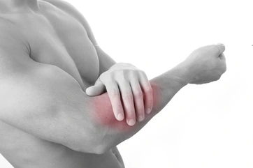
One of the most common causes of back and neck pain in older persons is facet joint osteoarthritis (FJ OA). As people age, facet-mediated pain becomes more common in clinical populations, indicating that FJ OA may play a significant role in treating spinal pain in elderly folks.
A positive diagnostic facet joint block may suggest that chronic spinal pain is coming from the facet joints. For these people, certain treatments like neurolysis using radiofrequency or cryoablation may help remove facet joint pain. According to a 2013 study that examined the evidence supporting lumbar facet joint injections and physiotherapy therapies, the injections produce a brief duration of pain relief. For patients with chronic lower back pain, physiotherapy therapies such as soft tissue massage and land-based lower back mobility exercises may be helpful during this period to enhance long-term results.
Facet joint arthritis
All joints experience degenerative changes with age, largely because of the mechanical nature of their function. The facet joint in particular and other joints in the spine are especially affected by this.
Facet joint arthritis or facet arthropathy are the frequent names for this condition. The degenerative process might cause the joint to expand, just like with any other type of arthritis. Small alterations to the facet joint may cause the intervertebral foramen to narrow, potentially putting pressure on the spinal nerve roots inside. Severe inflammatory reactions in the Z-joint, similar to a swollen arthritic knee, may occur in more severe cases.
Summary
The stability and movement of the spine depend heavily on the facet joints. The facet joints, like other synovial joints, experience a series of degenerative characteristics, such as osteoporosis, articular cartilage thinning, subchondral bone sclerosis, cartilage fibrillation, joint space narrowing, and the formation of juxtafacet cysts.
Facet tropism, gender, rising BMI, and anomalies of the ligamentum flavum are additional potential contributors to facet joint degeneration, along with lower spinal level, age, sagittal orientation, and IVD degeneration.
Facet joint degeneration can be detected and its severity evaluated using a variety of pathologic and imaging grading systems, each with unique advantages and disadvantages. The natural history of facet joint degeneration and its correlation with clinical symptoms should be further investigated to detect and quantify its severity.
A deeper comprehension of the facet joints, how they interact with other spinal structures, and how pain and impairment develop can help to improve the precision of spine patient management and treatment.
FAQs
Which joints are zygapophyseal?
The superior articular process of one vertebra and the inferior articular process of the vertebra directly above it form a synovial joint that is also referred to as the zygapophyseal or apophyseal joint. In every spinal motion segment, there are two facet joints.
What kind of joint does the facet joint belong to?
The articular facets of the vertebrae are joined by facet joints, which are symmetrical synovial-lined joints with a fibrous capsule. The inferior I facet of the vertebra above articulates with the superior facet of the lower vertebra.
Are joints with facets gliding?
You can bend and twist your body thanks to the facet joints of your spine, which slide and glide. As we age, our facet joints may develop adhesions that limit our range of motion. Adjustments aid in dissolving adhesions around the facets, restoring your range of motion and reducing pain.
What is the facet joint’s true name?
True synovial joints, facet joints—also referred to as zygapophyseal or apophyseal joints—can experience degenerative changes in a manner akin to that of other synovial joints.
What is the total number of facet joints?
The facet joints, which are a group of synovial, plane joints between the articular processes of two neighboring vertebrae, are also known as zygapophysial joints, zygapophyseal, apophyseal, or Z-joints. Each spinal motion segment has two facet joints, and the recurrent meningeal nerves innervate each facet joint.
A zygapophyseal joint is what kind of synovial joint?
Zygapophyseal joints, which make up the postero-lateral articulation between vertebral levels, are the only synovial joints in the spine. They are composed of a joint capsule, a synovial membrane, and hyaline cartilage covering the subchondral bone.
What is the facet joint’s size?
For the central zone, the facet joint space width was 1.93±0.51 (mean ± standard deviation) mm, the superior zone was 1.75±0.48 mm, the inferior zone was 1.63±0.49 mm, the medial zone was 1.48±0.44 mm, and the lateral zone was 1.65±0.48 mm. The right and left facet joints did not differ much.
What is a facet joint’s typical angle?
The articular surfaces of the facet joint, in the sagittal plane, often have an inclination angle between 20° and 78° in the cervical region, 55° and 80° in the thoracic region, and 82° and 86° in the lumbar region.
What features distinguish facet joints?
A capsule containing a trace amount of synovial fluid envelops the joint. To lessen friction between rubbing bones, the synovial fluid serves as an extra lubricant. In addition to providing support for the spine, healthy facet joints permit a great deal of bending and twisting.
What form does a facet take?
A facet is a characteristic of a polyhedron, polytope, or other analogous geometric structure in geometry that is typically one dimension smaller than the structure itself. More precisely, any polygon whose corners are the polyhedron’s vertices but are not a face is referred to as a facet of a polyhedron in three-dimensional geometry.
Which ligaments make up the facet joint?
Along the spine’s length, the facet joints are located on either side of the spinous processes on the back of each vertebra. The ligamentum flavum and the FCL surround the joint, which is made up of two facet surfaces from neighboring vertebrae. They help control joint motion by limiting synovial fluid flow.
In what location are facet joints found?
The facet joints serve as the articular pillars that give the entire vertebral column structural stability. They are located between the pedicle and lamina of the same vertebra.
What is the significance of facet joints?
These joints prevent the back from twisting excessively or slipping too far forward while allowing the spine to flex and twist. When these joints experience tension and damage from an injury, normal wear and tear, or disc degeneration, facet joint syndrome develops.
Why is a facet joint used?
The facet permits mobility while stabilizing the spine. Due to damage or “wear and tear” (degenerative change), these joints may become uncomfortable. The facet joints are the source of pain, which might spread.
References
- Wikipedia contributors. (2024c, June 2). Facet joint. Wikipedia. https://en.wikipedia.org/wiki/Facet_joint#:~:text=The%20facet%20joints%20(also%20zygapophysial,by%20the%20recurrent%20meningeal%2
- Zygapophyseal joints. (2023, October 30). Kenhub. https://www.kenhub.com/en/library/anatomy/zygapophyseal-joints