Coccygeal Nerve
The coccygeal nerve is the 31st and final spinal nerve, arising from the coccygeal region of the spinal cord. It primarily contributes to the coccygeal plexus, which provides sensory innervation to the skin over the coccyx and motor fibers to parts of the pelvic floor muscles. It plays a minor role in overall nerve function but is involved in sensations around the tailbone area.
Introduction
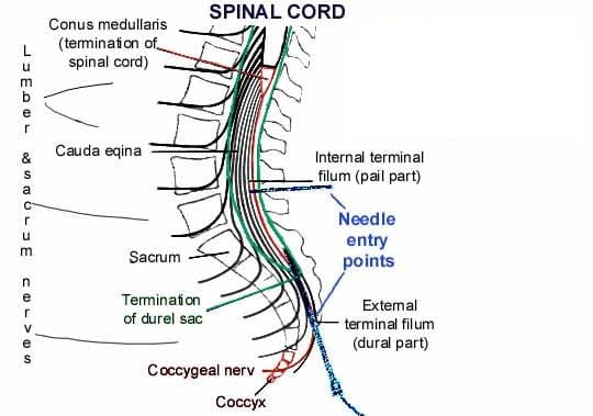
The final and smallest pair of spinal nerves are the coccygeal nerves (also known as tailbone nerves or spinal nerves Co). They come from the distal portion of the vertebral column, which is home to the conus medullaris, the final region of the spinal cord.
The sacral canal is where the coccygeal nerves leave the spinal column after descending inside the cauda equina. As an alternative, the sacral hiatus is where the nerves leave. The skin covering the tailbone (coccyx) is sensory innervated by the coccygeal nerve, which also contributes to the coccygeal plexus.
Course
They arise from the spinal cord’s terminal, the conus medullaris. The lumbar, sacral, and coccygeal spinal nerves descend to the cauda equina.
Through the sacral hiatus, the coccygeal nerve leaves the sacral canal and emerges underneath the first coccygeal segment. After circling the sacrum’s lateral edge, its anterior (ventral) ramus pierces the coccygeus muscle.
Branches
The coccygeal nerve finishes in a bifurcation, sending forth two terminal branches:
- Anterior (ventral) ramus of coccygeal nerve
- Posterior (dorsal) ramus of coccygeal nerve
Once the nerve leaves the sacral hiatus, the anterior ramus rises. Together with the anterior rami of the S4 and S5 spinal nerves, the coccygeal nerve’s anterior ramus helps form the coccygeal plexus. The anococcygeal nerve originates in the coccygeal plexus. Both the skin covering the tailbone and the sacrotuberous ligament are supplied by this nerve.
The skin at the back of the tailbone is supplied by the posterior ramus of the coccygeal nerve, which has a connecting branch from the S5 spinal nerve.
Examination
- X-ray.
- CT (computed tomography) scan.
- Your healthcare professional may do an examination to look for tumors, abscesses, or inflammation.
- MRI (magnetic resonance imaging).
- Bone scan.
Clinical Importance
Coccydynia or tailbone pain
Tailbone pain, also known as coccydynia, is a common clinical condition characterized by pain in the coccygeal region. Patients typically present with sharp or burning pain that gets worse when they are physically active.
This condition is typically caused by bone injuries that occur during sports like mountain biking, prolonged sitting, or even during the last trimester of pregnancy. Treatment for this condition varies depending on the cause, and it may even involve a coccygeal nerve block to relieve the pain.
Surgical Importance
In very unusual cases, your provider could suggest:
Partially removing your coccyx is known as a partial coccygectomy.
removal of your complete coccyx, or total coccygectomy.
Following a coccygectomy, recovery may take many months. Even if a surgeon removes the bone, there is no guarantee that your pain will go away. As a result, physicians only recommend coccygectomy when no other course of therapy works.
FAQs
What is the coccygeal nerve?
The Coccygeal Nerve consists of several branches and is part of the sacral plexus. It provides sensory innervation to the skin and muscles around the coccyx (tailbone) and the perianal region. The nerve transmits pain and touch sensations from these areas to the brain.
What signs indicate injury to the coccygeal nerve?
Dull (achy) or sharp (piercing) tailbone pain. ache in the tailbone that gets worse when you rise after sitting. Pain when you poop.
What is the function of the coccyx?
Background. Despite its small size, the coccyx serves several vital purposes. In addition to serving as the point of insertion for several muscles, ligaments, and tendons, it is one leg of the tripod, along with the ischial tuberosities, that supports a person’s weight when they are seated.
What negative consequences might a coccygeal nerve block cause?
This procedure is considered to be safe, and it is a minimally invasive procedure that does not require an overnight stay. But there are hazards associated with the needle insertion, including bleeding, numbness, nerve injury, and infection.
What is the Dermatome of the coccygeal nerve?
On the buttocks, in the vicinity of the coccyx, is the dermatome that corresponds to the coccygeal nerves.
What are the side effects of Coccydynia injection?
A coccyx injection for tailbone pain may have the following side effects:
Soreness: Injection site pain, edema, or soreness
Facial flushing: A temporary flushing of the face
Nausea: A temporary feeling of nausea
Abdominal cramps: A temporary feeling of mild abdominal cramps
Menstrual cycle: For certain women, there may be a brief change in the menstrual cycle.
Blood sugar: A temporary increase in blood sugar levels for people with diabetes
Serious side effects are rare but include: Rectal perforation, Hemorrhage, Infection, and Dural puncture.
You should tell your doctor if you have an allergy or have had a reaction to a steroid in the past.
Other information
If symptoms reappear, the injection can be repeated. The effects of the injection may persist for weeks, months, or even years.
References:
- Coccygeal nerve. (2022, December 5). Kenhub. https://www.kenhub.com/en/library/anatomy/coccygeal-nerve
- Tailbone pain (Coccydynia). (2025, February 7). Cleveland Clinic. https://my.clevelandclinic.org/health/diseases/10436-coccydynia-tailbone-pain