Chest Muscles
The chest muscles, also known as the pectoral muscles, play a crucial role in the upper body’s strength and functionality. These muscles are primarily responsible for movements of the shoulder joint, including flexion, adduction, and rotation of the arm.
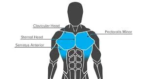
The pectoral region is found on the anterior chest wall. The serratus anterior, subclavius, pectoralis major, and pectoralis minor are the four muscles that apply force to the upper limb. “Pecs” is the term for the chest muscles.
Chest muscles connect the front of the human chest to the upper arm and shoulder bones. The pectoral region contains four muscles that control movement in the upper limbs or ribs.
Pectoralis Major.
The muscle closest to the skin in the pectoral region is the pectoralis major. It is large and fan-shaped, consisting of a sternal and clavicular head:
- Attachments:
The anterior surface of the medial clavicle is the source of the clavicular head.
The anterior surface of the sternum, the superior six costal cartilages, and the external oblique muscle’s aponeurosis combine to form the sternocostal head.
Both heads have a distal attachment to the humerus’ intertubercular sulcus. - Function: Adducts and medially rotates the upper limb, drawing the scapula anteroinferiorly. The clavicular head also functions independently to flex the upper limb.
- Innervation: the lateral and medial pectoral nerves.
- Blood supply: The pectoral branch of the thoracoacromial trunk provides arterial supply to the pectoralis major muscle.
Pectoralis Minor.
The pectoralis minor muscle is located beneath its larger counterpart, the pectoralis major. Both muscles are located in the anterior wall of the axilla.
- Attachments: Comes from the third to fifth ribs and inserts into the coracoid process of the scapula.
- Function: The scapula is stabilised by drawing it anteriorly against the thoracic wall.
- Innervation: medial pectoral nerve.
- Blood supply: The pectoral branch of the thoracoacromial trunk provides arterial supply to the pectoralis minor as well.
Serratus Anterior
The medial border of the axilla region is formed by the serratus anterior, which is situated more laterally in the chest wall.
- Attachments: The lateral aspects of ribs 1–8 are the starting points for the multiple strips that make up the muscle. They attach to the scapula’s medial border’s costal (rib-facing) surface.
- Function: Rotates the scapula, allowing the arm to be raised more than 90 degrees. It also protracts the scapula, securing it against the rib cage.
- Innervation: The long thoracic nerve.
- Blood supply: The serratus anterior receives arterial supply from the lateral thoracic artery, superior thoracic artery (upper half), and thoracodorsal artery (lower half).
Subclavius
The subclavius is a small muscle located directly underneath the clavicle and running horizontally. It offers the underlying neurovascular structures some minimal protection (e.g., in cases of trauma such as clavicular fractures).
- Attachments: Originates at the junction of the first rib and its costal cartilage. It attaches to the inferior surface of the middle third of the clavicle.
- Function: Secures and depresses the clavicle.
- Innervation: Nerve to the subclavius.
- Blood supply: The subclavius’ arterial supply comes from the clavicular branch of the thoracoacromial trunk.
Embryology
Skeletal muscle tissue is formed from the mesoderm of the original three germ layers. From here, the mesoderm differentiates into the paraxial and lateral plate mesoderm. The paraxial mesoderm, specifically those organised in the trunk, is divided into individual tissue blocks called somites. These somites, which are concentrated more dorsolaterally, aggregate into the dermatomyotome and are induced into myoblasts.
The modification employed by a family of basic Helix-Loop-Helix transcription factors, most notably MyoD1, is thought to be responsible for the mesoderm’s differentiation to eventual myoblast formation. The continuous expression of MyoD transcription factors is required for the genetic expression of genes involved in muscle development.
The cells of the dermatomyotome subdivide, and one of the resulting structures is the hypomere. The hypomeres differentiate into three layers: external intercostals, internal intercostals, and innermost intercostals, also known as the transverse thoracic muscle. A portion of the hypomere-derived myoblasts give rise to the anterior chest’s pectoral muscles, which include the pectoralis major, pectoralis minor, serratus anterior, and subclavius.
Exercises for chest muscles
Strengthening exercises
Incline pushups
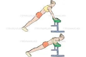
This is a good warmup to get the chest ready for work. According to research, a dynamic warmup before training can help prevent injury.
- Begin by placing your hands on the wall or a counter-height surface. Walk your feet back so that your body forms a roughly 45-degree angle with the floor.
- Keep your body straight and your spine neutral, then lower your chest to the surface you’re leaning against.
- Take a brief break, then go back to where you were before.
- Make sure the resistance is light enough for up to 20 repetitions. To make it easier, step closer to your hands; to make it more difficult, step further away.
Flat bench press.
- Lie back on the bench, knees bent and feet flat on the floor. Hold the barbell with your palms facing your feet and your thumb encircling it. To lift the weight off the rack, press your arms straight up towards the ceiling.
- Move the weight to the chest level.
- Slowly lower the weight to your chest while keeping your elbows bent at a 45-degree angle. Keep the bar roughly in line with your nipples.
- Pause for a moment, then return the weight to the starting position.
- Finish the three sets of eight to twelve repetitions.
Incline bench press.
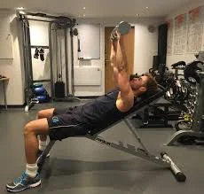
- Lie back on the incline bench, knees bent and feet flat on the floor. Hold the barbell with your palms facing your feet and your thumb encircling it. To lift the weight off the rack, press your arms straight up towards the ceiling.
- Place the weight above your collarbone.
- Slowly lower the weight to your chest, roughly mid-chest to just above your nipples.
- After stopping, move the weight back to its initial position.
- Finish the three sets of eight to twelve repetitions.
- As with the flat bench, keep your back and feet flat throughout the movement. Again, it is strongly advised that you complete this exercise with someone spotting you.
Pushup
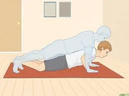
- Start on your hands and knees, then step back into a high plank position. Your arms should be slightly wider than your shoulders, and your legs should be straight with your quads. Your spine should be neutral and your hamstrings should be tight.
- Bend your elbows at a 45-degree angle to lower your chest to the floor while keeping your core tight. Match your body from head to toe.
- Aim to go as low as possible while maintaining core support and spine and pelvic alignment.
- Push your chest away from the ground until your elbows are straightened.
- Repeat for 8-12 repetitions. Do three sets.
Resistance band pullover
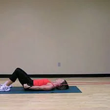
- Anchor the band to something solid. Then, lie on your back, head facing the anchor point. The band should be approximately 1-2 feet higher than your head.
- Grasp the band overhead with slight tension. Keep your thumbs pointing upward and your palms facing away from each other.
- Keep your core tight and your elbows straight as you pull the band towards your hips. Return carefully and slowly to your starting posture.
- Do three sets of eight to twelve repetitions.
Stretching exercises
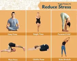
Child’s Pose.
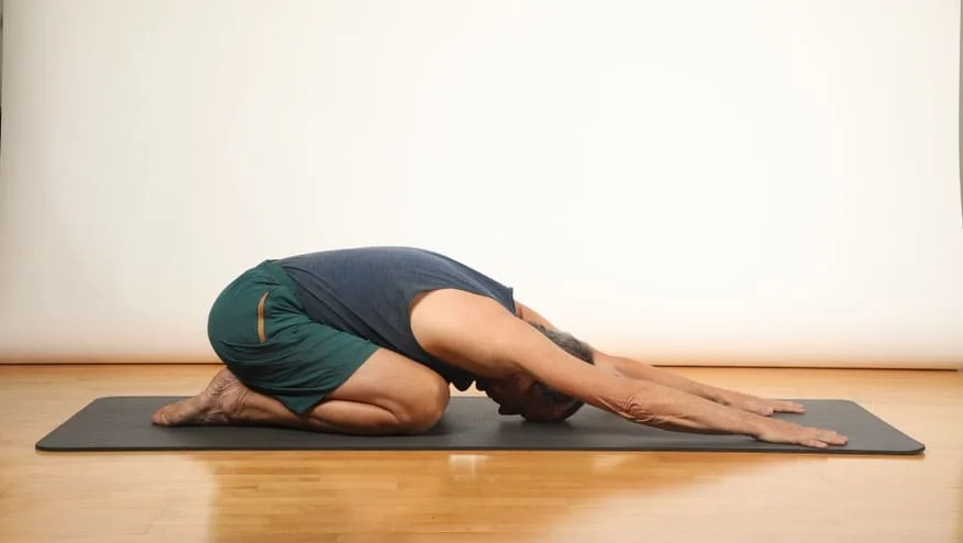
Child’s Pose is a great way to gently stretch the chest wall while maintaining proper alignment. To do this stretch:
- Consider a position on the table by putting yourself on your hands and knees.
- Ensure that the wrists are directly beneath the shoulders and the knees are beneath the hips.
- Gently walk your arms forward until your forehead rests on the mat, then sit your buttocks back on your heels.
- Hold for a few seconds, as directed by the physiotherapist.
- To exit, take a deep breath in and return your hands to your knees.
Chest Opener Stretch
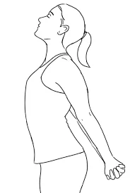
The chest opener stretch targets the chest and front shoulder muscles and is used by physiotherapists to relieve muscle tension and increase flexibility. There are several variations of this stretch, but the basic one can be performed as follows:
- When you sit or stand, keep your shoulders relaxed and your back straight.
With your palms pointing inside, interlace your fingers behind your back. - Lift your arms gently, keeping them straight and your chest open, and feel the stretch in your chest and shoulders.
- Keep your neck and jaw relaxed.
- Release the stretch slowly and repeat as directed by the physiotherapist.
This stretch is especially beneficial for those with muscle tension in the front of their shoulders and chest.
Doorway Stretch
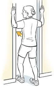
Physiotherapists commonly prescribe the doorway stretch to stretch both the pectoralis major and minor muscles, resulting in increased chest mobility. To do this stretch:
- Stand in a doorway, feet together.
- Bend your elbows to a 90-degree angle and place your forearms on the doorframe, keeping your elbows at shoulder height.
- Take a small step forward with one foot, gently leaning into the doorway until you feel a comfortable stretch across your chest.
- Keep your spine neutral and avoid excessive arching in the lower back.
- Repeat the stretch on the opposite side, stepping forward with the other foot.
Stretch your pectoralis major muscle.
The pec major is the larger of the two chest muscles, and it consists of two muscles:
Clavicular Portion
The pec major is the larger of the two chest muscles, and it is composed of two muscles: the clavicular portion on top and the sternal portion on the bottom. Stand in a room corner, palms and forearms against each wall, to stretch the clavicular portion. Slide your arms up so that your elbows are at 90-degree angles and align with your armpits. Lean forward to feel a stretch.
Sternal Portion
The pectoral major sternal portion is larger than the clavicular portion. The clavicular stretch is more common than the sternal stretch, but both are necessary. To perform this stretch, stand in the corner with your arms against the walls, just like in the other stretch. Open the forearms outward, pointing at 45-degree angles away from the body. Lean forward until the stretch is felt, then hold.
Summary
The pectoral region on the anterior chest wall consists of four muscles: pectoralis major, pectoralis minor, serratus anterior, and subclavius. These muscles control movement in the upper limbs or ribs. The pectoralis major is the largest and fan-shaped muscle closest to the skin, while the pectoralis minor stabilizes the scapula.
The serratus anterior forms the medial border of the axilla region and rotates the scapula. The subclavius is a small muscle beneath the clavicle, providing minimal protection to underlying neurovascular structures. Strengthening exercises for chest muscles include incline pushups, flat bench presses, pushups, and resistance band pullovers.
FAQs
What muscle surrounds the chest?
The pectoralis major muscles are fan-shaped muscles that connect your armpits to the centre of your breastbone, or sternum. The pectoralis minor muscles are smaller than the pectoralis major and run along your ribs just below your collarbone.
What purpose do chest muscles serve?
Your chest contains many of the structures required for life, such as your oesophagus, windpipe, lungs, and heart. The chest muscles move the arms across the body as well as up and down.
How do you stretch your chest?
Raise the left arm to shoulder height and place the palm and inside of the arm against the wall surface or doorway.You should have a 90-degree bend in your elbow. To feel the stretch, gently press the chest through the open space. Moving the arm higher or lower allows you to stretch different areas of the chest.
What is a chest stretch?
Through the palms facing up, clasp your hands behind your back. Pull your hands down and bring your shoulder blades together. Your chest should stand out. Hold for 10-20 seconds. The chest area and upper arms should feel stretched.
References:
- Muscles of the Pectoral Region – Major – Minor – TeachMeAnatomy. (2024, January 18). TeachMeAnatomy. https://teachmeanatomy.info/upper-limb/muscles/pectoral-region/
- Baig, M. A., & Bordoni, B. (2023, August 28). Anatomy, Shoulder and Upper Limb, Pectoral Muscles. StatPearls – NCBI Bookshelf. https://www.ncbi.nlm.nih.gov/books/NBK545241/
- Mpt, T. E. P. (2023, March 29). 7 Top Chest Exercises for a Strong and Functional Upper Body. Healthline. https://www.healthline.com/health/fitness-exercise/best-chest-exercises#8-best-exercises
- Jey, T. (2023, August 9). The 4 Best Stretches For A Strained Chest Muscle | Physiotherapists in Toronto | Yorkville Sports Medicine Clinic. Physiotherapists in Toronto | Yorkville Sports Medicine Clinic. https://www.yorkvillesportsmed.com/blog/the-four-best-stretches-for-a-strained-chest-muscle
- Egypt, C. E. L. C. (n.d.). IPC Physical Therapy Center. ipcphysicaltherapy.com. https://www.ipcphysicaltherapy.com/Exercise-full.asp?ExercisesID=357#:~:text=Slide%20the%20arms%20up%20so,until%20you%20feel%20a%20stretch.&text=The%20pec%20major%20sternal%20portion,stretch%2C%20yet%20both%20are%20important.

19 Comments