Radiation Therapy
Introduction
Radiation therapy (Radiotherapy) is a medical treatment that uses high doses of radiation to destroy cancer cells. It is one of the most common treatments for cancer and can be used alone or in combination with other treatments like surgery and chemotherapy.
This technique proposes to help eliminate or prevent the growth of malignant cells. Usually, a linear particle accelerator delivers it. If cancer has only progressed to one portion of the body and is confined there, radiation treatment may be curative for several cancer types.
To stop a tumor from growing back following surgery to remove a primary malignant tumor, it can also be used as adjuvant therapy (for example, early stages of breast cancer). Because radiation therapy complements chemotherapy, it has been utilized before, during, and following the drug in cases of cancer that are particularly susceptible to it.
Because radiation therapy can limit cell reproduction, it is commonly employed to treat malignant tumors.
Ionizing radiation damages cancerous tissue’s DNA, which results in cell death. Shaped radiation beams are focused from many angles of exposure to intersect at the tumor, producing a significantly bigger absorbed dosage there than in the surrounding healthy tissue, thus safeguarding healthy tissue (like skin) or organs that radiation treatment must enlarge to treat the tumor. In addition to the actual tumor, the radiation fields may also encompass the lymph nodes that drain from it if the cancer is clinically or radiologically associated with them, or also there is a suspected risk to get subclinical malignant spreading. These issues are frequently linked to both internal movement (breathing, for example, and bladder filling) and outward skin mark movement surrounding the tumor location.
Unlike radiology, which uses radiation for medical imaging and diagnosis, radiation oncology is a medical profession that deals with prescription drugs. Regarding treating cancer, a radiation oncologist regularly recommends radiation therapy in addition to chemotherapy. Palliative care (when a cure is not possible and the objective is for local illness management or symptomatic relief) or treatment (where the therapy has survival advantages and can be curative). With the plus point of immunotherapy, hormone therapy, chemotherapy, surgery, or a mix of the four, radiation treatment is frequently employed. Radiation therapy is an effective therapeutic option for the vast majority of common cancer types.
The particular motive of treatment – curative, adjuvant, neoadjuvant therapeutic, or palliative – depends on the dependency of the general health of the patient and the type, location, and stage of the tumor. For the body ready for a bone marrow transplant, whole body irradiation, or TBI, is one kind of radiation treatment. The other difference of radiation therapy that reduces exposure to healthy tissue during operations to treat malignancies of the breast, prostate, and other organs is called brachytherapy, There is a radioactive source inside or close to the area that requires consideration.
Radiation therapy can be used to treat a variety of non-cancerous disorders, including pterygium, pigmented villonodular synovitis, auditory neuromas, trigeminal neuralgia, severe thyroid eye disease, and to prevent keloid scar formation, vascular restenosis, and heterotopic ossification. Advances in radiotherapy for the management of non-malignant diseases have been unapproachable by concerns about radiation-induced cancer.
Cancer that has spread throughout the body cannot be cured by radiation treatment given that most of them don’t reach every part of the body.
What are the goals of radiation therapy?
Radiation is frequently the therapy of choice for the following reasons, however, it’s vital to keep in mind that every cancer and every individual is unique.
To cure or shrink early-stage cancer
Certain tumors are extremely radiation-sensitive. In some situations, radiation therapy alone may be utilized to reduce or eradicate the malignancy. In other cases as well, chemotherapy or other anticancer drugs may be used initially. For some malignancies, radiation treatment can be used either before surgery to reduce the tumor (pre-operative therapy, also known as neoadjuvant therapy) or following surgery to help prevent the cancer from returning (adjuvant therapy).
Radiation therapy may be the recommended course of action for certain tumors that are curable only with radiation therapy or with surgery. This is because radiation therapy may result in less harm and a higher chance of the affected body part functioning normally following treatment.
For some cancer forms, a combination of chemotherapy, radiation therapy, or other anti-cancer drugs may be employed. A class of medications known as radiosensitizers increases the sensitivity of cancer cells to radiation. Studies have demonstrated that in certain cancer cases, the combination of radiation therapy and anti-cancer medications can enhance the effectiveness of each treatment when administered alone. However, one disadvantage is that when taken in combination, side effects are frequently greater.
To prevent cancer from returning (recurring) in another area
From its original site, cancer can spread to different bodily areas. Medical professionals often assume that a small number of cancer cells may have already spread, even in cases where cancer cells are not apparent on imaging tests such as CT or MRI scans. Now and again radiation treatment is utilized to obliterate malignant growth cells before they form into cancers in the locale where the sickness spreads most rapidly.
For example, because some types of lung cancer frequently spread to the brain, patients with these diseases may get radiation therapy to the head even in cases when no cancer is known to exist. By carrying out this, cancer is prevented from ever spreading to the skull. Radiation therapy for cancer treatment and cancer prevention can sometimes be administered concurrently, particularly if the tumor’s potential site of metastasis is nearby.\\
Handle the advanced cancer’s symptoms and indicators.
There are instances when cancer has spread too far to treat. To improve the patient’s condition, some of these tumors can still be treated to reduce in size. Radiation therapy may be able to treat symptoms such as pain, dysphagia, dyspnea, and intestinal obstructions that may be brought on by advanced cancer. We refer to this as palliative radiotherapy.
For the treatment of cancer that has been resurfaced
Radiation therapy may be used to treat an individual’s cancer if it has returned, or recurred, or to alleviate the symptoms brought on by advanced cancer. The use of radiation following a recurrence is dependent upon several circumstances. For example, administering more radiation therapy in the same location may not be feasible if the cancer has returned to a portion of the body that has already received radiation treatment.
The amount of radiation that was previously utilized determines this. In other situations, radiation treatment could be given to the same area of the body or a separate one. For some cancers, radiation therapy may not be necessary even if they reoccur because of their poor response to it.
Technique
Mechanism of action
Radiotherapy disrupts cancer cells’ mitosis by destroying their DNA. A charged particle or a photon is the source of energy that causes DNA damage. The atoms that make up the DNA strand ionize directly or indirectly due to this damage. The dissociation of water produces independent free radicals, particularly hydroxyl radicals. These radicals produce indirect ionization, which damages DNA.
Free radicals are mainly responsible for radiation effects in photon therapy. Cells can repair both DNA impairments. On the other hand, double-stranded DNA breaks are far more difficult to repair and can result in significant genetic losses and chromosomal abnormalities. The likelihood that cells will die rises when double-strand breaks are targeted. Generally speaking, cancer cells are more stem cell-like and less differentiated than most healthy differentiated cells. They also multiply more often and are less able to repair damage that is not fatal. Cell division continues to perpetuate single-stranded DNA damage, which eventually leads to the accumulation of damaged DNA in cancer cells and their slower death or proliferation.
The lack of oxygen that photon radiotherapy causes in solid tumor cells is one of its biggest disadvantages. When solid tumors grow larger than their blood supply, hypoxia, a condition of low oxygen, occurs. Because oxygen produces free radicals that damage DNA, it is a powerful radiosensitizer that can increase the effectiveness of certain radiation exposures. Tumor cells in a hypoxic environment can be as much as three times greater resistant to radiation damage than those in an oxygen-rich environment.
The use of high-pressure oxygen tanks, hyperthermia therapy (heat therapy that dilates blood vessels to the tumor site), blood substitutes that carry increased oxygen, hypoxic cell radiosensitizer medications like metronidazole and misonidazole, and hypoxic cytotoxins (tissue poisons) like tirapazamine are just a few of the strategies that have been extensively researched to overcome hypoxia. Preclinical and clinical studies on the usage of an oxygen diffusion-enhancing substance such as trans sodium crocetin as a radiosensitizer are among the more recent research methodologies being examined.
Protons, boron, carbon, and neon ions are examples of charged particles that can directly damage the DNA of cancer cells through a high LET (linear energy transfer). The onwards particles also have an antitumor effect which is independent of tumor oxygenation, because they work specifically by direct energy transfer, which usually results in double-stranded DNA breaks.
Protons and other charged particles have low lateral side dispersion in the tissue because of their relatively high mass; as a result, the beam does not expand much, has minimal effect on the local tissue, and is centralized as the form of the tumor. They also use the Bragg peak effect to target the tumor more precisely. For an excellent illustration of the differences between charged particle treatment and intensity-modulated radiation therapy (IMRT), see proton therapy.
This process establishes a restricted range for tissue impairment once the tumor has occurred and lessens harm to healthy tissue between the charged particle radiation source and the tumor. This damage already done has little therapeutic value, raises the risk of adverse drug reactions, and raises the possibility of developing cancer again. This distinction is critical in situations when the proximity to other organs renders any stray ionization extremely harmful (such as head and neck tumors). Children’s bodies are still developing, thus they are more vulnerable to the negative effects of X-rays. Depending on several conditions, children are about ten times more likely to develop secondary malignancies after radiation therapy than adults.
Dose
For solid epithelial cells, the usual dose for patients in remission is 60-80 Gy; the usual dose for lymphomas is 20–40 Gy. When choosing a dosage, radiation oncologists take into account several additional criteria, such as the patient’s comorbidities, whether chemotherapy is being received before or after surgery, and the surgical success rate.
Treatment planning determines how a given dosage will be delivered (part of dosimetry).
Treatment planning is often performed using sophisticated software on specialized computers.
The total dose required can be calculated from several different angles or sources depending on the mechanism of radiation delivery. The planner will make an effort to create a strategy that reduces the dosage to the nearby healthy tissues while providing the tumor with a consistent prescribed dose.
With radiation therapy, gel dosimetry is a dosimetry technique that may be used to examine three-dimensional dose distributions.//
Fractionation
There are numerous key reasons why the entire dosage is fractionated, or spread out over time. Normal cells can recuperate more slowly due to fractionation, however, tumor cells are often less adept at repairing themselves between fractions. Similarly, tumor cells can reoxygenate between fractions, increasing tumor cell death if they have been either acutely or chronically hypoxic (and therefore radioresistant).
Radiation therapy facilities and even individual medical facilities have variable fractionation schedules. Medical facilities in North America, Australia, and Europe follow a conventional fractionation regimen of 1.8-2 Gy per day, five days every week. It is preferable to finish radiation therapy for some cancer types, such as head-and-neck and cervical squamous cell cancers, within a specific time frame since prolonging the dosing schedule strong dose might allow the tumor to start repopulating. Smaller fraction sizes are linked to lower incidence, hence a normal fraction size for kids would be 1.5 to 1.8 Gy per day, as in normal tissues, lower fraction sizes are linked to a lower frequency and severity of late-onset adverse effects.
When a treatment course is coming to a close, two fractions each day may be administered in some situations. This regimen, often called hyperfractionation or simultaneous acceleration, is applied to cancers that grow faster to a smaller size. This trend is particularly seen in head and neck malignancies.
Patients receiving palliative radiation therapy for simple painful bone metastases should not receive more than one portion of radiation therapy. A single treatment is ideal for improving patient comfort in patients with low life expectancy since it offers equivalent pain reduction and morbidity results to multiple-fraction treatments.
Schedules for fractionation
Hypofractionation is one fractionation schedule that is being utilized more and more and is still being researched. Standard doses for different cancer types range from 2.2 Gy/fraction to 20 Gy/fraction; the latter is more common in treatments that use stereotactic techniques, These include stereotaxic radiosurgery (SRS). Hypofractionation occurs because of the necessity to take advantage of some cancers’ radiosensitivity while simultaneously lowering the likelihood of a local recurrence by depriving clonogenic cells of the time they need to proliferate. Rather than regularly disrupting the process of clonogenic cell division (apoptosis) as with radiation therapy.
Estimation of dose based on target sensitivity
The susceptibility of various cancer forms to radiation varies. Genomic signals of intracellular radiosensitivity have been shown to correlate with clinical outcomes when used to predict radiation exposure in individual patients, although sensitivity is difficult to predict from biopsy proteomic or genomic analysis. samples The discovery that non-enzymatic compounds of manganese and small organic molecules provide radiation protection to bacteria offered an alternative approach to genomes and proteomics. It was discovered that manganese concentration and variation, which can be measured by electron paramagnetic resonance, are reliable indicators of radiosensitivity. This conclusion holds for human cells as well. The total cellular manganese concentrations and their fluctuation were shown to be associated with clinically inferred radio responsiveness in various tumor cells. This discovery should be helpful for more accurate radio doses and better cancer patient therapy.
Types
In the past, radiation treatment has been divided into three primary categories:
Brachytherapy is as well known as sealed source radiation therapy, and external beam radiation therapy, also known as XRT EBRT or teletherapy.
Variations include the location of the radiation source: external radiation is given externally, brachytherapy uses sealed radioactive devices precisely placed in the area to be treated, and systemic radioisotopes are administered orally or by infusion. Radioactive devices and sources can be placed by variations either permanently or temporarily during brachytherapy. The reload method is usually used to add temporary sources. Backloading is the surgical placement of a hollow tube or applicator into the organ to be treated. The sources are then loaded into the application. These included techniques that lead to more customized treatments, techniques that enhance the therapeutic ratio, an emphasis on patient satisfaction, and a list of areas that need more research.
External beam radiation therapy
This comes in the next three parts.
Traditional external beam radiation treatment
International standard source holder (usually lead), retaining ring, and remote cure “source” consisting of two stainless steel capsules welded to two stainless steel covers surrounding a shielded interior and cylinder of radioactive source material (usually made of cobalt-60) form a remote treatment radiation capsule. The specific diameter in measure of the “source” is about 30 mm.
Historically, medical linear accelerators that produce high-energy X-rays, kilovoltage therapy X-ray units, or devices that resemble linear accelerators but employ a sealed radioactive source, such as the one above, are used to deliver standard external beam radiation therapy (2DXRT). In 2DXRT, a single radiation beam is predominantly employed as well as sides./
Conventional suggests how the treatment is scheduled or simulated using a specially calibrated diagnostic X-ray device called a simulator. This type of equipment replicates the radiation beam arrangements that are typically well-established to achieve a desired plan, as well as the actions of a linear accelerator, sometimes by eye. Accurately targeting or localizing the volume to be treated is the goal of the simulation. This method is tried-and-true, usually efficient and dependable. The fear is that radiation poisoning to surrounding healthy tissues may limit the efficacy of some high-dose treatments.
Tumor control may not be readily achieved due to the sensitivity of the surrounding rectum. Physicians and physicists had little idea how much radiation was applied to healthy and malignant tissue before the development of CT scans. Because of this, practically all tumor locations now get 3-dimensional conformal radiation therapy as the standard of care.
Stereotactic radiation
It uses detailed imaging scans with beams of radiation focused on a well-defined tumor. When treating cancers in the brain or spine, radiation oncologists use stereotactic techniques, sometimes in collaboration with neurosurgeons.
Stereotactic radiation comes in two varieties. Stereotactic radiosurgery (SRS) is when doctors use one or more stereotactic radiation therapies to treat the brain or spine. Stereotactic body radiation therapy, or SBRT, refers to a variety of stereotactic radiation treatments administered to the body, including the lungs.
Stereotactic therapies, according to some doctors, have the advantage of delivering the optimum amount of radiation to the cancer in a shorter period than normal treatments, which can take six to eleven weeks. Moreover, treatments are carried out with extreme accuracy, which ought to lessen the radiation’s negative effects on healthy tissues. The fact that stereotactic therapies are limited to certain tiny tumors presents a challenge.
Because many institutions refer to stereotactic treatments by the manufacturer’s name rather than SRS or SBRT, it might be confusing. Axesse, Cyberknife, Gamma Knife, Novalis, Primatom, Synergy, X-Knife, Tomo Therapy, Trilogy, and True Beam are most of the brand names associated with these treatments. As equipment producers continue to create new, specialized technology to treat tumors, this list is subject to change.
Three-dimensional conformal radiation treatment and virtual simulation
The ability to employ specialist CT and MRI scanners in conjunction with planning software to generate three-dimensional pictures of tumors and surrounding normal structures has changed radiation therapy treatment planning.
The simplest type of planning, virtual simulation, places radiation beams more precisely than conventional X-rays, which frequently makes it impossible to preserve normal tissues and difficult to analyze soft-tissue structures.
3-dimensional conformal radiation therapy (3DCRT) is a virtual simulation enhancement approach that combines a multi-leaf collimator (MLC) and a variable number of beams to shape each radiation beam’s profile to match the profile of the target from the beam’s eye view (BEV). A larger dosage of radiation may be administered to the tumor than would be possible with traditional procedures when the treatment volume is shaped to fit the shape of the tumor. This is a result of the radiation’s lower relative toxicity to the normal tissues in the vicinity.
Intensity-modulated radiation therapy (IMRT)
It is delivered using a Varian True Beam Linear Accelerator. Additionally, IMRT enhances the capacity to tailor the treatment volume to concave tumor forms, which is advantageous in situations when the tumor is around a major organ or blood nutrition or spinal cord, two particularly sensitive structures.
Accurate radiation dosages are applied to malignant tumors or certain regions inside the tumor by the use of computer-controlled X-ray accelerators. Utilizing specialized computer programs, the radiation delivery pattern is established to carry out treatment simulation and optimization (Treatment Planning). By proportionating the strength of the radiation beam, the dose of radiation is of the three-dimensional form of the tumor. When the radiation exposure to the normal region of the cell is avoided or reduced, radiation dose intensity can be increased around the gross tumor volume. Compared to 3DCRT, this results in improved tumor targeting, fewer side effects, and better treatment outcomes.
While IMRT is increasingly used in more complicated body sites such as the CNS, head and neck, prostate, breast, and lung, 3DCRT is still commonly used in many other areas. Unfortunately, IMRT is limited by the requirement that qualified medical practitioners commit more time to it. This is because, in contrast to 3DCRT preparation, doctors have to manually outline each tumor along the whole disease location, one CT picture at a time. Then, to create a viable treatment plan, medical physicists and dosimetrists must be contacted. Even in the most cutting-edge cancer facilities, IMRT technology has only been utilized commercially since the late 1990s, thus radiation oncologists who did not learn about it during their residency programs will need to locate other educational resources before putting IMRT into practice.\
For many tumor locations, there is increasing evidence that any of these two approaches offers a superior survival advantage than traditional radiation treatment (2DXRT); yet, the capacity to lower toxicity is widely acknowledged. According to many important experiments led by Professor Christopher Nutting of the Royal Marsden Hospital, this is particularly true for cancers of the head and neck. Both approaches allow for dose escalation, which might improve usefulness. Concerns about the increased radiation exposure of normal tissue and the potential for secondary malignancy have been raised, particularly about IMRT.
Overconfidence in imaging accuracy can lead to a higher risk of missing lesions that migrate between or during a treatment (e.g., because of breathing or insufficient patient immobilization) or something that cannot be identified during design and therefore is not included in the treatment plan. To better regulate this uncertainty, new strategies are being developed. One such technique is to combine real-time imaging with real-time therapeutic beam formation.
One or more tiny implantable electric devices placed inside or adjacent to the tumor can be tracked and localized in real time using this method. For this, a variety of implanted medical devices are employed. One such solution is a magnetic transponder, which measures the magnetic field produced by many transmitting coils and relays the results back to the positioning system to pinpoint the location. A small wireless transmitter that generates an RF signal that is detected by a sensor array and used for tumor localization and real-time tracking is an alternative option for the implanted device.
The well-studied “tongue and groove effect” in IMRT refers to the phenomenon where radiation from overlapping MLC (multileaf collimator) leaves travels between their extended tongues and grooves, resulting in unwanted underdosing. While various techniques have been proven to either completely remove or significantly reduce the TG impact, their efficacy varies depending on which IMRT technique is used, and some of them incur additional costs.
Certain literature differentiates between “tongue and groove error” and “tongue or groove error” based on whether one or both sides of the aperture are blocked.
Volumetric modulated arc therapy (VMAT)
A radiation approach called volumetric modulated arc treatment (VMAT) was launched in 2007 and can create highly conformal dose distributions on target volume coverage while sparing normal tissues.
Three factors are changed while the patient is undergoing therapy, which accounts for the specificity of this procedure. To provide radiation, VMAT rotates the gantry (which is often a 360° rotating field with one or more arcs), modifies the beam’s speed and form using a multileaf collimator (MLC, or “sliding window” moving system), and increases the medical linear accelerator’s fluence output rate (dose rate). as treating patients, VMAT offers the benefit of shorter radiation delivery periods as compared to traditional static field intensity-modulated radiation therapy (IMRT). Depending on the kind of cancer, different amounts of healthy tissues and Organs at Risk (OAR) are spared during VMAT and traditional IMRT.
When treating oropharyngeal, hypopharyngeal, and nasopharyngeal carcinomas, VMAT offers comparable or superior organ at risk (OAR) protection. OAR protection yields mixed outcomes when it comes to treating prostate cancer; some studies recommend IMRT, while others promote VMAT.
Temporally feathered radiation therapy (TFRT)
The goal of temporally feathered radiation treatment (TFRT), which was first proposed in 2018[87], is to spare normal tissues without changing the dosage given to the tumor by making use of the inherent non-linearities in normal tissue healing. In 2021, a small clinical experiment showed that the technique was viable, albeit its effectiveness has not yet been extensively investigated.
Automated planning
Radiation therapy treatment plans at the moment include automated treatment planning. Generally speaking, computerized planning uses two techniques.
1) Knowledge-based planning: Using a library of well-designed plans, the treatment planning algorithm forecasts the target and dose-volume histogram of the organ at concern.
2) The alternative strategy is known as protocol-based planning; in this method, the treatment planning system attempts to imitate an expert treatment planner and assesses the plan quality iteratively by comparing it to the protocol.
Particle therapy
In particle therapy, intense ionizing particles such as carbon ions or protons are directed at the target tumor. As the particle enters the tissue, the dosage rises to a maximum (the Bragg peak) that appears close to the particle’s range, after which it falls to (almost) zero. This energy deposition profile has the benefit of depositing less energy into the healthier tissue that surrounds the target tissue.
Auger therapy
Applied thermotherapy (AT) is distinct from conventional radiation therapy in a few ways. Firstly, it does not rely on radioactive nuclei to damage cells at the cellular level. Second, it does not give dosage to the intended region at a dose lower than the specified tissue/organ locations by using numerous external pencil beams pointing in different directions. Instead, the aim of the in situ delivery of a very high dose at the molecular level using AT is to alter molecules in situ through molecular reorganizations and breakages, such as altering stacking structures, and by cellular metabolic processes linked to the aforementioned molecular structures.
Motion compensation
In many kinds of external beam radiation, motion can cause target tissue to migrate out of the planned beam path or bring other healthy tissue into it, which can negatively impact the way that therapy is delivered. Certain types of immobilization are frequently used to keep patients from moving their bodies in substantial ways while receiving treatment, but they cannot stop all motion, such as breathing. To promote these motions, several strategies have been developed.
Deep inspiration breath-holds, or DIBHs, are commonly used during breast treatments when it is imperative to keep radiation away from the heart. After inhaling, the patient in DIBH holds their breath to create a stable position for the treatment beam to be turned on. An external monitoring system, such as a spirometer or a camera with markers, can be used to perform this automatically.
For respiratory-gated therapy, in which the patient breathes freely and the beam is only activated at specific times throughout the breathing cycle, the same monitoring techniques as well as 4DCT imaging can be used. Other techniques include using 4DCT imaging to schedule treatments with motion-accounting margins and actively moving the treatment couch, or beam, to track motion.
Contact X-ray brachytherapy
Contact X-ray brachytherapy, often known as “CXB,” “electronic brachytherapy,” or the “Papillon Technique,” is a kind of radiation treatment used to treat rectal cancer. It uses low-intensity (50 kVp) kilovoltage. A therapy applicator is inserted into the tumor through the anus into the rectum and placed against the diseased tissue after an endoscopic examination has been performed to locate the tumor in the rectum.
Finally, the treatment tube is inserted into the applicator to deliver high-dose (30Gy) X-rays to the tumor outright, three times a week at intervals of two weeks. It is usually used to treat individuals with early-stage rectal cancer who might not be surgical candidates. According to a 2015 NICE study, the most common adverse effects were radiation-induced ulcers (occurring in 27% of cases) and bleeding (occurring in about 38% of cases).
Brachytherapy (sealed source radiotherapy)
A SAVI brachytherapy device
The delivery of brachytherapy involves putting a radiation source(s) within or close to the region that is to be treated.
Brachytherapy is a well-liked and effective treatment for tumors in a variety of body parts; it may also be used to treat cancers of the skin, breast, prostate, and cervical areas.
Radiation sources are positioned precisely in which the malignant tumor is in brachytherapy. This indicates that the radiation only impacts a relatively small region and that healthy tissues farther from the sources are exposed to less radiation. Brachytherapy has some benefits over external beam radiation therapy. First, it allows for the treatment of tumors with very high doses of targeted radiation while lowering the risk of needless harm to nearby healthy tissues. Brachytherapy typically takes less time for it to finish than other forms of radiation therapy. By doing this, the chance that cancer cells may divide and multiply in the interval between doses of radiation therapy will be reduced.
One instance of the confined character of breast brachytherapy is the way the radiation dosage is delivered via the SAVI device, which has numerous individually controllable catheters. When compared to previous iterations of breast brachytherapy and external beam radiation treatment, this technique reduces the exposure of healthy tissue and the associated adverse effects.
Radionuclide therapy
Radionuclide treatment, also known as systemic radioisotope therapy, radiopharmaceutical therapy, or molecular radiotherapy, is a type of targeted therapy. The chemical characteristics of the isotope, such as radioiodine, which the thyroid gland particularly absorbs 1,000 times better than other body organs, maybe the reason for targeting. To target a radioisotope is to link it to an antibody or a distinct molecule and let it forward to the desired region. Ingestion or infusion (into the circulatory system) are two methods of delivering the radioisotopes.
Examples include the use of oral iodine-131 to treat thyroid cancer or thyrotoxicosis, and the infusion of metaiodobenzyl guanidine (MIBG) to treat neuroendocrine cancers using radioactive peptide receptors.
Another instance is the radioembolization of liver tumors or liver metastases with the hepatic artery injection of radioactive yttrium-90 or holmium-166 microspheres. One treatment approach that uses these microspheres is selective internal radiation therapy. The microspheres, which have a diameter of around 30 μm, or roughly one-third of a human hair, are inserted straight into the artery that supplies blood to the tumors.
The first step in these therapies is inserting a catheter into the leg’s femoral artery, traveling to the intended target location, and starting the therapy. In contrast to traditional systemic chemotherapy, a more focused approach is possible since the microspheres will be delivered directly to the tumor by the blood nourishing it. SIR-Spheres, TheraSpheres, and QuiremSpheres are the three types of microspheres that are now available.
Malignant bone metastases are one of the main conditions treated with systemic radioisotope therapy. Radioisotopes ignore healthy, undamaged bone and target particular areas of sick bone. Often employed isotopes for treating bone metastases include radium-223, strontium-89, samarium (153Sm), and lexidronam.
The anti-CD20 monoclonal antibody ibritumomab tiuxetan (Zevalin) was authorized by the US Food and Drug Administration (FDA) in 2002. It is conjugated with yttrium-90. The tositumomab/iodine (131I) tositumomab regimen (Bexxar), which consists of an unlabeled and an iodine-131 labeled anti-CD20 monoclonal antibody, was authorized by the FDA in 2003. These medications, which were approved to treat refractory non-Hodgkin’s lymphoma, were the forerunners of the modern field of radioimmunotherapy.
Intraoperative radiotherapy
Applying therapeutic doses of radiation to a target location, such as a cancerous tumor, while the area is exposed during surgery is known as intraoperative radiation therapy or IORT.//
Rationale
It accurately targets a high radiation dosage while exposing the surrounding tissues—which are either protected or moved throughout the procedure—to as little radiation as possible. After the tumor is surgically removed, conventional radiation treatments like external beam radiation therapy (EBRT) have several disadvantages: Even with contemporary radiation planning, the difficult localization of the wound cavity sometimes results in the missing tumor bed where the maximum dosage should be delivered. Furthermore, the standard waiting period between EBRT and surgical tumor removal may allow for tumor cell repopulation. By accurately targeting the targeted organs with radiation, it is possible to prevent these potentially detrimental consequences and immediately sterilize any remaining tumor cells. Tumor cell stimulation by wound fluid is an additional characteristic.
How is radiation therapy given?
Three methods are available:
It is carried performed during outpatient stays at medical facilities. It is in most cases administered throughout several weeks, however occasionally it is given twice daily for a few weeks. A person exposed to radiation from the outside world is not radioactive and does not need to take extra safety measures at home.
Internal radiation: Inside the body, a radioactive source is placed in or near a tumor. Certain forms of brachytherapy may include injecting radiation into the body and allowing it to do its job. Periodically, it is inserted into the body and then taken out. For a while, certain safety measures are required for this kind of radiation. It’s important to realize, though, that internal radiation eventually ceases to be radioactive within the body.
Systemic radiation: Certain cancers are treated with radioactive medications that are ingested or injected into veins.
Then, these medications circulate throughout the body. After taking these medications, you may need to take extra care at home for a while.
The kind and location of your cancer are going to influence what kind of radiation you may get. Several types are employed in specific situations. Regarding the kind of radiation prescribed for you, how it affects your body, and any necessary precautions, your cancer care team can provide precise answers.
Types of cancer it treats
According to the National Cancer Institute (NCI) Trusted Source, clinicians routinely utilize external beam radiation to treat the following kinds of cancer:
- breast cancer
- lung cancer
- prostate cancer
- colon cancer
- cancers of the head or neck
According to the National Cancer Institute, brachytherapy may be a highly successful treatment for malignancies in specific regions of the body, including:
- cervix
- vagina
- uterus
- rectum
- head and neck
- eye
In addition, a physician could advise brachytherapy for malignancies of the:
- prostate
- brain
- lung
- skin
- breast
- esophagus
- anus
- bladder
Side effects of radiation therapy
Everyone reacts differently to radiation treatment. It is wise to know that side effects are possible, albeit you may not experience all or any of them.
The intensity and amount of side effects may vary according to the location, kind of cancer, and overall health status. Any pre-existing problems you had before being diagnosed with cancer may have an impact on how you respond to treatment.
Adverse effects might happen during or just after therapy. However, you may experience long-term negative effects for months or years after receiving radiation. Consult your doctor in advance to discuss and make plans for these possible side effects. They can assist in identifying how to avoid or control side effects when feasible.
The most prevalent adverse effects of radiation treatment may include:
Fatigue
Radiation might leave you feeling exhausted since some of your healthy cells may be harmed by the malignant ones. As you continue with treatments, you may get increasingly exhausted.
The duration and intensity of radiation fatigue can vary depending on the type of treatment. Around the third week of treatment, most radiation therapy patients start to feel tired. Fatigue frequently worsens during the sixth week of medication and may last until the end of the specified course of action.
Inform your doctor if you suffer from any fatigue-related symptoms. They’ll want to keep an eye on your health and could recommend particular aids.
Skin irritation
Your skin may begin to alter around the radiation location. The intensity of the alteration might vary from slight redness to the development of sores.
Radiation dermatitis is a typical treatment-related response. You may develop dryness and flaky skin. Radiation-induced tiredness might vary in length and intensity depending on the type of therapy received. Typically, people receiving radiation therapy start to feel weary during the third week.
Source of therapy. Fatigue typically worsens during the sixth week of treatment and might last until the treatment is completed.
If you are experiencing tiredness symptoms, contact your doctor. They will want to monitor your condition and may recommend particular tactics to help.
To prevent radiation dermatitis, your physician could advise:
- Proper skin care entails using only warm water and mild soap.
- Avoid using oil-based lotions and creams.
- Wear loose-fitting garments to avoid sun exposure and extreme temperatures. Apply steroid cream or gel-like hydrocortisone.
- It is vital to advise your doctor of any skin changes you are experiencing to reduce discomfort and track the healing process. Sometimes the problems resolve themselves after you’ve finished therapy.
Certain parts of your body may also expand. For example, if you are undergoing breast cancer therapy, the rays may cause fluid collection in your breasts, a disease known as lymphedema.//
How do you preserve your skin during radiation therapy?
Skin changes are a side consequence of radiation exposure, and you may need to take special precautions to preserve it. Some ways to accomplish this include:
- Avoid wearing tight garments or elastic over the region being treated.
- Avoid using adhesive tapes on the affected region. Instead, use paper tape.
- Avoid scraping, scouring, or rubbing the surface.
- Before applying a heating pad or an ice pack to the affected area, consult your doctor.
- Consult your doctor about using sunscreen on the area to further protect it from exposure.
- Instead of scratching, use mild soap and warm water to clean the area.
- Consult your doctor before shaving the area.
- Before using any lotions, fragrances, or deodorants on the spot, see your doctor.
Hair loss
If you undergo radiation therapy in hairy areas of your body, you may have hair loss around the treatment site. In some cases, you can lose the majority of your hair if you have radiation therapy to the head.
Your hair may regrow following therapy. You should take some precautions to protect your scalp and the area around it from radiation. For example, if you wear a wig, ensure that the lining does not touch or irritate your scalp. Wearing a scarf or cap to shield your skin from the sun might also be useful.
Low blood cell counts
Radiation kills cancerous cells, but it can also harm healthy cells that aid in wound healing and infection defense. If your blood cell count drops too low, your doctor may suspend therapy until it reaches a certain threshold.
Pain
Radiation therapy causes edema and the loss of healthy cells, both of which might hurt your body. Your cancer doctor may recommend pain relief options, such as medication and other therapy.
Radioprotective drugs
Radioprotective medications, which shield healthy tissue from radiation, may be prescribed by your doctor. These medications only work for specific types of radiation and regions of the body, although they may help reduce adverse effects.
Site-specific side effects
You may have extra site-specific side effects if radiation is applied to particular physiological locations, such as the neck and brain. These side effects may include:
Brain
- hair loss
- headaches
- nausea
- vomiting
- hearing loss
- seizures
- Brain fog and forgetfulness
Head and neck
- mouth and throat sores
- dry mouth
- trouble swallowing
- changes in taste
- earache
- tooth decay
Breast
- skin changes include irritation, dryness, and color
- breast soreness
- breast swelling
Chest
- sore throat
- cough
- shortness of breath
- heart complications
- radiation pneumonitis
- chest pain
- early coronary artery disease
Abdomen
- nausea
- vomiting
- cramps
- diarrhea
- constipation
Bladder
Symptoms may include discomfort or burning during urination, difficulty urinating, blood in urine, and increased frequency of urination.
Urinary incontinence
Radiation treatment accidents
Strict measures are in place to reduce the possibility of patients being accidentally overexposed to radiation therapy. However, mistakes do occur; for example, between 1985 and 1987, the radiation treatment machine Therac-25 was involved in at least six mishaps in which patients were given up to one hundred times the authorized dose; one or two individuals died immediately as a result of the radiation overdoses. From 2005 to 2010, a Missouri hospital overexposed 76 patients (the majority of whom had brain tumors) for five years due to poorly installed new radiation equipment.
Although medical mistakes are extremely uncommon, radiation oncologists, medical physicists, and various other radiation therapy treatment team members are attempting to eradicate them.
In 2010 American Society of Radiation Oncology (ASTRO) established Target Safely, a safety effort that, among other things, intended to document mistakes across the country so that clinicians could learn.
Use in non-cancerous diseases
Quick view of the radiation portal on the surface of the hand, including the machine portal of the head shield opening. Ledderhose syndrome and early-stage Dupuytren’s disease are treated with radiation therapy. When Dupuytren’s disease is at the nodules and cords stage or when fingers have a minor distortion of fewer than 10 degrees, radiation treatment is utilized to prevent the illness from progressing further. Radiotherapy is also used in certain cases after surgery to prevent the progression of the disease. Low doses of radiation are utilized, often three grays for five days, followed by a three-month interval and again three grays for five days.
Can radiation therapy cause cancer?
It is one of the potential adverse effects of treatment that doctors must consider while weighing the advantages and dangers of each option. Although the risk of getting a second cancer as a result of these therapies is usually small and exceeded by the advantages of curing the illness, it is nonetheless present. This is one of the many reasons why each situation is unique, and each individual must be involved in determining which type of treatment is best for them. The danger varies depending on where the radiation therapy will be administered in the body.
If the team treating your cancer recommends radiation treatment, it is because they feel the benefits exceed the risks. Nonetheless, you decide to make. Finding out as much as you can about the potential advantages and disadvantages of radiation therapy may help you make an informed decision about whether it’s right for you.//
Does radiation therapy affect pregnancy or fertility?
Females: It can damage the developing fetus. If there is a risk you will become pregnant, see your doctor about birth control choices.
If you are or may be pregnant, notify your doctor straight away.
If the region of your body exposed to radiation contains the ovaries, the radiation dose may cause the ovaries to stop working (sterility), rendering you unable to bear children. It is critical to understand the risks associated with this possibility before undergoing radiation therapy. If you are considering radiation therapy that will impact your ovaries, discuss with your doctor how this may influence your ability to have children in the future.
Males: As a result, physicians frequently advise men not to conceive a woman during or shortly after therapy. To find out additional information regarding this, connect with your physician.
If the radiation-exposed region involves the testicles, the dosage may cause the testicles to stop working (sterility), rendering you unable to bear children. No clear studies have been done on how sperm exposed to radiation affects future babies. If you are considering radiation therapy to the testicles, talk to your doctor about how it may affect your ability to have children in the future.
Outlook
Radiation therapy may be sufficient to cure certain early-stage malignancies.
However, according to the NCI, research shows that cancer treatment results are higher if a patient receives both radiation and chemotherapy after surgery.
It is crucial to remember that a human cannot receive an infinite quantity of radiation. As a result, doctors decide therapy to a particular area of the body and limit the overall quantity a person can receive during their lifetime.
Although radiation therapy is normally painless, it can create unpleasant side effects. If a person experiences pain, he should notify his medical team.
Radiation therapy can impair a person’s ability to have children. Before beginning therapy, you should consult with your doctor about this possibility.
According to the ACS, radiation treatment may modestly enhance a person’s chances of developing another cancer.
Risks
Radiation therapy may produce adverse consequences, or it could perhaps not. The radiation dose and the component of your body that gets exposed to it both have a role. In the course of therapy, any negative effects can be treated. The majority of negative effects will subside during therapy.
- Part of the body is being treated. Common side effects.
- Possible side effects of the medication include irreversible hair loss, skin discomfort, and exhaustion.
- Head and Neck Dry mouth, thicker saliva, trouble swallowing, sore throat, changes in the way food tastes, nausea, mouth ulcers, tooth decay
- Chest difficulty swallowing, cough, shortness of breath
- AbdomenNausea, vomiting, and diarrhea.
- Pelvis Diarrhea, bladder discomfort, frequent urination, and sexual dysfunction.
- Sometimes side effects appear after therapy. These are known as late side effects. New cancer can appear years or decades after cancer treatment, although this is very unusual. This can be caused by radiation or other treatments. Staging into another form of primary cancer. Inquire with your doctor about any potential short- and long-term negative effects from therapy.
Questions about Radiation Therapy
Before starting the treatment, you will be asked to sign a consent form, where it is written that your doctor has described the help of radiation therapy, the possible dangers, the type of radiation used, and the alternative. . treatments. and your alternative treatment choices. Before completing the permission form, be sure you’ve got all of your questions addressed.
- To kill or reduce the tumor? To prevent or halt cancer spread? To reduce the possibility of cancer recurrence?
- What is the likelihood of the cancer spreading or returning if I have – or do not receive – radiation therapy?
- What form of radiation therapy will I receive?
- Are there any other therapeutic options?
- What can I do to prepare for treatment?
- Is it okay for me to eat before treatment, or should I avoid particular foods?
- Do I have to follow a certain diet while undergoing treatment?
- What will radiation therapy be like?
- How frequently is it given? How long will each therapy take? How long will I be undergoing radiation?
- What should I do if I cannot obtain treatment due to transportation issues or inclement weather?
- How will I feel when receiving treatment? Will I be able to work? Go to school? Take care of my family.
- What side effects will I likely experience, when will they occur, and how long will they persist?
- Will any of these adverse effects alter how I eat and drink, exercise, or work?
- Will the therapy and/or adverse effects affect how I look?
- What long-term adverse effects can I experience?
- Are there any additional precautions I should take during or after treatment?
Overview
This radiation therapy kills cancer cells by using high-energy lasers. Proton radiation is one alternate type of radiation treatment, though.
Radiation technologies used nowadays are quite accurate. They target beams at the malignancy while shielding healthy tissues from high levels of radiation.
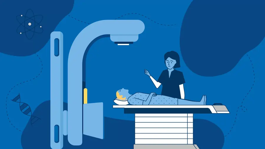
Radiation therapy can be administered within or outside of the body. The utter prevalent type is external beam radiation treatment. This therapy employs a big piece of equipment known as a linear accelerator. During the process, high-energy beams are directed from the gadget to a specific location on your body.
Brachytherapy is a type of internal radiation treatment. Brachytherapy is a frequent cancer treatment.
Radiation treatment harms cells by damaging their genetic information. Genetic material determines how cells develop and divide. Radiation treatment may cause harm to both healthy and malignant cells. However, healthy cells are more capable of self-repair than malignant cells. The goal of radiation therapy is to treat cancer with the lowest possible level of damage to healthy cells.
FAQs
Is radiation therapy safe?
Is radiation safe and effective? Yes, radiation is a well-known, safe, and efficient method of treatment. Every year, millions of people are safely treated with radiation to cure or reduce the symptoms of malignancies including head and neck, brain, breast, cervical, prostate, and skin cancer.
What are the side effects of radiation treatment?
Inquire with your healthcare staff about any side effects.
Sore skin. …
Tiredness. …
Hair loss. …
Feeling sick. …
Problems eating and drinking. …
Diarrhea. …
Stiff joints and muscles. …
Sex and fertility issues.
How does a patient’s life proceed following radiation therapy?
Don’t be shocked if you get fatigued, lose energy, or feel weak over time. It might take some time or several weeks for you to get more accurate after treatment. You may need to speak with your employer about changing your schedule, lowering the number of hours you work, or working from home (if possible).
How long does radiation therapy take?
You will be in the treatment room for approximately 15 to 30 minutes for each external radiation therapy session, but only one to five minutes of that time will be spent receiving radiation.
Which is safer, chemo or radiation?
Radiation treatment is administering high dosages of radiation beams directly into a tumor. The tumor shrinks or dies as a result of the ionizing radiation altering its DNA structure. Because it only targets a single area of the body, this curative therapy has fewer side effects than chemotherapy.
References
- Radiation therapy – Mayo Clinic. https://www.mayoclinic.org/tests-procedures/radiation-therapy/about/pac-20385162
- Radiation therapy. Wikipedia. https://en.wikipedia.org/wiki/Radiation_therapy
- Ways where Cancer Is Processed Using Radiation Therapy. (n.d.). American Cancer Society. https://www.cancer.org/cancer/managing-cancer/treatment-types/radiation/basics.html
- What to know about radiation therapy. https://www.medicalnewstoday.com/articles/types-of-radiation-therapy#types