Pronator Quadratus Muscle
Introduction
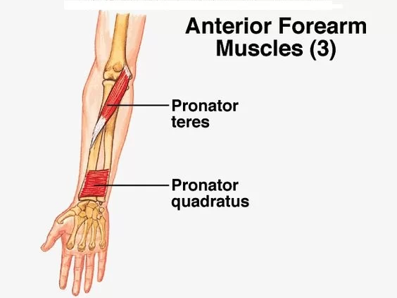
The pronator quadratus is a quadrangular, thin, short, and flat muscle located in the anterior compartment of the forearm. It is a member of the deep group of forearm flexors, together with flexor digitorum profundus and pollicis longus. The superficial group of forearm flexors overlays these three muscles.
The pronator quadratus connects the distal regions of the radius and ulna. As the name suggests, the primary function of this muscle is forearm pronation.
Structure
Its fibres are perpendicular to the arm’s path, travelling from the most distal quarter of the anterior ulna to the distal quarter of the radius. It has two heads: the superficial head emerges from the anterior distal portion of the ulna’s diaphysis (shaft) and inserts into the radius’s anterior distal diaphysis and metaphysis.
The deep head has the same origin, but it inserts closer to the ulnar notch. It is the only muscle that connects the ulna at one end and the radius at the other. The anterior interosseous artery carries arterial blood.
Origin
The pronator quadratus is a flat, short, quadrilateral muscle that originates on the anterior aspect of the distal shaft of the ulna and is partially covered by an aponeurosis.
Insertion
Superficial muscle fibres extend laterally and distally to the anterior surface of the distal shaft of the radius, where they also insert. Deeper fibres enter superiorly into the ulnar notch of the radius.
Blood supply
The anterior interosseous artery, which originates in the common interosseous artery, supplies arterial blood to the pronator quadratus. The latter is a branch of the ulnar arteries.
Nerve Supply
The anterior interosseous nerve, a branch of the median nerve, innervates the pronator quadratus muscle.
The anterior interosseous artery and the anterior interosseous nerve pass through the forearm’s interosseous membrane, which is situated between the ulna and radius bones. The anterior interosseous nerve originates from the median nerve at elbow level.
Along with other forearm muscles and structures, including the flexor pollicis longus and the radial part of the flexor digitorum profundus, the nerve nourishes the pronator quadratus muscle.
The electrical impulses that enable the muscle to contract and cause forearm pronation are sent via the nerve.
Function
The pronator quadratus causes forearm pronation by acting on the radioulnar joints. During this action, the head of the radius pivots around the ulna, rotating the palm posteriorly or inferiorly, depending on whether the forearm is flexed. Pronator quadratus motion is assisted by pronator teres and brachioradialis muscles.
This muscle’s position across the distal forearm suggests that it has a protective role. When weight-bearing activities require upward pressure, the pronator quadratus keeps the distal extremities of the radius and ulna together, protecting and stabilising the distal radioulnar joint. It also protects the interosseous membrane during forceful and fast forearm rotations by dispersing the stresses acting on it.
Examination
A combination of physical examination and imaging investigations is usually used to diagnose a pronator quadratus muscle injury or disease. The following are some typical techniques for identifying pronator quadratus muscle issues:
Physical examination:
A medical professional could examine the forearm physically to look for signs of weakness, discomfort, and soreness.
They could also evaluate the stability of the distal radioulnar joint and test the patient’s forearm rotation.
Imaging studies:
The pronator quadratus muscle and associated components can be seen using imaging tests including MRI scans, ultrasounds, and X-rays.
These investigations can assist in locating tendon ruptures, muscle rips, and other anomalies that might be contributing to symptoms.
Electromyography (EMG):
EMG is a diagnostic examination that measures nerve and muscle electrical activity.
It may be utilised to evaluate the pronator quadratus muscle’s function and detect any dysfunction or injury to the nerves.
Provocative tests:
To evaluate the pronator quadratus muscle’s function, a medical professional may use particular provocative tests.
The patient is asked to oppose the examiner’s effort to rotate their forearm as part of the resisted pronation test, for instance.
A pronator quadratus muscle issue might be indicated by pain or weakness during this test.
When diagnosing issues with the pronator quadratus muscle, a medical professional would often use a thorough process that includes the patient’s medical history, symptoms, physical examination results, and any pertinent imaging or diagnostic testing.
Clinical Importance
Myofascial Trigger point (TrP)
The Pronator Quadratus muscle has two major referred pain types of the TrP. Pain typically extends to the medial part of the forearm, both distally and proximally. In many cases, the discomfort travels proximally to the medial epicondyle and distally to the fifth digit of the hand. The pain also extends to the third and fourth fingers, which is the second most prevalent pattern.
Pronator Spasticity
Neurolytic drugs such as botulinum toxin, phenol, or alcohol are injected into the motor point of the pronator quadratus to treat pronator spasticity in stroke patients.
Anterior Interosseous Nerve (AIN) lesions
The electrophysiologic diagnosis of AIN lesions is performed utilising the Pronator Quadratus muscle, which is the essential muscle for this diagnostic technique.
Distal Radius Fracture
Volar plating is the most common approach for treating this fracture, and the consequence is flexor tendon rupture, which can be avoided by mending the pronator quadratus muscle during surgery. Fixing the distal radius fracture while retaining the pronator quadratus muscle can result in greater pronation movement, radioulnar joint stability, a strong grip, and pain relief, all of which lead to improved wrist function in the early postoperative period.
Carpal tunnel syndrome
The median nerve is squeezed as it travels through the wrist in this situation.
It may impact the pronator quadratus muscle and result in hand and wrist discomfort, numbness, and weakness.
Tennis elbow
The tendons on the outside of the elbow are impacted by tennis elbow, a typical overuse condition. The pronator quadratus muscle may be impacted by the forearm discomfort and weakness it causes.
Radial tunnel syndrome
This kind of nerve compression condition can also result in wrist and forearm discomfort and weakness.
It happens when the radial nerve is squeezed as it travels through the pronator quadratus muscle and other forearm muscles.
Pronator quadratus muscle pain
Overuse, repetitive wrist motions, or strain—often observed during typing, lifting, or racket sports—can cause Pronator quadratus muscle pain. Deep, agonizing pain close to the wrist and trouble rotating the forearm are common symptoms.
Treatment
Stretching
When the pronator quadratus muscle is tight, it might mimic Carpal Tunnel Syndrome. Many musicians maintain their forearms in pronation for extended periods, resulting in muscular tension.
For a right-hand Pronator Quadratus stretch, supinate your right hand (little finger) towards the sternum. Press the right hand towards the abdomen with the other hand (left) to enhance wrist flexion, and press the right elbow forward if supination is severe. The stretch should include wrist flexion and supination movements.
Wrist Supination Stretch
- With your arm extended in front of you, palm down, you can either sit or stand.
- Gently move your palm upward (supination) with your other hand.
- Repeat two to three times for each arm after holding for 20 to 30 seconds.
Active Supination Stretch with Stick
- With your elbow bent at a 90-degree angle, hold a light stick or hammer in your hand.
- Rotate your forearm slowly so that the palm is facing up.
- Repeat two to three times on each side after holding for 20 seconds.
Forearm Stretch Against a Wall
- With your fingers pointed downward, place your hand on a wall.
- To extend the forearm, gently press your hand against the wall.
- Hold for 20–30 seconds, then do it two or three times.
Strengthening
Muscles can be strengthened with theraband, flex-bars, weight cuffs, and dumbbells. First, supinate the hand and grip the end of the flex-bar before pronating the forearm. Resistance is utilised based on the state of the patients/clients.
Pronation/supination with resistance band:
To begin, fasten a resistance band to a fixed item, such as an anchor point or doorknob.
With your elbow bent at a 90-degree angle and your palm pointing down, grasp the opposite end of the band.
Keeping your elbow still, slowly turn your wrist and forearm so your palm is facing up.
After a few seconds of holding this posture, carefully go back to the beginning position, palm down.
After ten to fifteen repetitions, switch to the opposite arm.
Wrist curls with dumbbell:
With your elbow resting on a bench or other sturdy surface and your palm facing up, hold a dumbbell in your hand.
Curl your wrist upward to elevate the weight, and then slowly drop it back down.
After ten to fifteen repetitions, switch to the opposite arm.
Forearm twists with resistance band:
A resistance band should first be fastened to a stationary object.
With your elbow bent at a 90-degree angle and your palm pointing down, grasp the opposite end of the band.
Turn your wrist so that your palm is facing up, then turn it back to the beginning position so that it faces down.
After ten to fifteen repetitions, switch to the opposite arm.
Finger extensions with rubber band:
With your palm pointing down, wrap a rubber band over your thumb and fingers.
Stretch the band by slowly extending your fingers, then return to the beginning position. Repeat several times.
Starting with a lightweight or resistance band is crucial, and as the muscle gets stronger, progressively increase the weight or resistance. Before starting any new fitness regimen, speak with a licensed physical therapist or other healthcare professional, particularly if you have a history of wrist or forearm pain or injury.
FAQs
What does the pronator quadratus do?
Functions. The pronator quadratus produces forearm pronation by acting on the radioulnar joints. During this movement, the head of a radius pivots around the ulna, turning the palm posteriorly or inferiorly if the forearm is flexed. The pronator teres and brachioradialis muscles support this pronator quadratus motion.
What causes pronator quadratus pain?
Compression of the median nerve at the elbow can cause discomfort and numbness in the distal distribution, as well as weakness in the flexor pollicis longus, flexor digitorum profundus of the index finger, and pronator quadratus.
What is the origin and insertion point of the pronator?
The pronator teres muscle originates from the medial epicondyle and medial supracondylar ridge of the humerus and inserts on the lateral side. It helps in forearm pronation and elbow flexion on the surface of the radius.
Here’s a more detailed breakdown:
Origin- Humeral Head: The medial supracondylar ridge of the humerus.
Ulnar Head: Coronoid process of the ulna.
Insertion: Lateral surface of the radius (near the centre).
Action: Pronation of the forearm at the proximal radioulnar joint helps to flex the forearm at the elbow joint.
Innervation: Median nerve (C6 and C7)
How do you treat pronator quadratus injury?
Some of the nonsurgical pronator syndrome treatments your specialist may first recommend include the following:
Rest and Activity Modification.
NSAIDs (e.g., Motrin, Advil, Aleve)
Corticosteroids (to Reduce Inflammation and Ease Discomfort)
Splints.
What is the pain pattern of pronator quadratus?
There were two major pain patterns identified. Pain extended distally and proximally from the injection site along the forearm’s medial side was the most prevalent pattern (57%). In half of these patients, the discomfort spread to the medial epicondyle proximally and the fifth digit distally.
References
- Pronator quadratus muscle. (2023, September 19). Kenhub. https://www.kenhub.com/en/library/anatomy/pronator-quadratus-muscle
- Wikipedia contributors. (2024h, December 18). Pronator quadratus muscle. Wikipedia. https://en.wikipedia.org/wiki/Pronator_quadratus_muscle
- Thakkar, D. (2023a, March 28). Pronator quadratus muscle – origin, insertion, function- samarpan. Samarpan Physiotherapy Clinic. https://samarpanphysioclinic.com/pronator-quadratus-muscle/