Olfactory nerve
Introduction
The first and smallest cranial nerve is the olfactory nerve. This nerve is one of two that do not connect to the brainstem. Your olfactory nerve anatomy is part of the central nervous system.
The process begins in the brain and ends in the upper nose. It is also part of your autonomic nervous system, which controls bodily functions.
The olfactory nerve, or first cranial nerve, is responsible for your sense of smell. The olfactory nerve is composed of multiple sensory nerve fibres. This nerve is essential for using your senses to appreciate your favourite smells.
Each nostril contains approximately six to ten million olfactory sensory neurons. That means there’s a lot of work going on inside your nostrils to help you smell your surroundings or the food you’re eating.
Anatomical Course of Olfactory nerve
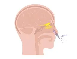
The anatomical course of the olfactory nerve describes how special sensory information is transmitted from the nasal epithelium to the brain’s primary olfactory cortex.
Nasal epithelium
Olfactory receptors are found in the nasal epithelium and detect smell. These axons (fila olfactoria) form small bundles of true olfactory nerves that travel by small foramina within the cribriform plate in the ethmoid bone into the cranial cavity.
Olfactory Bulb
Once inside the cranial cavity, the fibres enter the olfactory bulb, which is located in the olfactory groove of the anterior cranial fossa.
The olfactory bulb is an ovoid structure made up of specialised neurons known as mitral cells. The olfactory nerve fibres synapse with mitral cells, forming synaptic glomeruli. Second-order nerves originate in the glomeruli and travel posteriorly into the olfactory tract.
Olfactory Tract
The olfactory tract travels posteriorly along the inferior surface of the frontal lobe. In the tract approaches the anterior perforated material (at the level of the optic chiasm), it splits in medial and lateral stria:
- The lateral stria connects axons to the primary olfactory cortex in the temporal lobe’s uncus.
- The medial stria transports axons from the anterior commissure’s medial plane to the olfactory bulb on the opposite side.
Numerous other brain regions, such as the piriform cortex, amygdala, olfactory tubercle, and secondary olfactory cortex, receive nerve fibre transmission from the primary olfactory cortex. These areas are involved in the memory and perception of olfactory stimuli.
Function of Olfactory nerve
CN 1 It allows you to detect scents, odours, aromas, and more. Smelly substances emit small molecules. Inhaling transports these molecules to your nose. These molecules are detected by specialised cells called olfactory receptors. The receptors send this information to your brain via the olfactory nerve, allowing you to perceive smell.
The olfactory system provides a sense of smell in two ways:
- Nostrils: Smelly substances emit small molecules that can stimulate olfactory receptors. Receptors work in specific combinations, allowing you to recognise various types of smells.
- Back of your throat: Chewing food or sipping a drink releases molecules that aid in smell. These molecules travel up your throat to the olfactory receptors at the back of your nose.
The olfactory mucosa contributes significantly to your ability to smell. This membrane is located in the upper part of your nasal cavity and contains a variety of cells.
- Olfactory receptor cells support two processes: the dendritic process and the central process. Dendritic processes direct cells to tiny hairs in the olfactory mucosa, where they stimulate olfactory cells. Central processes move cells in the opposite direction.
- Sustentacular cells provide support for nearby tissue.
- Basal cells are the precursors to olfactory receptor cells and sustentacular cells.
Embryology
Embryologically, the face develops from five swellings derived from the first and second pharyngeal arches known as facial prominences that appear in the fourth week of development. These include the frontonasal prominence, as well as the mandibular and maxillary prominences. The olfactory placodes, a thickened ectoderm area on each side of the frontonasal prominence, appear around the fourth week of embryonic development.
The olfactory placodes grow in size until the sixth week when the centres of each placode invaginate to form the nasal pits. The nostril pits ultimately develop into the olfactory epithelium, which houses olfactory nerves and is divided into two nasal processes: medial and lateral. The nose, philtrum, and primary palate then develop.
Conditions and Disorders Related to Olfactory Nerve
Symptoms of Olfactory nerve
- Anosmia is the complete loss of smell.
- Dysosmia, also known as phantosmia, is the spontaneous occurrence of unpleasant or strange odours.
- Hyposmia is a partial loss of smell.
- Parosmia refers to a distorted sense of smell. For example, familiar foods could smell like chemicals or mould.
Causes of Olfactory nerve
The following medical conditions and situations may affect olfactory nerve function:
- Sinusitis and nasal polyps.
- Tobacco use.
- Poor oral hygiene.
- Environmental toxins and chemicals, such as insecticides.
- Severe brain injuries, including concussions.
- Medications such as antibiotics.
- Head and neck cancer.
- Diabetes.
- Alzheimer’s disease.
- Brain tumour.
- Parkinson’s disease.
- Epilepsy.
Post-viral olfactory loss
The common cold is the most common condition that affects the olfactory nerve, but other viral illnesses can cause the same effect.
You’re probably aware that when nasal congestion fills your sinuses, it can cause a loss of smell that returns once the congestion has cleared.
Still, it might take a while to heal completely. This is known as post-viral olfactory loss (PVOL), and everyone has likely experienced it at some point. Researchers aren’t sure why this happens, but they suspect it’s because certain viruses, such as the common cold and influenza, cause damage to the mucous membrane and olfactory epithelium.
Post-traumatic olfactory loss
A head injury can cause anosmia or hyposmia, also known as post-traumatic olfactory loss (PTOL). The loss is related to both the severity of the injury and the part of the head that was damaged. Injuries to the back of the head are most likely to cause loss of smell.
That may seem odd given that the olfactory nerves are located in the front of the brain. The brain may thrust forward and strike the inside front of the skull, precisely where the olfactory nerve is located, in the event of a blow to the back of the head. Then, as the brain recovers, it tugs on the delicate nerve fibres, which can snag on the rough edges of the tiny holes in the skull through which they emerge.
The olfactory nerves can be severed in this way, but most smell loss is caused by bruising of the olfactory bulb.
PTOL can also result from facial trauma, such as a blow to the nose.
Assessment
The olfactory nerve can easily be assessed at the bedside or in a clinic. To test the integrity of the olfactory nerve, pinch or block one nostril while the patient is blindfolded or with the eyes closed, and then have the patient smell aromatic materials like coffee, cinnamon, vanilla, and more.
Avoid using strong or noxious-smelling substances like alcohol or ammonia. Request that the patient identify the substance by describing its smell. Next, continue the same action on the other nostril. This is done to compare the ability to smell on both sides.
Treatment
Medical treatment
Corticosteroids
The most common treatment for OD, especially after upper respiratory tract infection, is topical and oral corticosteroids, which are effective in around 25-50% of cases. The administration of nasal spray is less effective than oral and nebulized treatments, respectively.
Antioxidant
Oxidative stress, caused by a disruption in the balance of reactive oxygen species and protective antioxidants, is a major pathogenic factor in a wide range of diseases, including viral infections.
Zinc
Zinc, an important immune trace element, has a variety of effects on immune response and infection.
Intranasal insulin.
Insulin has been found to play a direct role in the modulation of olfactory signals. The OB has a high concentration of central insulin receptors, so central insulin is converted there. Increased central insulin resistance is linked to a variety of OD-causing diseases.
Intranasal insulin works nicely in treating postinfectious OD because it has no side effects, does not increase blood glucose levels, and enhances olfactory function.
Nasal saline irrigation.
Nasal saline irrigation is a traditional method of respiratory or nasal care. Nasal saline rinsing removes dust particles, allergens, and air pollutants from the nasal cavity, improving mucosal ciliary oscillation, reducing mucosal edoema, promoting local blood circulation, and improving mucosal clearance.
A saline wash also hydrates the mucosal mucus layer, raises the rate of ciliary oscillation, and decreases the production of local inflammatory mediators, all of which are particularly beneficial to enhancing mucociliary dysfunction and mucus stagnation caused by viral infections.
Both hypertonic and isotonic saline can reduce nasal inflammation of the mucous membrane and help heal damaged nasal mucociliary epithelium. Multiple research studies have shown that nasal saline irrigation can effectively improve the symptoms of rhinitis and sinusitis caused by bacteria or viruses, including OD.
Surgical Treatment
Surgical treatments such as septoplasty, rhinoplasty, and endoscopic sinus surgery aim to eliminate
Nasal obstruction and the removal of inflamed mucosa or polyps. Secondary benefits of these surgeries may include improved olfactory function. Delank and Stoll found that surgery improved olfactory function for 25% of hyposmia and 5% of anosmia.
A longitudinal MRI study found that patients with CRSwNP had increased olfactory bulb volume following endoscopic sinus surgery. Nasal surgery can occasionally result in olfactory loss due to synechia, crusting, and damage to the epithelium, despite being generally effective.
For severe and debilitating unilateral phantosmia, selective olfactory bulb removal or endoscopic olfactory mucosa removal can be performed on the affected side.
Olfactory training.
Olfactory training may enhance olfactory function, despite limited research. Olfactory receptor neurons regenerate after functional deficits and olfactory cues
may influence this regenerative process. Olfactory training is a 12-week programme where patients are exposed twice daily to four strong odours (rose, eucalyptus, lemon, and cloves).
Summary
The olfactory nerve, the first and smallest cranial nerve, is responsible for the sense of smell. It is part of the central nervous system and helps detect scents, odours, and aromas. The nerve is composed of multiple sensory nerve fibers and is responsible for detecting smells in the nose and throat. The olfactory system provides a sense of smell through the nose and the back of the throat.
The olfactory mucosa contributes significantly to the ability to smell. Symptoms of olfactory nerve disorders include anasmia, dysosmia, hyposmia, and parosmia. The olfactory nerve is involved in the autonomic nervous system, which controls bodily functions.
Olfactory nerve function can be affected by various medical conditions and situations, including sinusitis, tobacco use, poor oral hygiene, environmental toxins, severe brain injuries, medications, head and neck cancer, diabetes, Alzheimer’s disease, brain tumours, Parkinson’s disease, and epilepsy. Post-viral olfactory loss (PVOL) occurs when the common cold or influenza causes damage to the mucous membrane and olfactory epithelium. Post-traumatic olfactory loss (PTOL) occurs when a head injury causes anosmia or hyposmia. The olfactory nerve can be assessed using aroma tests.
Treatment options include corticosteroids, antioxidants, zinc, intranasal insulin, nasal saline irrigation, surgical treatments like septoplasty, rhinoplasty, endoscopic sinus surgery, and olfactory training. However, these treatments may sometimes result in olfactory loss due to synechia, crusting, and damage to the epithelium.
FAQs
What caused damage to the olfactory nerve?
The olfactory nerve (ON) is the only cranial nerve that is exposed to the surrounding environment. As a result, it is susceptible to damage from head trauma, viral infection, inflammatory stimulation, and chemical toxins, all of which can cause olfactory dysfunction.
What kind of sense is olfactory?
Olfaction, or the sense of smell, detects and discriminates between odours as well as social cues, which influence our innate responses.
What is the treatment for the olfactory nerve?
The current best evidence for treating one common type of olfactory loss, rhinosinusitis, is systemic and/or topical corticosteroid therapy. If no other intervention is provided, an oral steroid taper can provide initial but usually temporary relief in these patients.
What olfactory nerve is responsible for smell?
The first cranial nerve (CN I) is the olfactory nerve. It is also part of your autonomic nervous system, which controls bodily functions. This nerve activates your sense of smell. Cranial nerve 1 is the shortest sensory nerve in the body.
What are olfactory cells?
The olfactory epithelium is made up of several different types of cells. The most significant of these is the olfactory receptor neuron, a bipolar cell that transmits olfactory information centrally through a small-diameter, unmyelinated axon on its basal surface.
What is unique about the olfactory nerve?
The olfactory nerve, which emerges from the embryonic nasal placode, differs from cranial nerves in how it regenerates itself if damaged. The olfactory nerve is sensory and arises from the olfactory mucosa in the upper part of the nasal cavity.
References:
- Dellwo, A. (2021, August 23). The Anatomy of the Olfactory Nerve. Verywell Health. https://www.verywellhealth.com/olfactory-nerve-anatomy-4686024
- Helwany, M., & Bordoni, B. (2023, August 14). Neuroanatomy, Cranial Nerve 1 (Olfactory). StatPearls – NCBI Bookshelf. https://www.ncbi.nlm.nih.gov/books/NBK556051/
- Olfactory Nerve. (n.d.). Physiopedia. https://www.physio-pedia.com/Olfactory_Nerve
- The Olfactory Nerve (CN I) – Pathway – Anosmia – TeachMeAnatomy. (2018, December 24). TeachMeAnatomy. https://teachmeanatomy.info/head/cranial-nerves/olfactory-cni/
- Professional, C. C. M. (n.d.). Olfactory Nerve. Cleveland Clinic. https://my.clevelandclinic.org/health/body/23081-olfactory-nerve
- Hu, B., Gong, M., Xiang, Y., Qu, S., Zhu, H., & Ye, D. (2023, November 17). Mechanism and treatment of olfactory dysfunction caused by coronavirus disease 2019. Journal of Translational Medicine. https://doi.org/10.1186/s12967-023-04719-x

2 Comments