
Dorsal Scapular Nerve
Introduction
The dorsal scapular nerve originates from the root of spinal nerve C5, which is one of the brachial plexus’s lateral branches. On rare occasions, it could originate from the brachial plexus’ superior trunk.
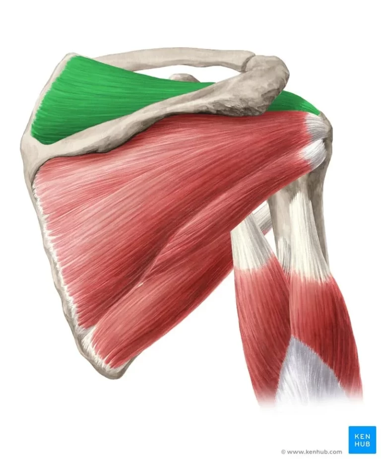
The dorsal scapular nerve is a motor neuron that innervates the levator scapulae, rhomboid major, and rhomboid minor muscles. These muscles work together to raise and lower the scapula.
Structure
The dorsal scapular nerve is a motor neuron that innervates the levator scapulae, rhomboid major, and rhomboid minor muscles. These muscles are regarded as periscapular stabilizing muscles and function both collectively and dynamically. They have the ability to raise and retract the scapula on their own.
Although cadaveric and anatomical investigations have indicated varying contributions to the DSN from any or all combinations of C4 to T1 nerve roots, the dorsal scapular nerve lacks sensory fibers. Although proprioceptive fibers are part of the motor system and innervate muscles for proprioception, they are not formally considered sensory fibers.
For instance, proprioceptive fibers from cervical segments are found in the sternocleidomastoid and trapezius muscles; nevertheless, the spinal accessory nerve, the eleventh cranial nerve, does not strictly have a sensory component.
Function
The rhomboid major and rhomboid minor muscles receive motor innervation from the dorsal scapular nerve. It also nourishes the branches that emerge from spinal nerves C3 and C4, as well as the levator scapulae muscle. The levator scapulae raises the scapula, while the rhomboids pull it posteriorly medially towards the spinal column.
Course
The dorsal scapular nerve emerges from the proximal portion of the anterior ramus of the C5 spinal nerve, directly above the clavicle. It passes through the scalenus medius muscle and descends between the serratus posterior superior and scalenus posterior muscles on one side and the levator scapulae on the other. It then follows the anterior boundary of the rhomboid muscles over the medial scapular area.
The deep branch of the transverse cervical artery or the dorsal scapular artery typically accompanies the dorsal scapular nerve.
Muscle Supply
The levator scapulae muscle attaches to the superior angle of the scapula after beginning at the transverse processes of C1–C4. When applied to the scapula, this muscle pulls upward and marginally aids in a downward rotation. The cervical spine can also be drawn towards the same side shoulder with the help of the levator scapulae.
The spinous processes of T1-4 for the rhomboid major and C6-7 for the minor are the origin of the rhomboid major and minor muscles. Both muscles attach to the scapula’s medial edge; the rhomboid major usually inserts under the scapula’s spine, while the rhomboid minor usually inserts above it. These muscles work together to stabilize the scapula while the upper extremity is in functional use. The rhomboid major and minor can retract the scapula on their own.
Embryology
The anterior (ventral) main rami of C5 innervates the levator scapulae, rhomboid major, and rhomboid minor muscles, which are derived from a paraxial mesoderm origin. The cervical region’s anterior motor roots originate in the spinal cord’s basal plate.
Each cervical spinal neuron is formed by the union of these anterior roots with the sensory (posterior) roots, which are produced from neural crest cells. The spinal nerve swiftly splits into an anterior primary ramus and a posterior primary ramus after leaving the intervertebral foramen. The anterior ramus of the C5 spinal nerve gives rise to the dorsal scapular nerve.
Anatomical Variation
A dual innervation from the anterior rami of both C5 and C6 has also been observed frequently, and other variations of the dorsal scapular nerve innervation have been identified when It was discovered that this nerve and the long thoracic nerve share a connecting branch. Other variations of the dorsal scapular nerve innervation have been described as originating from a prefixed C4 anterior rami contribution joining with the C5 anterior ramus to form the dorsal scapular nerve.
Only 30% of the levator scapulae were innervated by the dorsal scapular nerve, according to a 1997 study of 30 cadaver neck dissections; the bulk of innervations originated in the cervical plexus (C3–4).
According to a 2016 cadaver research, in 48% of instances, the levator scapulae alone received innervation from the dorsal scapular nerve, and in 52% of samples, the levator scapulae and rhomboids received innervation from the same dorsal scapular nerve.
Examination
Examining the function of the rhomboids and levator scapulae can help detect DSN.
Clinical Importance
The most frequent cause of dorsal scapular nerve entrapment is middle scalene muscle hypertrophy. Clinical manifestations of DSN compression/entrapment neuropathy might include a variety of conditions:
- Pain
- Muscle spasms/tightness (involving the levator and/or rhomboids)
- Midscapular dysesthesia
Scapular winging caused by atrophic alterations and muscular denervation in a chronic context
By providing a firm platform to pull against during functional tasks like lifting the arm upward, a secure scapula benefits the entire upper extremity. Rhomboid muscles that are in good working order are crucial for scapular stability. According to a number of studies, a weak rhomboid often causes the scapula to flare out posteriorly due to mild winging and an inability to retain it firmly.
Entrapment of the dorsal scapular nerve is a frequent cause of rhomboid paralysis. The most common location for a dorsal scapular nerve injury is an entrapment connected to the middle scalene muscle. Patients with dorsal scapular nerve disease often have myofascial scapular pain syndrome.
The symptoms of these instances, which are often misdiagnosed, include discomfort on the scapula’s medial border and the potential for pain to radiate to the forearm and lateral arm. Clinicians must keep in mind the dorsal scapular nerve as a potential cause when patients report interscapular discomfort since the absence of a sensory distribution linked to this motor neuron makes it more difficult to link the two.
Surgical Importance
For patients with impaired pulmonary function, an anterior approach entails the danger of an iatrogenic phrenic nerve block, which might have major repercussions. As a result, patients with a history of severe COPD are frequently given an alternative anesthetic modality. A typical anesthetic technique for regional surgery encompassing the proximal humerus, shoulder joint, and lateral clavicle is an interscalene brachial plexus block.
This danger is reduced by the posterior approach, particularly when ultrasound guidance is used. The dorsal scapular nerve is encountered in 75% of patients in investigations of this ultrasound-guided treatment. This often affects not just the dorsal scapular nerve but also the long thoracic nerve (21%). There are also instances (24%), in which both nerves are affected at the same time.
FAQs
What are the symptoms of dorsal scapular nerve damage?
Signs and symptoms
Abnormal and/or reduced shoulder movement.
Pain in the lower neck, upper/mid back, and shoulder areas.
Winging of the shoulder blade (i.e.Tilting the blade away from the rib cage.)
Difficulty with drawing shoulders backward and together.
What is the mechanism of damage to the dorsal scapular nerve?
The etiology of damage to the DSN is likewise extremely broad ranging from postural to overuse in overhead labour and activity. Other disorders in this region, including CD, NP, SICK scapula, and a posterolateral arm pain pattern exhibit a remarkable similarity to DSN neuropathy.
Can the dorsal scapular nerve cause chest pain?
There may be concurrent anterior chest wall discomfort and tenderness along the sternocostal boundary, as well as poorly localized ipsilateral arm pain that is significantly more pronounced than the interscapular pain. DSN entrapment may manifest as an “atypical” thoracic outlet syndrome.
What causes scapular weakness?
Scapular dyskinesis can be caused by muscle weakness, imbalance, tightness, or even detachment (in rare cases). Damage to the nerves that feed the muscles. Injuries to the scapula’s supporting bones or to the shoulder joint itself.
Can stress cause scapular pain?
Stress can manifest in many ways, including pain between shoulder blades. When we are stressed, our muscles tense up, which can result in painful knots and spasms. As a response to stress, our bodies may also release certain hormones that can cause inflammation and discomfort in the back.
References
- Dorsal scapular nerve. (2023, November 3). Kenhub. https://www.kenhub.com/en/library/anatomy/dorsal-scapular-nerve
- Dorsal scapular nerve. (2023, July 30).StatPearls.https://www.ncbi.nlm.nih.gov/books/NBK459343/
One Comment