Cuboid Bone
Introduction
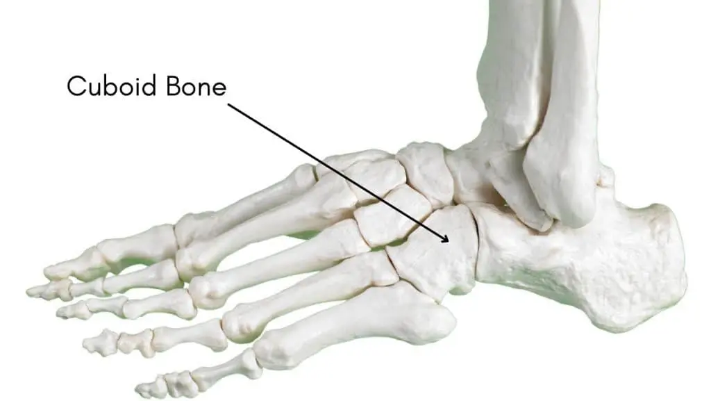
The cuboid bone is a cube-shaped tarsal bone located in the foot, playing a crucial role in the structure and function of the lateral arch. Situated on the outer side of the foot, it connects with the calcaneus (heel bone) posteriorly and the fourth and fifth metatarsal bones anteriorly.
It is a component of the midfoot and aids in giving the foot support and stability when moving. Cuboid syndrome, cuboid subluxation, and cuboid fractures are common disorders or injuries related to the cuboid bone. When the cuboid bone misaligns, it can lead to cuboid syndrome, which is painful and uncomfortable.
Cuboid syndrome is typically treated with physical therapy, immobilization, and rest to realign the bone. Repetitive stress or trauma can cause cuboid fractures, which can be treated with surgery, immobilization, or a mix of the two.
To guarantee an accurate diagnosis and course of treatment, it is crucial to get medical help if you think you may have a cuboid fracture or any other foot injury. The cuboid bone’s anatomy and function are covered in this article. It also discusses related conditions and potential treatment needs.
What is Cuboid Bone?
- There are seven tarsal bones on the exterior of the foot, and the cuboid bone is framed by just one of them. The foot and ankle are joined by this rectangular in shape bone.
- After the fourth and fifth (pinky) toes, this sophisticated cuboid bone sits between the metatarsals and the calcaneus, or heel bone.
- Additionally, it gives the foot stability. The cuboid acts as a pulley to help guide your foot downward and as a point of attachment for muscles. Additionally, it facilitates movement in the foot’s lateral (outside) column.
Anatomy of the Cuboid Bone
- The cuboid bone is a tiny, cube-shaped bone found in the foot. Outside the foot, between the calcaneus and the fourth and fifth metatarsals, is the cuboid, the smallest of the seven tarsal bones.
- There is a gap on the superior surface of the cuboid bone known as the peroneal sulcus. The tendons of the brevis and peroneus longus muscles are located in the gap. Plantarflexion, or pointing the foot downward, and eversion, or turning the foot outward, are assisted by the peroneus longus tendon, which passes through this groove. This groove is also traversed by the peroneus brevis tendon, which aids in the foot’s eversion.
- The cuboid bone’s rough lateral surface serves as attachment points for ligaments that provide foot stability when bearing weight. The calcaneocuboid ligament attaches the cuboid bone to the calcaneus, while the lateral plantar calcaneocuboid ligament connects it to the calcaneus’s lateral plantar side.
- The muscles and ligaments responsible for preserving the foot’s arches attach to this protrusion. Attached to this tuberosity is the plantar calcaneocuboid ligament, which maintains the foot’s longitudinal arch. The tibialis posterior muscle tendon also attaches to the tuberosity, which helps in arch stability and inversion (turning the foot inward).
- The navicular bone and the medial part of the cuboid bone have connected to create a joint. The midfoot region can move and be more flexible because of this joint, which is called the naviculocuboid joint. This joint is stabilized and the foot’s arches are maintained by ligaments such as the plantar calcaneonavicular ligament, sometimes referred to as the spring ligament.
Muscle attachment to the cuboid bone
Numerous muscles and tendons involved in foot movement and arch support join to the cuboid bone. These muscle attachments are essential to the foot’s general stability and functionality. The primary tendons and muscles that connect to the cuboid bone are as follows:
- Tibialis Posterior: The tibialis posterior tendon bonds to the cuboid bone’s tuberosity. This tendon travels along the back of the leg and ankle, starting from the posterior surface of the tibia, or inner lower leg bone. Supporting the arch and inversion, or bending the foot inward, depends on the tibialis posterior tendon. It offers stability when bearing weight and aids in maintaining the foot’s medial longitudinal arch.
- Flexor Digitorum Longus: The cuboid bone’s tuberosity is where the flexor digitorum longus tendon is also attached. This tendon exits the posterior region of the tibia and loops around the ankle and rear of the leg. Flexing (curling) the toes downward is the function of the flexor digitorum longus tendon, which also helps with foot stability and grip during walking and running.
- Flexor Hallucis Longus: Although it does not directly link to the cuboid bone, the flexor hallucis longus tendon runs next to it. This tendon begins at the rear surface of the fibula and travels along the back of the leg and ankle. The big toe is flexed (curled) downward by the flexor hallucis longus tendon, which also helps with balance and foot stability.
- Proper foot mechanics and function depend on these muscle attachments to the cuboid bone. When engaging in weight-bearing exercises, they enable stability, arch support, and controlled movement. Foot pain, instability, and changed gait patterns can result from damage or dysfunction to various muscles and tendons.
Blood supply of the Cuboid Bone
- Branches of the anterior, posterior, and peroneal tibial arteries are among the several sources of blood flow to the cuboid bone. These arteries give the bone oxygenated blood, which enables it to be maintained and fed.
- To deliver blood to the downward portion of the foot, the distal metatarsal arteries diverge from the anterior tibial artery. Additionally, these arteries branch off to supply blood to the dorsal portion of the cuboid bone.
- Blood transits from the posterior tibial artery to the somewhere It forms the lateral plantar artery, which provides blood to the plantar part of the bone, and ultimately branches to the cuboid bone.
- besides that, blood gets delivered to the cuboid bone via the peroneal artery. It produces branches that provide blood to the cuboid bone and the lateral side of the foot.
Lymph supply of the Cuboid Bone
- Along with artery supply, the cuboid bone receives venous drainage through corresponding veins. Alongside the arteries, these veins aid in the removal of blood that has lost oxygen from the bone.
- The cuboid bone also contains lymphatic veins. These vessels help remove waste materials and extra fluid from the bone tissue. The lymphatic vessels, like the blood vessels, pass through the bone.
- All things considered, the cuboid bone’s blood and lymphatic supply are essential to its health and functionality. While lymphatic drainage aids in maintaining a healthy environment by eliminating waste materials and extra fluid, adequate blood flow guarantees that the bone tissue is properly nourished and oxygenated.
Function of the Cuboid Bone
In the foot, the cuboid bone performs several crucial roles.
- Weight-bearing and stability: When engaging in weight-bearing activities like walking, running, and jumping, the cuboid bone aids in stabilizing the foot. It creates joints with the nearby bones, such as the navicular bone, fourth and fifth metatarsals, and calcaneus (heel bone). These joints preserve the foot’s structural integrity while enabling regulated movement.
- Muscle attachment: Several tendons and muscles involved in arch support and foot movement attach to the cuboid bone. On the superior surface of the cuboid bone, the peroneal sulcus is where the tendons peroneus longus, and brevis pass through. These tendons aid in eversion, or turning the foot outward, and plantarflexion, or pointing the foot downward. Attached to the cuboid bone’s tuberosity, the tibialis posterior tendon aids in inversion, or turning the foot inward, and supports the arch.
- Arch support: The cuboid bone helps to keep the foot’s arches—more especially, the longitudinal arch—strong. The longitudinal arch is supported by the plantar calcaneocuboid ligament, which is attached to the cuboid bone’s tuberosity. The naviculocuboid joint is stabilized and the foot’s arches are maintained by ligaments like the plantar calcaneonavicular ligament, commonly known as the spring ligament.
- Flexibility and joint movement: The cuboid bone joins with other bones, such as the navicular and calcaneus, to form joints. The midfoot area can move and be more flexible thanks to these joints, which include the calcaneocuboid and naviculocuboid. This facilitates shock absorption during exercises and enables adaption to uneven surfaces.
- Nerve and blood supply: The cuboid bone receives blood and nutrition from the posterior tibial artery’s subunits. The oxygen and nutrients required for the bone’s health and function are supplied by these blood veins. The cuboid bone is additionally innervated by nerves, which give the surrounding structures motor control and sensory data.
All things considered, the cuboid bone is essential for giving the foot support, stability, and mobility. Its many roles are influenced by its physical characteristics and interactions with nearby bones, muscles, ligaments, and blood vessels. To diagnose and treat illnesses or injuries that may impact the cuboid bone’s function in foot mechanics, it is critical to comprehend its functions.
Disorders affecting the cuboid bone
The cuboid bone may be impacted by several related disorders. These ailments can include everything from inflammatory disorders to catastrophic injuries. The following are a few of the most prevalent cuboid bone-related conditions:
- Cuboid syndrome: This condition is represented by the cuboid bone being misaligned or displaced. It is also referred to as cuboid subluxation or lateral plantar neuritis. Trauma, excessive use, or repeated foot stress can also cause this. Cuboid syndrome is characterized by swelling, soreness, and tenderness on the side of the foot, as well as trouble walking or bearing weight on the affected foot.
- Cuboid fracture: Cuboid fracture refers to a break or cracks in the cuboid bone. A direct impact or trauma to the foot, such as a fall or an accident sustained during sports, is typically the cause of this kind of fracture. Pain, swelling, bruising, trouble walking or bearing weight on the injured foot, and deformity are all signs of a cuboid fracture.
- Tarsal coalition: The term “tarsal coalition” refers to a disorder in which two or more foot bones, particularly the cuboid bone, fuse abnormally. This disease can exhibit appearances at birth or later in life. In the affected foot, tarsal coalition may result in discomfort, stiffness, and a restricted range of motion. It might also make you more likely to get other foot disorders like arthritis or flat feet.
- Arthritis: The development of inflammation and joint degradation are the hallmarks of arthritis. Two types of arthritis that can harm the cuboid joint include rheumatoid arthritis and osteoarthritis. Pain, stiffness, swelling, and trouble moving the foot are all symptoms of cuboid joint arthritis.
- Tendinitis: The inflammation of a tendon, the tissue that connects muscles to bones, is known as tendinitis. Overuse or recurrent stress on the foot can cause inflammation in the tendons surrounding the cuboid bone. Pain, edema, and trouble moving the foot may result from this.
- Stress fractures: Repetitive stress or overuse can result in these microscopic cracks or breaks in a bone. Stress fractures can occur in the cuboid bone, particularly in those who run or jump or perform other activities that cause repetitive contact on the foot. Pain, swelling, and soreness on the lateral side of the foot are signs of a stress fracture in the cuboid bone.
If you have any symptoms or suspect that you may have any related cuboid bone disorders, you should get medical help. A medicine expert can make a precise diagnosis and suggest suitable courses of action, such as rest, immobilization, physical therapy, medication, or, in extreme situations, surgery.
Treatment for Cuboid Bone Condition
Cuboid bone rehabilitation is contingent upon the degree of the damage and the particular underlying condition. An outline of the rehabilitation procedure for a few typical conditions is provided below:
- Cuboid syndrome: The goals of rehabilitation for cuboid syndrome are to improve foot function, reduce pain, and restore normal alignment. RICE (rest, ice, compression, and elevation) and the use of shoe inserts or orthotics to support the foot are possible treatment options. To increase stability and strengthen the muscles surrounding the foot, physical therapy activities are frequently recommended. Calf stretches, toe curls, ankle range-of-motion exercises, and balance training are a few examples of these activities.
- Cuboid fracture: To allow the bone to heal, the foot is usually immobilized with a cast, boot, or splint during the rehabilitation process. After the fracture has healed, physical therapy could be suggested to regain function, strength, and range of motion. This could involve moderate weight-bearing activities, foot and ankle muscle strengthening exercises, and mild stretching exercises.
- Tarsal coalition: Conservative treatments for tarsal coalition may include rest, immobilization with a boot or cast, and physical therapy to strengthen the surrounding muscles and increase the range of motion. To separate the fused bones and restore normal foot function, surgery can be required in certain situations. Pain control, wound healing, and a gradual return to weight-bearing activities will be the main goals of post-operative rehabilitation.
- Arthritis: The goals of cuboid joint rehabilitation are to lessen pain, increase joint mobility, and improve total foot function. Medication to control pain and inflammation, physical therapy exercises to increase joint strength and flexibility, assistive devices like braces or orthotics to support the foot, and lifestyle changes to lessen joint stress are some possible treatment choices.
- Tendinitis: To lessen discomfort and inflammation, rehabilitation for cuboid tendinitis requires rest, ice, compression, and elevation. Exercises used in physical therapy are frequently recommended to increase ankle and foot stability, strength, and flexibility. A gradual return to activities or sports, strengthening exercises for the foot and ankle muscles, and stretching exercises for the calf and plantar fascia are some examples of this.
- Stress fractures: Rest and immobilization are usually part of the rehabilitation process for cuboid bone stress fractures so that the bone can recover. To restore function, strength, and flexibility soon after the fracture has healed, physical therapy may be recommended. Gentle stretching techniques, increasing weight-bearing activities, and a gradual return to full activity or sports are a few examples of this.
For a customized rehabilitation plan based on your unique condition and requirements, you must speak with a doctor or physical therapist. To guarantee a secure and efficient recovery, they can offer advice on suitable exercises, activity adjustments, and rehabilitation progression.
Summary
The cuboid bone is a cube-shaped tarsal bone located in the foot, playing a crucial role in the structure and function of the lateral arch.
It aids in supporting and stabilizing the foot’s outside border. Walking is made easier by the muscle that is connected to the cuboid, which helps you point your foot downward. Cuboid fractures and cuboid syndrome are medical diseases that can impact the bone. Conservative care, such as physical therapy, is typically advised for both disorders. Surgery may be necessary for fractures in some situations.
FAQs
Can someone who has a cuboid fracture walk?
It may be sufficient to treat cuboid fractures with little pain or edema with an elastic bandage or a fracture boot and walk with some weight bearing until the symptoms have subsided to a reasonable degree. It is advised to wear a brief walking cast for four to six weeks if there is significant initial pain.
For what purpose is a cuboid used?
The cuboid acts as a pulley to help guide your foot downward and as a point of attachment for muscles. Additionally, it facilitates movement in the foot’s lateral (outside) column. Although cuboid fractures are rare, they can happen occasionally under certain conditions.
What does pain in your cuboid bone mean?
The cuboid bone may move too far as a result of some strong motions or extended postures, interfering with its natural position or motion. Immediate foot pain results from this, and it may get worse when standing or walking on the foot.
How much time does it take for a cuboid fracture to heal?
Pain management, altered activity, physical therapy, and a protective boot with arch support for four to six weeks with weight bearing as tolerated are the mainstays of treatment for a non-displaced cuboid fracture. Surgery and internal fixation with wires, plates, screws, and bone may be necessary for a displaced fracture.
References
- The Healthline Editorial Team. (2018b, January 24). Cuboid. Healthline. https://www.healthline.com/human-body-maps/cuboid-bone#2
- Wikipedia contributors. (2024b, August 18). Cuboid bone. Wikipedia. https://en.wikipedia.org/wiki/Cuboid_bone
- Ocs, T. P. D. (2023, August 21). Anatomy of the Cuboid Bone. Verywell Health. https://www.verywellhealth.com/cuboid-bone-anatomy-5089150
- Cuboid Bone | Complete Anatomy. (n.d.). www.elsevier.com. https://www.elsevier.com/resources/anatomy/skeletal-system/appendicular-skeleton/cuboid-bone/18696
- Patel, D. (2023b, July 11). Cuboid Bone – Anatomy, Function, Related Muscles. Samarpan Physiotherapy Clinic. https://samarpanphysioclinic.com/cuboid-bone-anatomy/

