Costovertebral joints
The costovertebral joints are synovial joints that connect the ribs to the thoracic vertebrae. Each rib articulates with the vertebral body at two points: the costocorporeal joint (between the rib head and the vertebral bodies) and the costotransverse joint (between the rib tubercle and the transverse process). These joints facilitate rib movement during respiration and provide stability to the thoracic cage.
Introduction
The term “costovertebral joints” refers to two sets of synovial plane joints that enclose the thoracic cage from the back by joining the proximal end of the ribs with their respective thoracic vertebrae.
- The head of the rib is connected to the vertebral bodies by articulating the rib head.
- The costotransverse junction joins the rib and the vertebral transverse processes.
The purpose of these motions is to allow the ribs to be raised and lowered during breathing. This ultimately leads to the thorax’s lateral diameter increasing and the lung parenchyma expanding as air is inhaled.
Anatomy
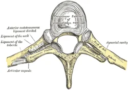
The head of the rib (a normal rib has two facets, each with a distinct synovial joint divided by a ridge) makes up the costovertebral joint. For every rib, the head articulates with:
- The upper costal facet of the same vertebra connects with the lower rib facet
- The upper facet connects with the lower facet of the vertebral body situated above.
- The first rib connects exclusively to the T1 vertebra, while the three lowest ribs connect solely to their respective vertebral bodies.
Structure
Costovertebral joints, which are synovial in nature, link the ribs with the vertebral column. They are essential for the movement of the rib cage when breathing. These joints are made up of two primary articulations:
Joints of heads of ribs (costocorporeal joints)
Articular surfaces
The costocorporeal joints link the concave costal facets of thoracic vertebrae with the convex articular facets located on the heads of all ribs;
- There are two costal facets on vertebrae T1–T10: an inferior and a superior one. Often called costal demi facets, these facets are broken by the superior or inferior borders of the vertebral body.
- The superior costal facet of the numerically comparable vertebra and the inferior costal facet of the vertebra directly above it are the two adjacent vertebrae that the heads of ribs 2–9 articulate with (For instance, rib 2 articulates with T1 and T2 vertebrae). As a result, the ribs touch the intervertebral disc between the appropriate vertebrae.
- One continuous costal facet on each thoracic vertebra (T1, T10, T11, and T12) accommodates ribs 1, 11, and 12.
The simple synovial joints are the first, tenth, eleventh, and twelfth costocorporeal joints. The complicated and compound joints are those that have intra-articular ligaments that divide the synovial cavity into two sections. These are joints 2 through 9.
Ligaments and joint capsule
A fibrous capsule encloses costocorporeal joints, which are further strengthened by intra-articular, radiating, and capsular ligaments. Every joint has a fibrous capsule that runs between the edges of the vertebral and costal articulating surfaces. The costotransverse ligaments merge with the back edges of fibrous capsules.
From the anterior surface of each rib head to the nearby vertebral bodies and intervertebral disc, radiate ligaments extend. They extend in three different directions.
- The superior connection between the rib and the vertebra directly above
- Horizontally, to attach the IV disc to the rib head
- Secondly, attach the rib to the appropriate thoracic vertebra (rib 2 to vertebra T2).
The second to tenth complicated costocorporeal joints are affected by this. The radiate ligaments are what attach the first, eleventh, and twelfth ribs to the appropriate vertebra and the vertebra above.
The only complicated costocorporeal joints that have intra-articular ligaments are those of the ribs 2–9. This ligament extends horizontally from the crest on the rib head to the nearby intervertebral disc in each joint. The superior and inferior compartments of the joint cavity are thus separated by the intra-articular ligaments.
Costotransverse joints
The connections between the articular facet on the rib’s tubercle and the transverse process of its numerically equal vertebra are known as costotransverse joints. The only ribs that engage in these joints are the top 10, which articulate with the corresponding vertebrae’s transverse processes.
The costotransverse joints are absent on T11 and T12 levels because the eleventh and twelfth ribs lack articular surfaces and tubercles.
Articular surfaces
The connections between the articular facet on the rib’s tubercle and the transverse costal facet on the vertebra’s transverse process at the same numerical level are known as costotransverse joints.
On the tubercles of ribs 1–6, the articular surfaces are vertically convex, while on the transverse processes, the corresponding costal facets are concave. The articular facets of ribs 7–10 are orientated posteriorly and inferomedially, facing the superior surfaces of the transverse processes, and are almost flat.
Ligaments and joint capsule
Attached to the edges of the articular facets are the fibrous capsules that encapsulate costotransverse joints. Each capsule’s inner surface is lined with a synovial membrane that encloses the joint’s synovial cavity.
The costotransverse, superior and lateral costotransverse, and accessory ligaments all serve to support the joints:
- The costotransverse ligament fills the little space between the vertebral transverse process and the rib neck. From the rear of the rib neck to the anterior surface of the neighboring transverse process, its numerous short, horizontally oriented fibers make this link.
- The superior surface of the rib neck and the inferior surface of the transverse process of the vertebra directly above are joined by the superior costotransverse ligament. Except for the first costotransverse joint, it is present in all of them. The external intercostal muscle fibers divide the ligament’s two layers, which are anterior and posterior. The course of the posterior layer is superomedial, whereas the route of the anterior layer is superolateral. The posterior layer unites with the external intercostal muscle, while the anterior layer fuses with the internal intercostal membrane on their spinal attachments.
- The transverse process’s tip and the lateral, non-articular portion of the rib tubercle are joined by the lateral costotransverse ligament.
- Located medially to the superior costotransverse ligament, an accessory ligament is isolated from it by the thoracic spinal nerve’s posterior ramus.
Innervation of the Costovertebral Ligaments
The posterior rami of spinal nerves C8–T11 innervate both kinds of costovertebral joints through their lateral branches.
- There are segments in the innervation.
- Both the level above and a spinal nerve of its numerically comparable level supply the fibers to each joint.
Blood supply
The First-10th posterior intercostal arteries and the supreme intercostal arteries are the branches of the thoracic aorta that supply the costovertebral joints.
Movements
The motions occurring at these joints are referred to as ‘pump-handle’ or ‘bucket-handle’ movements, and they involve only a small degree of gliding and rotation of the rib head.
- These movements serve to allow the ribs to be lifted upward and outward while breathing.
- Ultimately, this leads to an increase in the thorax’s lateral diameter and an expansion of the lung parenchyma during inhalation.
An important element of the biomechanics of chest wall movement is the costovertebral complex. The actions of the costovertebral joints and intervertebral movement are made possible by the costovertebral ligaments. The ligaments serve to:
- At the costovertebral joint, affix and stabilize the ribs on the thoracic vertebra while permitting some movement. They assist in the load-bearing, protective, postural, and scaffolding functions of the thoracic cage due to its stabilizing properties.
- Permit and restrict movement of the ribs at the transverse joint to enable maximum expansion of the thoracic cavity as required for respiratory demand. Their behavior regarding the costovertebral and intervertebral complexes permits lateral bending and axial rotation.
Muscles That Act on the Costovertebral Joints
The muscles responsible for breathing are the main movers of the costovertebral joints.
- Respiratory diaphragm
- Intercostal muscles.
All muscles that are connected to the ribs and classified as accessory respiratory muscles can, however, move these joints;
Sternocleidomastoid, scalene, serratus anterior, pectoralis major, pectoralis minor, latissimus dorsi und serratus posterior superior.
Clinical Significance
The costovertebral joints, which are articulations between the vertebral column and the ribs, are essential for spinal movement, respiratory mechanics, and thoracic stability. Among their clinical significance are:
1. Respiratory Function
- During respiration, these joints permit rib movement (such as pump-handle and bucket-handle motions).
- Breathing difficulties and ailments such as respiratory insufficiency in chronic diseases (e.g., COPD, ankylosing spondylitis) can result from dysfunction.
2. Pain Syndromes
- Costovertebral joint dysfunction: Often misdiagnosed as thoracic spine problems or cardiac pain, this condition can cause localized pain close to the spine and ribs.
- Sharp, localized pain that gets worse with deep breathing or movement is caused by rib subluxation.
- Referred pain: Pain that mimics other conditions and radiates to the chest, back, or abdomen can be a sign of dysfunction.
3. Trauma & Fractures
- Costovertebral joint disruption from rib fractures can cause pain and compromised lung function.
- Whiplash injuries can result in a strain on the costovertebral joint, which can lead to chronic pain after trauma.
4. Arthritis & Degeneration
- Osteoarthritis: Common in the elderly, this condition causes pain, stiffness, and decreased thoracic mobility.
- Fusing these joints can result from ankylosing spondylitis, which can cause severe spinal stiffness and restrictive lung disease.
5. Postural & Mechanical Issues
- Chronic pain and dysfunction can result from changes in joint biomechanics caused by kyphosis, scoliosis, or poor posture.
6. Medical Procedures & Considerations
- Costovertebral joint integrity must be taken into account during thoracic surgeries (such as rib resections and spinal surgeries).
- Nerve involvement: Joint inflammation may be a contributing factor to disorders such as complex regional pain syndrome (CRPS) due to its proximity to sympathetic nerves.
FAQs
Costovertebral joints: what are they?
The term “costovertebral joints” refers to two sets of synovial plane joints that enclose the thoracic cage from the back by joining the proximal end of the ribs with their respective thoracic vertebrae.
The costovertebral area is located where?
The 12th rib on your back, at the base of your rib cage, is the costovertebral angle (CVA). It’s the 90-degree angle that forms between your spine and that rib’s curve. The Latin words “costo” and “vertebra” mean “rib” and “joint,” respectively.
Is amphiarthrosis present in the costovertebral joint?
Since the costovertebral joint is categorized as a diarthrosis, it is a synovial joint that can move freely. A synovial cavity full of synovial fluid, which permits easy movement between the articulating bones, is a characteristic of diarthrosis.
What is the term “costovertebral” in medicine?
Costovertebral may refer to: The articulations that join the thoracic vertebrae’s bodies and rib heads called costovertebral joints.
Costovertebral joints are what?
The thoracic cage is enclosed from the back by two sets of synovial plane joints known as the costovertebral joints, which join the proximal end of the ribs with their matching thoracic vertebrae.
In what location is the costovertebral region?
The acute angle that forms between the vertebral column and the twelfth rib on either side of the human back is known as the costovertebral angle (Latin: arcus costovertebralis). The costovertebral angle is indicated in this rear view of the human skeleton.
Costovertebral: What is it?
Of or on a rib and the vertebra that it is attached to.
Which word is the root of costovertebral?
There is only one root in the term costovertebral. To elaborate, the prefix “costo” denotes the ribs, the root “vertebra” denotes the spine’s vertebrae, and the suffix “al” denotes “about.”
References
- Wikipedia contributors. (2024f, November 13). Costovertebral joints. Wikipedia. https://en.wikipedia.org/wiki/Costovertebral_joints
- Costovertebral and costotransverse joints. (2023, November 3). Kenhub. https://www.kenhub.com/en/library/anatomy/costovertebral-joints