Costochondral Joints
Introduction
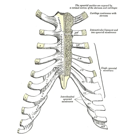
Costochondral joints are the cartilaginous connections between the ribs and the costal cartilage in the front of the ribcage. These joints provide flexibility and support for breathing movements. They are synchondroses, meaning they are immobile and composed of hyaline cartilage. Injuries or inflammation in these joints can lead to costochondritis, a common cause of chest pain.
They are primary cartilaginous joints, also known as synchondrosis, which are hyaline cartilaginous joints. Each rib features a cup-shaped depression with which the costal cartilage articulates. These joints typically don’t move at all. Plane synovial joints connect the sixth and ninth rib costal cartilages. Fibrous articulation separates the ninth and tenth ribs’ costal cartilages.
The periosteum holds the two costal cartilages together when their lateral ends are received into a depression in the sternal end of the rib.
Anatomy
The cartilaginous joints that join the ribs to the costal cartilages in the anterior thoracic wall are known as costochondral joints or synchondroses. They are essential for preserving the rib cage’s stability and flexibility during breathing.
Structure
The primary cartilaginous joints (synchondroses) that join the costal cartilages to the ribs are called costochondral joints.
Movement during breathing is made possible by these joints, which give the thoracic cage flexibility and structural integrity.
Articular surfaces
A rib’s link to its costal cartilage is known as the costochondral joint.
It happens when bone finishes and cartilage starts on the thoracic wall.
The rounded lateral end of the costal cartilage and the roughened cup-shaped anterior end of the ribs make up the joint’s two articular surfaces.
- Rib End: A thin layer of compact bone covers the spongy bone at the sternal end of the rib.
- Through the sternocostal joints, the hyaline cartilage structure known as the costal cartilage progressively moves from the rib to the sternum.
- Bone-Cartilage Interface: Without a synovial cavity, the periosteum of the ribs forms a continuous connection with the perichondrium of the costal cartilage.
Joint capsule and ligaments
These joints lack ligaments, a joint capsule, and a hollow. The rib’s periosteum connects the bone and cartilage by running continuously through the costal cartilage’s perichondrium.
The point where the ribs articulate with their corresponding costal cartilage is known as the costochondral joint. Given that it is a cartilaginous joint (synchondrosis), movement is limited to nonexistent.
The costochondral joint does not have actual ligaments to support its structure, in contrast to synovial joints. Rather, it is kept stable by:
- Periosteum and Perichondrium: The primary stabilizing elements. At the intersection, the periosteum (around bone) and perichondrium (around cartilage) combine to provide strength.
- Intercostal Muscles: These muscles support the rib cage and attach close to the internal and external costochondral joints.
- Connective tissue and surrounding fascia offer extra support but don’t work as separate ligaments.
The costochondral joint lacks the ligamentous components of the sternocostal or costovertebral joints, which are seen in synovial joints because it is a synchondrosis.
Innervation
The intercostal nerves are the anterior rami of the thoracic spinal nerves, and their branches supply the costochondral joints.
Blood supply
The anterior intercostal arteries’ branches provide the costochondral joints with arterial flow. These joints’ related anterior intercostal veins are responsible for their venous drainage.
Movements
The actual costochondral articulations are static joints that are incapable of moving. The front ends of the ribs can, however, be attached to the sternum with flexibility thanks to the costal cartilage, which can also bend and twist somewhat to let the thoracic diameters expand during breathing.
The costochondral joint, where the ribs join the costal cartilage, is a cartilaginous joint (synchondrosis). Because of its static and inflexible design, it does not permit much mobility.
Movement at the Costochondral Joint
- Minimal Flexibility: The hyaline cartilage that makes up the joint gives it a tiny quantity of flexibility, which permits mild expansion and bending.
- Passive Movements: The joint does not move actively as synovial joints do, however, it may gently distort in response to external stimuli, rib cage expansion, and breathing.
- Indirect Contribution to Breathing: During inhalation and exhalation, the rib cage expands and contracts, and the costochondral joints passively support these motions without producing their motion.
Muscles acting at the costochondral joint (junction)
The immobility of this joint means that no muscles can directly affect it. However, the anterior muscle fibers and the aponeurosis of the intercostal muscles have sites of attachment at the costochondral joints.
Clinical significances
The cartilaginous joints that connect the costal cartilage to the ribs are called costochondral joints. These joints are essential to the thoracic cage’s stability and flexibility. Their clinical importance consists of:
Costochondritis
- Costochondral joint inflammation causes chest pain that resembles heart problems.
- Frequently idiopathic, although infections, trauma, and excessive use can all cause it.
- ECG findings were normal and there were no cardiac risk factors, which distinguished it from myocardial infarction (heart attack).
- The costochondral joints swell locally in this uncommon inflammatory disease.
- The second or third costochondral junction is frequently impacted.
- Although self-limiting, it may result in chronic chest pain.
Rib Fractures & Trauma
- Costochondral joints are susceptible to blunt trauma, such as falls or auto accidents.
- Breathing difficulties and rib instability can result from fractures.
Arthritis and Degenerative Changes
- These joints may be impacted by diseases such as ankylosing spondylitis and rheumatoid arthritis, which can cause persistent pain and stiffness.
Slipping Rib Syndrome
- Causes sporadic pain and clicking sensations and is caused by hypermobility or dislocation of the costochondral joint.
- Occurs frequently in the eighth, ninth, and tenth ribs.
Surgical Considerations
- Maintaining costochondral integrity is essential during chest surgeries (such as rib resection and thoracic surgeries) to prevent long-term pain and respiratory issues.
- In reconstructive surgery, costochondral grafts are occasionally employed, particularly in mandibular reconstruction.
FAQs
What does the term “costochondral” mean?
Involving or connecting costal cartilage and ribs.
What is the number of costochondral junctions?
The third or fourth ribs are more commonly afflicted, however any of the seven costochondral connections might become inflamed. Injury, recurrent minor trauma, and extraordinary physical activity are the most likely causes.
What kind of joint is a costochondral joint?
The joints of the thoracic wall that unite the sternal ends of the ribs and their corresponding costal cartilages are called costochondral joints. They are categorized anatomically as synchondrosis, or primary cartilaginous joints, where hyaline cartilage connects the bones.
Which joint is the first rib’s costochondral?
The upper eight costochondral junctions form a tough joint with very little movement, similar to the costosternal joint. These joints are called hyaline cartilaginous joints, and they resemble the synchondrosis.
What role does the costochondral junction play?
The point where the costochondral cartilage and bone connect is known as the costochondral junction. The costochondral joint, a hyaline cartilaginous junction, connects the ribs and the sternum. Joints move more easily thanks to hyaline cartilage.
References
- Wikipedia contributors. (2024a, February 12). Costochondral joint. Wikipedia. https://en.wikipedia.org/wiki/Costochondral_joint
- Costochondral joint (junction). (2023, October 30). Kenhub. https://www.kenhub.com/en/library/anatomy/costochondral-joint-junction
One Comment