Calf Muscles
Introduction
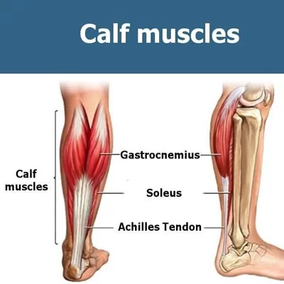
The calf muscles, located on the back of the lower leg, are a crucial part of the human body’s musculoskeletal system. These muscles, primarily consisting of the gastrocnemius and the soleus, play a vital role in various movements, including walking, running, and jumping.
The gastrocnemius is the most superficial muscle, with two heads: medial and lateral. The two heads of the gastrocnemius come together to create a confluent muscular belly.
The lateral head develops from the lateral surface of the lateral femoral condyle, while the medial head develops from the posterior, non-articular aspect of the medial femoral condyle. The calcaneal tendon, sometimes referred to as the Achilles tendon, is formed when the gastrocnemius muscle belly joins the soleus muscle distally and attaches to the posterior calcaneus.
The calf muscle, which plantarflexes the ankle joint, is innervated by the tibial nerve. Located deep within the gastrocnemius is the large, flat muscle known as the soleus. The tendon portion of the plantaris, a small muscle, is incredibly long. It is easy to confuse the tendinous portion of a nerve.
The plantaris muscle originates in the lateral supracondylar line of the femur and is completely absent in up to 10% of the population. The muscle descends medially and eventually forms a tendon that runs down the leg, between the gastrocnemius and soleus. The calcaneal tendon joins forces with this tendon.
Embryology
Upper limbs typically develop before lower limbs. Lower limb development begins in week 4 of gestation and is well differentiated by weeks 8 to 10. The limb buds begin to develop following the activation of mesenchymal cells in the lateral plate mesoderm.
Gastrocnemius muscle
The gastrocnemius is a large muscle in the posterior leg. The calf’s bulk is formed by the most superficial of the leg’s muscles, located posteriorly. The name derives from the Greek words γαστήρ (gaster), meaning stomach or belly, and κvήμη (knee), meaning leg. The combination of the two words refers to the “belly of the leg” or the bulk of the calf.
It is part of the triceps surae, a three-headed muscle group that includes the soleus muscle. They participate in a variety of basic activities, including walking, running, and leaping.
Origin
The gastrocnemius muscle has two heads that originate from the femur. The medial head is formed by the popliteal surface of the femoral shaft and the posterior surface of the medial condyle.
The lateral head develops from a facet on the upper posterolateral surface of the lateral condyle of the femur, where it joins the lateral supracondylar line. Both heads originate from the capsule of the knee joint.
Insertion
At the inferior margin of the popliteal fossa, the two heads meet and fuse to form a single, elongated muscle belly. This accounts for the majority of the soft tissue bulge on the posterior leg, also known as the calf.
The fleshy part of the muscle extends to about the middle of the calf. In the lower leg, the gastrocnemius muscle fibres form a broad aponeurosis over time. The aponeurosis gradually narrows and fuses with fibres from a deeper muscle, the soleus, to form the calcaneal (Achilles) tendon. The calcaneal tendon connects the posterior surface of the calcaneus in the foot.
Innervation
Tibial nerve (S1, S2)
Functions
The gastrocnemius is a strong plantar flexor in the foot at the talocrural joint. It also bends the leg at the knee. The gastrocnemius’ actions are typically considered part of the triceps surae group, which also includes the soleus. These are the primary plantar flexors of the foot. The muscles are typically large and powerful. The gastrocnemius muscle is responsible for propulsion when walking, running, or jumping.
Soleus muscle
The soleus muscle is a broad, flat leg muscle located on the posterior leg. It extends from just below the knee to the heel and is immediately adjacent to the gastrocnemius. These two muscles, along with the plantaris, are part of the superficial posterior compartment calf muscles. Soleus contraction causes strong plantar flexion. It also helps us maintain an upright posture because of its function as an antigravity muscle.
Origin
The soleus muscle originates from the soleal line on the dorsal surface of the tibia, medial border of the tibia, head of the fibula, and posterior border of the fibula. A portion of the fibres originate from the soleus’ tendinous arch, which spans the tibia and fibula and arches over the popliteal vessels and tibial nerve.
Insertion
The soleus muscle runs alongside the gastrocnemius muscle and connects to the posterior surface of the calcaneus via the calcaneal tendon. The calcaneal tendon, also known as the Achilles tendon, is the strongest in the human body. It is visible and palpable at the heel.
Innervation
Tibial nerve (S1, S2)
Function
The soleus muscle functions similarly to the gastrocnemius muscle. They work together to form a primary plantar flexor; their contraction causes plantar flexion of the upper ankle joint, allowing the heel to be lifted against gravity when walking or jumping. The soleus muscle is one of several antigravity muscles (along with the leg extensors, gluteus maximus, and back muscles) that help humans maintain an upright posture.
Blood supply of calf muscles
The blood supply to the calf muscles comes from the popliteal artery, which divides into the anterior and posterior tibial arteries. The fibular (or peroneal) artery is derived from the posterior tibial artery. The posterior tibial artery travels alongside the tibial nerve and enters the plantar aspect of the foot via the tarsal tunnel. The anterior tibial artery connects the tibia and fibula via a gap in the interosseous membrane. It runs the entire length of the leg and into the foot, becoming the dorsalis pedis artery.
Lymphatic drainage of calf muscles
The venous supply to the calf is divided into superficial and deep veins. The superficial veins include the greater saphenous vein and the small saphenous vein. The deep veins consist of the popliteal vein, anterior tibial vein, posterior tibial vein, and fibular vein. The greater saphenous vein is the body’s longest vein, extending the entire length of the lower extremity.
Cardiothoracic surgeons frequently use this vein for coronary artery bypass grafting. The small saphenous vein is a relatively large vein that runs along the posterior aspect of the calf, passing between the gastrocnemius muscle heads before draining into the popliteal vein.
The popliteal vein is formed when the anterior and posterior tibial veins converge. When the popliteal vein enters the femoral region, it becomes the femoral vein. The anterior tibial veins are responsible for the drainage of the knee, ankle, tibiofibular, and anterior leg joints. The lateral and medial plantar veins supply blood to the posterior tibial vein, which drains the lower leg’s posterior muscles and the plantar surface of the foot. The fibular veins, also called peroneal veins, transport blood from the leg’s lateral compartment to the posterior tibial vein.
Importance of Calf Muscle
The calf muscles are responsible for the initial propulsion of movements such as running, walking, and cycling. Most people train calves for aesthetic reasons. However, calf muscle serves a purpose other than to look good. The calf muscle is commonly referred to as the pseudo or periphery heart due to its critical function of returning blood from the leg and foot to the heart. Blood flow from the lower body to the heart must overcome gravity, so the contraction of the calf muscle generates external pressure that propels the blood through the veins to the heart.
The gastrocnemius muscle is used more during dynamic, high-force activities, whereas the soleus muscle is more active during postural and static contractions. The position of the knee during the plantar flexion resistance exercise influences the activity of the gastrocnemius muscle, which crosses the knee and ankle. At 90 degrees of knee flexion, the gastrocnemius experiences passive insufficiency and thus becomes less active. During the calf raise exercise, keep your knee 90 degrees flexed to target the soleus and zero degrees flexed to target the gastrocnemius.
The calves also serve as a deceleration mechanism for the body. When sprinting and stopping or changing directions quickly, the calves absorb up to 10 to 12 times the body weight. To avoid injury during the eccentric phase of any exercise, trained calves must bear this load and ensure that deceleration occurs safely.
The calves also help to stabilise the knees, which is important for jumping exercises because unstable knees can lead to poor form and injuries. The joints are protected by a strong set of calves.
Well-trained calves produce more vertical jumping power. The gastrocnemius, which is mostly made up of fast-twitch muscle fibres, allows the calves to perform quick and explosive movements like high jumps, squat jumps, and sprints. Although genetics determine how many fast-twitch muscle fibres each person has, strengthening the calves allows everyone to perform these powerful movements.
Clinical importance of calf muscles
- Muscle strain is the most common type of calf injury. It happens when your muscle fibres overstretch or tear. It is usually the result of strenuous exercise or overuse. Running and sports involving jumping or sudden stops and starts, like football, volleyball, basketball, and soccer, are common causes of this injury.
- Leg cramps and muscle spasms in the calves can be extremely painful. Leg cramps may happen at any time of day or night. They can be caused by a variety of factors, including pregnancy, dehydration, certain medications, and health conditions.
- Tennis leg: This type of muscle strain injury involves the gastrocnemius muscle. Tennis leg is a term used by providers to describe what happens when your leg extends while your foot flexes. Tennis players place their legs in this position when serving a tennis ball and “push off” into motion. But it can occur in any sport.
- Compartment syndrome is a serious, life-threatening condition that occurs when pressure builds up within a muscle. Pressure reduces the flow of blood and oxygen. The injury could be caused by trauma (such as a fracture) or strenuous exercise.
- Calf muscle tear: All shin muscle strains cause the tearing of some muscle filaments. More serious injuries can cause a partial or complete tear of the muscle.
- A calf muscle strain is commonly referred to as a pulled shin muscle. Pulling the muscle involves stretching the shin muscle.
- Calf muscle rupture: A complete gash to the shin muscle, resulting in severe pain and inability to walk. The shin muscle may collapse, resulting in a lump that can be seen and felt through the skin.
- Rhabdomyolysis typically affects several muscles throughout the body. Shin muscle breakdown can be caused by prolonged pressure, medication side effects, or a serious medical condition.
- Calf muscle myositis is inflammation of the shin muscle. Infections or autoimmune conditions (caused by the vulnerable system incorrectly attacking the body’s tissues) are usually to blame, though shin muscle myositis is unusual.
- Calf muscle cancer: Cancer of the shin muscle is rare. The excrescence can begin in the shin muscle (sarcoma) or spread to the shin muscle from elsewhere (metastasis).
Exercises for calf muscles
Following are the Best Calf Exercises:
Strengthening Exercises
Double-leg calf raise.
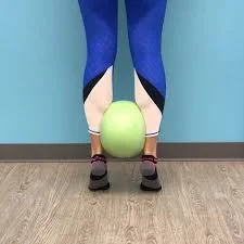
Calf raises are a classic calf-strengthening exercise. They use your body weight to strengthen and tone the gastrocnemius and soleus muscles.
Starting position: Stand near a wall for balance. To protect your joints, space your feet hip-width apart and keep your ankles, knees, and hips vertically aligned.
Action: To raise your body, press down on the balls of both feet. Keep your abdominal muscles pulled in so that you can move straight up rather than forward or backward.
Single-Leg Calf Raise.
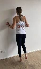
You can increase the intensity of the calf raise by performing it on one leg. This allows you to further strengthen your calf muscle.
Starting position: Stand with one leg near a wall for balance and the other bent behind you. To protect the joints, ensure that the ankle, knee, and hip of the leg being worked on are vertically aligned.
Press down into the ball of your foot to lift your body. Keep your abdominal muscles pulled in to avoid shifting forward or backward
Seated Calf Raise.
Start by sitting in a firm, sturdy chair with your feet flat on the floor. Keep your knees directly over your feet. Refrain from bending or extending your knees. Lean forward, place your hands on your thighs near your knees, and push down to add resistance.
Action: Slowly press down on the balls of your feet to raise your heels as high as possible. Finally, slowly lower your heels. Repeat.
Stretching exercises
Towel Stretch
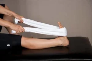
- While seated with your legs straight, wrap a towel around the ball of your foot.
- Keeping your knee straight, pull the towel’s ends towards you.
- Hold for 30 seconds. Repeat three times per side. Three times a day.
Lunge stretch.
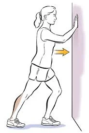
- Stand arm’s length from a wall or kitchen counter.
- Position one foot behind the other. Slowly bend your front knee while keeping your back leg straight and heel on the floor. Engage your core muscles.
- Hold for about 30 seconds and relax. Switch legs and repeat.
- Repeat this exercise three times on each side, three times a day.
Step Stretch
Hold something for support. Step both feet hip-width apart onto the bottom step of the stairs. Slide one foot back, leaving only the ball of the foot on the step. Keeping this knee straight, bend the opposite knee and lower the heel of the step until the calf tightens. Hold for 30 seconds, then relax. Repeat 3 times on each side, 3 times per day.
Summary
The calf muscles, including the gastrocnemius and soleus, are the largest in the posterior part of the lower leg. The gastrocnemius is the most superficial muscle, with two heads: medial and lateral. It is part of the triceps surae group and is responsible for propulsion during activities like walking, running, and jumping. The soleus muscle, located deep within the gastrocnemius, is a broad, flat leg muscle that forms a primary plantar flexor and helps maintain an upright posture.
The soleus muscle, along with the plantaris, is part of the superficial posterior compartment calf muscles and works together to form a primary plantar flexor. Blood supply to the calf muscles comes from the popliteal artery, which divides into the anterior and posterior tibial arteries. The venous supply to the calf is divided into superficial and deep veins, with superficial veins including the greater saphenous vein and the small saphenous vein.
The calf muscle is crucial for the initial propulsion of running, walking, and cycling. It is also known as the pseudo or periphery heart, as it returns blood from the leg and foot to the heart. The gastrocnemius muscle is used more during dynamic, high-force activities, while the soleus muscle is more active during postural and static contractions. The calves also serve as a deceleration mechanism for the body, absorbing up to 12 times the body weight during sprinting and stopping or changing directions. They help stabilize the knees, which are important for jumping exercises, and produce more vertical jumping power.
Clinical importance of calf muscles includes muscle strain, leg cramps, tennis leg, compartment syndrome, shin muscle tear, shin muscle rupture, rhabdomyolysis, shin muscle myositis, and shin muscle cancer. Strengthening exercises for calf muscles include double-leg calf raises, single-leg calf raises, seated calf raises, towel stretch, lunge stretch, and step stretch. Strengthening exercises can help prevent muscle strain, leg cramps, and other health issues.
FAQs
Which is the most important calf muscle?
The gastrocnemius muscle is a complex muscle essential for walking and maintaining posture. The gastrocnemius is the largest muscle in the back of the lower leg and is extremely powerful.
What’s the calf made of?
The calf is made up of muscles from the leg’s posterior compartment: the gastrocnemius and soleus (which make up the triceps surae muscle) and the tibialis posterior. The sural nerve supplies innervation.
What is the primary function of the calf muscle?
The calf muscles control plantarflexion of the foot and ankle. Activities involving the calf muscles include running and jumping.
How to work the calf muscles?
Calf raises.
Begin in a standing position, feet hip-width apart and core engaged. Squeeze your calf muscles and gradually raise your body, lifting your heels until you’re on your toes. Make sure to stand tall and straight. Then carefully lower your heels back to the floor.
What are the positive implications of the calf?
Strong calf muscles enable better-running mechanics, resulting in increased speed and endurance. Furthermore, maintaining strong calves can help to prevent calf pain and fatigue, which are common complaints among runners — so keeping your calves in good shape is essential to helping you run at your best.
References:
- Gastrocnemius muscle. (2023, October 30). Kenhub. https://www.kenhub.com/en/library/anatomy/gastrocnemius-muscle
- Binstead, J. T., Munjal, A., & Varacallo, M. (2023, May 23). Anatomy, Bony Pelvis and Lower Limb: Calf. StatPearls – NCBI Bookshelf. https://www.ncbi.nlm.nih.gov/books/NBK459362/
- Soleus muscle. (2023, September 19). Kenhub. https://www.kenhub.com/en/library/anatomy/soleus-muscle
- Physiotherapist, N. P. (2024, January 15). Calf Muscle: Origin, Insertion, Innervation & Exercise. Mobile Physiotherapy Clinic. https://mobilephysiotherapyclinic.in/calf-muscle-details-and-exercise/
- Professional, C. C. M. (n.d.). Calf Muscle. Cleveland Clinic. https://my.clevelandclinic.org/health/body/21662-calf-muscle
- Taylor, R. B. (2023, July 18). Calf-Strengthening Exercises. WebMD. https://www.webmd.com/fitness-exercise/strengthening-calf-muscles
- Calf Muscle Stretching – Move Better Gwent. (n.d.). Move Better Gwent. https://movebettergwent.nhs.wales/self_management/calf-muscle-stretching/

39 Comments