Calcaneofibular Ligament
Introduction
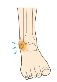
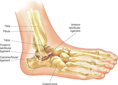
The calcaneofibular ligament (CFL) is a strong, fibrous band located on the lateral side of the ankle. It connects the fibula to the calcaneus (heel bone) and provides stability by preventing excessive inversion of the foot. It is commonly injured in ankle sprains, particularly in combination with the anterior talofibular ligament (ATFL).
Like the posterior talofibular ligament, ankle twisting or rolling can injure the calcaneofibular ligament, resulting in pain, swelling, and instability.
Depending on the extent of the injury, RICE, immobilization, physical therapy, or surgery may be used to treat a calcaneofibular ligament injury. Preventive steps such as wearing suitable shoes and using ankle supports during exercise can also reduce your risk of sustaining a calcaneofibular ligament injury.
Structure of the Calcaneofibular ligament
A robust band of connective tissue called the calcaneofibular ligament connects the lateral surface of the calcaneus bone in the foot to the lateral malleolus of the fibula bone. Together with the anterior and posterior talofibular ligaments, it is one of the three primary ligaments that support the ankle joint.
The calcaneofibular ligament has a slightly curved shape and is about 2-3 cm in length.
It is strong and stable because it is made up of dense collagen fibers that are oriented parallel to one another.
There are two sections to the ligament: the superficial portion and the deep portion. Stability during plantar flexion (pointing the toes downward) and inversion (rolling the ankle outward) is provided by the surface portion, which is situated on the outside of the ankle. Stability during dorsiflexion (raising the toes upward) and inversion are provided by the deep portion, which is situated deeper within the ankle joint.
Other structures in the ankle joint, such as the lateral talocalcaneal ligament, the lateral talocalcaneal interosseous ligament, and the lateral talocalcaneal articulation, are also connected to the calcaneofibular ligament.
Attachment of the Calcaneofibular ligament
The calcaneofibular ligament is connected to the lateral surface of the calcaneus bone and the lateral malleolus of the fibula bone in the ankle joint.
Part of the fibula bone, the lateral malleolus is a bony protrusion on the outside of the ankle. The calcaneofibular ligament is attached at the base of the lateral malleolus, where the ankle joint and bone meet.
The largest bone in the foot, the calcaneus, forms the heel. The calcaneus bone’s lateral surface—the side of the bone that faces away from the body’s midline—is where the calcaneofibular ligament is attached. The attachment site is located close to the back of the heel, close to the point where the Achilles tendon connects to the bone.
The calcaneofibular ligament is connected to additional ankle joint structures in addition to these two bones. Among these are the lateral talocalcaneal interosseous ligament, which joins the talus to the fibula bone, and the lateral talocalcaneal ligament, which joins the talus to the calcaneus bone. Additionally, the ligament is attached to the joint between the calcaneus and talus bones, known as the lateral talocalcaneal articulation.
The ankle joint is supported and stabilized during movement by the calcaneofibular ligament’s attachment to these different structures. Ankle joint pain, edema, and instability may arise from injury or trauma that disrupts one of these attachments.
Functions of the Calcaneofibular ligament
Stabilizing the ankle joint and limiting excessive foot rotation or movement are important functions of the calcaneofibular ligament. This ligament serves some vital purposes, including:
Limiting inversion: The calcaneofibular ligament helps in preventing excessive inversion or rolling of the foot inward. Ankle sprains frequently result in this mechanism of injury, and the ligament helps to restrict the degree of inversion that can happen.
Supporting lateral stability: Because the lateral side of the ankle joint is less stable than the medial side, it is especially prone to injury. By giving the ankle joint lateral stability, the calcaneofibular ligament helps to limit excessive rotation or movement.
Supporting dorsiflexion: Dorsiflexion is the upward motion of the foot toward the shin. This movement is supported by the calcaneofibular ligament, which gives the ankle joint stability and tension.
Preserving joint alignment: Additionally, the ligament helps maintain the proper alignment of the calcaneus and fibula bones, which is essential for the joint’s overall health and functionality.
In general, ankle joint stability and function are significantly affected by the calcaneofibular ligament. An injury to this ligament may cause the ankle joint to become less mobile, painful, and unstable.
Blood supply of the Calcaneofibular ligament
The fibula, one of the two bones in the lower leg, and the calcaneus, or heel bone, are joined by the calcaneofibular ligament, a robust, fibrous band. It serves to limit excessive lateral (outward) foot movement and is a crucial ankle joint stabilizer.
The calcaneofibular ligament receives blood from a number of sources. The peroneal artery, a branch of the posterior tibial artery, is the most significant of these. The muscles and other tissues in the lower leg and foot receive blood flow from this artery, which runs down the back of the leg.
The calcaneofibular ligament is supplied by smaller blood vessels in addition to the peroneal artery. These consist of the anterior tibial artery and branches of the lateral malleolar artery. The ligament receives extra blood flow from these arteries, which run along the front and outside of the ankle joint, respectively.
Maintaining the calcaneofibular ligament’s health and function depends on its blood supply. Ankle joint instability and pain may result from weakened or injured ligaments caused by poor blood flow. Injuries to the ligament, such as sprains or tears, may also restrict its function by blocking its blood flow.
Injuries of the Calcaneofibular ligament
The most frequent cause of injury to the calcaneofibular ligament is an ankle sprain, though there are other possible causes as well. The ligaments on the outside of the ankle stretch or tear when the foot twists or rolls inward, resulting in an ankle sprain. Activities that can cause this injury include:
Sports: Sports like basketball, soccer, or volleyball that require quick direction changes, jumping, or landing can strain the ankle joint and raise the risk of an ankle sprain.
Running: Ankle sprains and calcaneofibular ligament injuries can also be increased by running on uneven ground or in unsuitable footwear.
Accidents: Ankle sprains and ligament tears can also result from tripping, falling, or twisting the ankle in an accident.
High heels: Wearing high heels increases the risk of ankle sprains and ligament injury because they put a lot of pressure on the ankle joint.
Symptoms
Depending on the degree of injury, calcaneofibular ligament injury symptoms can vary in intensity. Typical symptoms include the following:
Pain: Pain is the most common sign of a calcaneofibular ligament injury. With movement or pressure, the pain could become dull or sharp.
Swelling: Ankle joint swelling is another typical sign of a ligament injury. The affected foot may be difficult to move due to mild or severe swelling.
Bruising: When a ligament injury occurs, the ankle joint may also sustain some bruises. The bruises can appear over a few days and can be mild or severe.
Walking difficulties: Walking or bearing weight on the injured foot may be difficult because of a ligament injury. This could be the result of ankle joint pain, swelling, or instability.
Instability: An ankle joint that feels loose or wobbly may result from a calcaneofibular ligament injury. Walking and other activities requiring balance may become very difficult as a result.
Following an ankle injury, you must seek medical attention if you experience any of these symptoms. Your physician can assess the degree of injury and suggest the best direction of action to help in your recovery.
Diagnosis
In the ankle joint, a band of tissue called the calcaneofibular ligament joins the fibula to the calcaneus, the heel bone. The ankle joint is stabilized by this ligament, which also keeps the foot from rolling inward too much.
A medical professional’s physical examination is usually the first step in diagnosing a calcaneofibular ligament injury. The doctor will check for pain, swelling, bruises, and instability in the ankle during the examination. Additionally, they might conduct particular tests to assess the ankle joint’s strength and range of motion.
To confirm the diagnosis and determine the degree of ligament injury, imaging tests like X-rays, MRIs, or CT scans may occasionally be prescribed. These examinations can assist in identifying any related ankle joint fractures or other injuries.
The severity of the injury will determine the available treatment options after a calcaneofibular ligament injury has been diagnosed. Rest, ice, compression, and elevation (RICE) therapy and over-the-counter painkillers can be used to treat mild injuries. Physical therapy, immobilization with a brace or cast, and occasionally surgery are necessary for more serious injuries.
In general, prompt diagnosis and treatment of a calcaneofibular ligament injury can facilitate quicker recovery and help stop additional ankle joint injuries. It is crucial to get medical help right away if you think you may have hurt your ankle or if you are exhibiting any signs of a ligament injury.
Treatment of the Calcaneofibular ligament
The medial talar tilt test
A physical examination method called the medial talar tilt test, sometimes referred to as the inversion stress test, is used to evaluate the stability of the ankle joint, particularly the calcaneofibular ligament. Ankle sprains frequently cause injury to this ligament, which joins the fibula and calcaneus bones on the outside of the ankle.
The patient lies on their back with their foot hanging off the edge of the examination table to perform the calcaneofibular ligament medial talar tilt test. To apply lateral strain to the foot, the examiner grasps the lower leg with one hand while holding onto the heel with the other. The ankle joint’s range of motion and stability are then evaluated by tilting the foot outward (eversion) and inward (inversion).
A goniometer, a device used to measure joint angles, is used to measure the tilt in degrees. With a maximum inversion angle of 5 degrees and a maximum eversion angle of 5–10 degrees, a typical ankle joint should move very little during the medial talar tilt test.
The medial talar tilt test may reveal a calcaneofibular ligament injury or another ankle joint injury if the ankle joint moves or becomes unstable excessively. To confirm the diagnosis and choose the best option for treatment, additional diagnostic procedures including imaging scans or physical exams could be required.
The medial talar tilt test for the calcaneofibular ligament is a simple, non-invasive technique that can help direct treatment decisions and provide essential information on ankle joint stability for patients with ankle injuries or chronic ankle instability.
Conservative treatment
Rest, ice, compression, and elevation (RICE) is a common conservative treatment for calcaneofibular ligament (CFL) injuries to minimize pain and swelling. For calcaneofibular ligament injuries, the following are some particular conservative therapy options:
Immobilization: To enable the calcaneofibular ligament to heal appropriately, the ankle joint may occasionally need to be immobilized using a brace or cast. This can encourage healing and stop more ligament injuries.
Physical treatment: To help regain ankle joint strength, flexibility, and range of motion when discomfort and swelling have decreased, physical therapy may be suggested. This could involve range-of-motion and stretching exercises, as well as exercises to strengthen the muscles surrounding the ankle.
NSAIDs (nonsteroidal anti-inflammatory drugs): Pain and inflammation related to calcaneofibular ligament injuries can be lessened with over-the-counter NSAIDs such as naproxen or ibuprofen.
Ice and rest: Applying ice to the injured ankle and allowing it to rest will help reduce swelling and pain. For the first several days following the accident, ice should be applied for 15 to 20 minutes at a time, multiple times each day.
Compression: Using an elastic bandage or brace to apply pressure can help support the ankle joint and reduce swelling.
Elevation: Raising the injured ankle above the level of the heart will assist improve blood flow to the area and lessen edema.
The fact that not all calcaneofibular ligament injuries may be treated conservatively is very important to remember, particularly if the injury is severe or involves other ankle joint components. Surgery may be required in some situations to rebuild or repair the injured ligament. It is crucial to consult a healthcare professional to determine the optimal treatment plan for each specific disease.
Physiotherapy treatment
Exercises and manual therapy methods are commonly used in physiotherapy treatment for calcaneofibular ligament (CFL) injuries to accelerate recovery, lessen discomfort and swelling, and recover ankle joint function. Specific physiotherapy interventions for calcaneofibular ligament problems include the following:
Range of motion activities: These exercises aim to increase the ankle joint’s flexibility. This could involve activities like ankle pumps or circles.
Strength training: The purpose of strength training is to increase the strength of the muscles surrounding the ankle joint. This could involve activities like ankle dorsiflexion or calf lifts.
Proprioception and balance training: This type of training aims to increase the ankle joint’s capacity to sustain stability and balance. This could involve activities like wobbleboard workouts or single-leg balancing.
Manual treatment: Manual therapy techniques like massage, joint mobilization, or soft tissue mobilization may be used to reduce pain and improve ankle joint range of motion.
Electrical stimulation: This technique can be used to lessen discomfort and encourage the healing of the injured area.
Bracing or taping: These techniques can be utilized to stabilize and support the ankle joint while engaging in activities that may worsen the condition.
The overall goals of physiotherapy for calcaneofibular ligament injuries are to encourage healing, lessen discomfort and swelling, and give the ankle joint its full range of motion. Working with a trained healthcare provider to create a customized treatment plan that meets your unique requirements and objectives is crucial.
Risk factors of the calcaneofibular ligament
In the ankle joint, a band of tissue called the calcaneofibular ligament (CFL) joins the fibula to the heel bone (calcaneus). There are several risk factors for calcaneofibular ligament injuries, such as:
Ankle sprains: The most frequent reason for injury to the calcaneofibular ligament is ankle sprains. They occur when the ankle bends or twists above its natural range of motion, injuring the ligaments.
Sports: Activities that require running, jumping, or abrupt direction changes increase the risk of calcaneofibular ligament injury. Soccer, tennis, and basketball are a few examples.
Past ankle injuries: Individuals who have experienced ankle injuries in the past are more at risk of calcaneofibular ligament injuries. Previous injuries can weaken the ligaments and make them more at injury risk.
Poor footwear: Ankle injuries, especially calcaneofibular ligament injuries, can be more likely to occur while wearing shoes that don’t provide enough stability or support.
Anatomical characteristics: By affecting how weight is transmitted across the foot and ankle, some anatomical characteristics, such as a high arch or flat feet, can raise the risk of calcaneofibular ligament injuries.
Age: People’s ligaments lose some of their flexibility as they get older, making them more at injury risk. In elderly adults, this may raise the risk of calcaneofibular ligament injury.
Obesity: Carrying too much weight affects the ankle joint, raising the possibility of ligament injury.
The risks of suffering a CFL injury can be raised by a number of risk factors. It’s critical to recognize these risk factors and take precautions to lower your risk, such as wearing suitable footwear and engaging in exercises that build stronger ankle muscles.
How to avoid calcaneofibular ligament injuries
By lowering the risk factors that can result in these injuries, calcaneofibular ligament (CFL) injuries can be prevented. To avoid calcaneofibular ligament injuries, follow these tips:
Put on suitable shoes: Wearing shoes with suitable stability and support may help you prevent calcaneofibular ligament issues by protecting your ankles. Try to find shoes with a non-slip sole, a firm heel counter, and good arch support.
To help support the ankle joint and lower the risk of injury, strengthen the muscles surrounding it. Exercises that work the muscles in the calf, ankle, and foot can be beneficial. Examples include toe curls, ankle circles, and calf lifts.
A healthy weight is important because too much weight strains the ankle joint and raises the possibility of ligament injury. Another way to lower this risk is to maintain a healthy weight.
Warm up before exercise: It’s crucial to warm up the joints and muscles before an exercise session. This could potentially prevent injuries.
Use suitable technique: It’s crucial to use the correct technique when playing sports or engaging in other physical activities to prevent twisting or turning the ankle beyond its natural range of motion.
After an ankle injury, it’s crucial to take some time off and let it heal completely before engaging in any physical activity again. The risk of re-injury can increase if you return too quickly.
The risk of injury can be decreased by using ankle braces or supports, which can give the ankle joint more stability and support.
All things considered, preventing calcaneofibular ligament injuries involves lowering the risk factors that may contribute to these injuries. CFL injuries can be avoided by wearing the right shoes, strengthening the muscles surrounding the ankle joint, keeping a healthy weight, warming up before exercise, using the right technique, resting and recovering from injuries, and thinking about ankle braces or supports.
FAQs
What leads to injuries to the calcaneofibular ligament?
A common cause of calcaneofibular ligament injuries is rolling or twisting the ankle, which can cause the ligament to tear or stretch.
What signs of a calcaneofibular ligament injury are present?
Pain, swelling, bruising, and trouble walking or bearing weight on the injured ankle are all signs of a calcaneofibular ligament injury.
How much time does it take to heal from an injury to the calcaneofibular ligament?
The severity of a calcaneofibular ligament injury can affect how long it takes to recover. While more serious injuries might take several months to completely heal, milder injuries might heal in a few weeks.
Is it possible to avoid calcaneofibular ligament injuries?
Yes, by lowering the risk factors that can result in these injuries—such as wearing suitable footwear, strengthening the muscles surrounding the ankle joint, and using proper technique when exercising—calcaneofibular ligament injuries can be avoided.
What features does the calcaneofibular ligament have?
Through its connection between the fibula and heel bones, the calcaneofibular ligament serves to stabilize the ankle joint.
How is an injury to the calcaneofibular ligament diagnosed?
Imaging tests like MRIs and X-rays, along with a physical examination and medical history, are usually used to diagnose calcaneofibular ligament injuries.
Is it possible for a calcaneofibular ligament injury to cause chronic ankle instability?
Injuries to the calcaneofibular ligament that are not properly treated and recovered may actually lead to chronic ankle instability and an increased risk of re-injury.
How does one define the calcaneofibular ligament?
From the tip of the fibula’s lateral malleolus downward and slightly backward to a tubercle on the calcaneus’ lateral surface, the calcaneofibular ligament is a thin, rounded cord.
References
- Patel, D. (2023h, August 22). Calcaneofibular ligament – Anatomy, structure, function. Samarpan Physiotherapy Clinic. https://samarpanphysioclinic.com/calcaneofibular-ligament/