Anatomical Snuff Box
Introduction
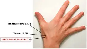
The anatomical snuffbox, or the radial fossa, is a triangular indent located on the outer side of the back of the hand. It can be found at the level of the wrist bones and is most noticeable when the thumb is extended.
Historically, this indent was used to hold snuff (finely ground tobacco) before inhaling through the nose, which is how it got the name ‘snuffbox.’
History and Etymology
When snuff was in vogue, users would take a small quantity of snuff from their container and place it into the “anatomical” snuffbox (in contrast to a physical snuffbox carried in one’s pocket, hence the term “anatomical”), then block one nostril with their index finger and inhale the snuff through the open nostril. It was a common practice to share snuff with those around you.
Detailed Anatomy
The anatomy of the anatomical snuffbox consists of its borders, base, roof, and contents.
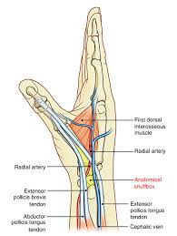
Borders: The lateral (radial) side is defined by the tendons of the abductor pollicis longus (APL) and extensor pollicis brevis muscles (EPB), while the medial (ulnar) side is defined by the tendon of the extensor pollicis longus muscle (EPL).
Base: It is made up of the distal edge of the extensor muscles retinaculum and where the tendons of the extensor pollicis longus and extensor pollicis brevis muscles attach. The base of the anatomical snuffbox contains several bony landmarks. From proximal to distal, they include the radial styloid, scaphoid, trapezium, and the base of the thumb metacarpal. These landmarks can be felt when the hand is in ulnar deviation with the thumb extended.
Roof: The roof is composed of skin and superficial fascia.
Contents: The radial artery crosses through the floor of the anatomical snuffbox at an angle, passing deep between the heads of the adductor pollicis muscles. Additionally, the cephalic vein and the superficial branch of the radial nerve can be found on the roof of the anatomical snuffbox.
Gross Anatomy
Boundaries
- Medial: Tendons of the extensor pollicis longus
- Lateral: Tendons of the extensor pollicis brevis and, more laterally, the abductor pollicis longus.
Floor
- Scaphoid and trapezium bones, along with the tendons of the extensor carpi radialis longus and brevis
- One may feel the radial styloid process proximally and the base of the first metacarpal distally.
Contents
- The radial artery, superficial branch of the radial nerve, and cephalic vein (which may vary).
Structures
Boundaries
The medial boundary (ulnar side) of the snuffbox is the tendon of the extensor pollicis longus. The lateral boundary (radial side) is formed by two closely aligned tendons, the extensor pollicis brevis and the abductor pollicis longus. Consequently, the anatomical snuffbox is most evident, exhibiting a more pronounced concavity, during thumb extension. The proximal border is delineated by the radius styloid process, and the distal border is approximated by the vertex of the isosceles triangle that represents the snuffbox.
The floor of the snuffbox varies based on wrist movement, but both the trapezium and primarily the scaphoid can be palpated.
Contents
The primary components of the anatomical snuffbox include the radial artery, a branch of the radial nerve, and the cephalic vein:
- The radial artery runs across the floor of the anatomical snuffbox, before turning inward to pass between the heads of the adductor pollicis muscle.
- In some cases, the radial pulse may be detected by placing two fingers on the upper portion of the anatomical snuffbox.
- The superficial branch of the radial nerve is located within the skin and subcutaneous tissue of the anatomical snuffbox, providing sensation to the dorsal surfaces of the lateral three-and-a-half digits, along with the corresponding area on the back of the hand.
- The cephalic vein originates from the dorsal venous network of the hand and traverses the anatomical snuffbox to ascend along the anterolateral side of the forearm.
To better understand the anatomical snuffbox’s contents, it helps to separate the structures into two categories: those located above the extensor retinaculum the tendons of the outcropping muscles, and those situated beneath these elements.
The extensor retinaculum is a thin fibrous connective tissue band that spans the posterior part of the distal forearm, functioning to prevent the tendons of the extensor and outcropping muscles, located beneath it, from bowing out during muscle contraction, which could place tension on the tendons.
Structures superficial to the extensor retinaculum and outcropping muscle tendons include the dorsal digital branches of the superficial radial nerve. The radial nerve descends from the posterior compartment of the arm and moves into the cubital region by crossing in front of the lateral epicondyle of the humerus. After that, it divides into superficial and deep branches. The superficial cutaneous branch proceeds down the forearm under the brachioradialis muscle. As it approaches the distal part of the forearm, it enters the roof of the anatomical snuffbox and divides into several dorsal digital branches. One of these branches extends into the anatomical snuffbox.
Structures located beneath the extensor retinaculum and the outcropping muscle tendons include:
Radial artery: The radial artery originates as a branch of the brachial artery as it traverses the cubital fossa. It descends along the outer side of the forearm, positioned deep to the brachioradialis muscle, and at the wrist, it lies lateral to the flexor carpi radialis tendon. Subsequently, it exits the forearm by moving posterolaterally to navigate obliquely along the base of the anatomical snuffbox.
Cephalic vein: The cephalic vein emerges medial to the digital nerve. It develops from the dorsal venous network that drains the dorsal side of the hand. The vein ascends from the outer side of the forearm and the arm, running superficially within the subcutaneous tissue.
Tendons of extensor carpi radialis longus and brevis: As the outcropping muscles emerge from beneath the extensor digitorum, they are located superficially to the tendons of the extensor carpi radialis longus and brevis. These tendons run medially to the radial artery along the floor of the anatomical snuffbox and attach to the base of the second and third metacarpals.
Neurovascular anatomy
Beneath the tendons forming the edges of the anatomical snuffbox lies the radial artery, which passes through the anatomical snuffbox on its path from the typical area for detecting the radial pulse to the proximal space between the first and second metacarpals, contributing to the superficial and deep palmar arches.
Within the anatomical snuffbox, the radial artery is nearby (within 2 mm) to the superficial branch of the radial nerve near the styloid process of the radius in 48% of cases, whereas in 24%, it is in close relation to the lateral cutaneous nerve of the forearm. The cephalic vein originates within the anatomical snuffbox, while the dorsal cutaneous branch of the radial nerve can be felt by gently stroking along the extensor pollicis longus with the back side of a fingernail.
Clinical significance
The radius and scaphoid articulate beneath the snuffbox, forming the foundation of the wrist joint. In a situation where a person falls onto an outstretched hand (FOOSH), this area absorbs the majority of the impact. Consequently, these two bones are the most commonly fractured in the wrist. If there is localized tenderness in the snuffbox, an understanding of wrist anatomy allows for a quick assumption that a fracture is probably present in the scaphoid.
This is logical since the scaphoid is a small, irregularly shaped bone that facilitates movement but does not provide stability to the wrist joint. Under excessive force applied to the wrist, this small scaphoid is likely to be the weakest point. Scaphoid fractures frequently lead to medico-legal issues.
An anatomical variation in the blood supply to the scaphoid indicates that blood first reaches this area distally. Thus, in the event of a fracture, the proximal portion of the scaphoid will lack a blood supply and, if not addressed, will suffer avascular necrosis within the snuffbox. Due to the scaphoid’s small size and shape, it can be challenging to ascertain, early on, whether it is fractured using an X-ray. Additional complications include carpal instability (ligament rupture) and fracture dislocations.
Clinical relevance
Scaphoid fractures: The anatomical snuffbox is crucial for evaluating scaphoid fractures. Within the anatomical snuffbox, the scaphoid and radius join to be part of the wrist joint. Following a blow to the wrist (such as falling on an outstretched hand), the scaphoid absorbs most of the force. If localized pain is detected in the anatomical snuffbox, a scaphoid fracture is likely the cause. Notably, tenderness in the snuffbox is highly sensitive to scaphoid fractures but lacks specificity. Given this lower specificity, patients with tenderness in this area must undergo radiographic examinations of the wrist. Failing to detect a fracture could impact quality of life due to the risk of non-union. Additionally, the location of this fracture plays a crucial role in determining treatment options to avoid avascular necrosis of the bone due to its unique blood supply.
Radial artery aneurysms are uncommon and are typically linked to trauma, including penetrating or iatrogenic causes. They occur less frequently than ulnar artery aneurysms, though the reasons for this difference are unclear. Cases following blunt trauma have been documented.
Venous Cannulation: The dorsal lateral (radial) section of the wrist is most frequently used for venous cannulation because the cephalic vein is more pronounced in that area.
De Quervain Tenosynovitis: The diagnosis of De Quervain’s Tenosynovitis is associated with the anatomical snuffbox. This condition affects the first dorsal compartment, which includes the muscles that form the lateral compartment of the anatomical snuffbox (APL and EPB). The diagnosis of De Quervain tenosynovitis is established through a comprehensive history and physical examination.
Mnemonic
If you’re looking for an easy way to recall the elements of the anatomical snuffbox, consider using the mnemonic provided below!
- CARTs
- Cephalic vein
- Artery (radial)
- Radial nerve (Superficial branch)
- Longus and brevis tendon attached to the extensor carpi radialis
Summary
The boundaries of the anatomical snuffbox and the structures that cross them are summarised below.
Borders
Proximal = radial styloid process
Distal = base of 1st metacarpal
Floor = scaphoid and trapezium
Medial = extensor pollicis longus
Extensor pollicis brevis & abductor pollicis longus are lateral.
Contents
Dorsal digital branches of the radial nerve and the cephalic vein are considered superficial.
Extensor carpi radialis longus and brevis tendons, deep = radial artery
FAQs
What three muscles form the anatomical snuffbox?
The abductor pollicis longus, extensor pollicis brevis, and extensor pollicis longus are the three thumb muscles that define the medial and lateral margins of the snuffbox.
What purpose does the snuffbox serve?
Anatomical Snuff Box
Injuries to the Scaphoid: The anatomical snuffbox is clinically important for evaluating scaphoid fractures. Within the anatomical snuffbox, the scaphoid and radius join to constitute a portion of the wrist joint. When a wrist experiences trauma (such as falling on an outstretched hand), the scaphoid absorbs the majority of the impact.
Why is it called the anatomical snuffbox?
The term “anatomical snuffbox” originates from the tradition of placing powdered tobacco or snuff in the depression and inhaling it through the nose.
What does pain over the anatomical snuffbox indicate?
Tenderness in the anatomical snuffbox is a highly sensitive indicator for scaphoid fractures, while pain during scaphoid compression and tenderness at the scaphoid tubercle are often more specific.
References
- Skalski, M., & Knipe, H. (2013). Anatomical snuff box. Radiopaedia.org.
- https://doi.org/10.53347/rid-22107
- Anatomical snuffbox. (2023, November 22). Kenhub.
- https://www.kenhub.com/en/library/anatomy/anatomical-snuffbox
- (n.d.)https://www.physio-pedia.com/Anatomical_Snuff_Box
- Wikipedia contributors. (2025, January 10). Anatomical snuffbox. Wikipedia.
- https://en.wikipedia.org/wiki/Anatomical_snuffbox
- TeachMeAnatomy. (2023, October 2). The Anatomical Snuffbox: Contents, Boundaries
- TeachMeAnatomy. https://teachmeanatomy.info/upper-limb/areas/anatomical-snuffbox/